NOD2 Antibody (2D9) - BSA Free
Novus Biologicals | Catalog # NB100-524
Key Product Details
Species Reactivity
Validated:
Cited:
Predicted:
Applications
Validated:
Cited:
Label
Antibody Source
Format
Product Specifications
Immunogen
Localization
Clonality
Host
Isotype
Theoretical MW
Disclaimer note: The observed molecular weight of the protein may vary from the listed predicted molecular weight due to post translational modifications, post translation cleavages, relative charges, and other experimental factors.
Scientific Data Images for NOD2 Antibody (2D9) - BSA Free
![Western Blot: NOD2 Antibody (2D9)BSA Free [NB100-524]](https://resources.rndsystems.com/images/products/NOD2-Antibody-2D9-Western-Blot-NB100-524-img0018.jpg "Western Blot: NOD2 Antibody (2D9)BSA Free [NB100-524]")
Western Blot: NOD2 Antibody (2D9)BSA Free [NB100-524]
NOD2-Antibody-2D9-Western-Blot-NB100-524-img0018.jpg![Immunohistochemistry-Frozen: NOD2 Antibody (2D9) - BSA Free [NB100-524]](https://resources.rndsystems.com/images/products/NOD2-Antibody-2D9-Immunohistochemistry-Frozen-NB100-524-img0016.jpg "Immunohistochemistry-Frozen: NOD2 Antibody (2D9) - BSA Free [NB100-524]")
Immunohistochemistry-Frozen: NOD2 Antibody (2D9) - BSA Free [NB100-524]
Immunohistochemistry-Frozen: NOD2 Antibody (2D9) [NB100-524] - Overlay of NOD2-DyLight 488 (green) with phase contrast of murine colon. Image from verified customer review.![Flow (Intracellular): NOD2 Antibody (2D9) - BSA Free [NB100-524]](https://resources.rndsystems.com/images/products/NOD2-Antibody-2D9-Flow-Intracellular-NB100-524-img0010.jpg "Flow (Intracellular): NOD2 Antibody (2D9) - BSA Free [NB100-524]")
Flow (Intracellular): NOD2 Antibody (2D9) - BSA Free [NB100-524]
Flow (Intracellular): NOD2 Antibody (2D9) [NB100-524] - An intracellular stain was performed on THP-1 cells with NOD2 (2D9) antibody NB100-524AF488 (blue) and a matched isotype control NBP2-27287AF488 (orange). Cells were fixed with 4% PFA and then permeablized with 0.1% saponin. Cells were incubated in an antibody dilution of 5 ug/mL for 30 minutes.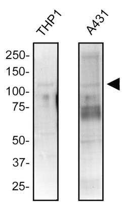
![Immunocytochemistry/ Immunofluorescence: NOD2 Antibody (2D9) - BSA Free [NB100-524]](https://resources.rndsystems.com/images/products/NOD2-Antibody-2D9-Immunocytochemistry-Immunofluorescence-NB100-524-img0020.jpg "Immunocytochemistry/ Immunofluorescence: NOD2 Antibody (2D9) - BSA Free [NB100-524]")
Immunocytochemistry/ Immunofluorescence: NOD2 Antibody (2D9) - BSA Free [NB100-524]
Immunocytochemistry/Immunofluorescence: NOD2 Antibody (2D9) [NB100-524] - NOD2 was detected in human monocytes using NOD2 antibody (2D9) [DyLight 488 (NB100-524G)]- with a concentration of 1:500 in PBS for 2 hours. Image from verified customer review.![Immunohistochemistry: NOD2 Antibody (2D9) - BSA Free [NB100-524]](https://resources.rndsystems.com/images/products/NOD2-Antibody-2D9-Immunohistochemistry-NB100-524-img0007.jpg "Immunohistochemistry: NOD2 Antibody (2D9) - BSA Free [NB100-524]")
Immunohistochemistry: NOD2 Antibody (2D9) - BSA Free [NB100-524]
Immunohistochemistry: NOD2 Antibody (2D9) [NB100-524] - Analysis of PFA fixed mouse sciatic nerve section using anti-NOD2 antibody. Image from verified customer review.![Flow Cytometry: NOD2 Antibody (2D9) - BSA Free [NB100-524]](https://resources.rndsystems.com/images/products/NOD2-Antibody-2D9-Flow-Cytometry-NB100-524-img0019.jpg "Flow Cytometry: NOD2 Antibody (2D9) - BSA Free [NB100-524]")
Flow Cytometry: NOD2 Antibody (2D9) - BSA Free [NB100-524]
Flow Cytometry: NOD2 Antibody (2D9) [NB100-524] - An intracellular stain was performed on THP-1 cells with NOD2 Antibody (2D9) NB100-524 (blue) and a matched mouse IgG1 isotype control (orange) MAB002. Cells were fixed with 4% PFA and then permeabilized with 0.1% saponin. Cells were incubated in an antibody dilution of 2.5 ug/mL for 30 minutes at room temperature, followed by Mouse IgG (H+L) Cross-Adsorbed Secondary Antibody, Dylight 550 (35503, Thermo Fisher).![Western Blot: NOD2 Antibody (2D9)BSA Free [NB100-524]](https://resources.rndsystems.com/images/products/NOD2-Antibody-2D9-Western-Blot-NB100-524-img0017.jpg "Western Blot: NOD2 Antibody (2D9)BSA Free [NB100-524]")
Western Blot: NOD2 Antibody (2D9)BSA Free [NB100-524]
Western Blot: NOD2 Antibody (2D9) [NB100-524] - Whole cell protein from THP-1 cells was separated on a 7.5% gel by SDS-PAGE, transferred to PVDF membrane and blocked in 5% non-fat milk in TBST. The membrane was probed with 2 ug/ml anti-NOD2 in 1% milk, and detected with an anti-mouse HRP secondary antibody using chemiluminescence.![Flow Cytometry: NOD2 Antibody (2D9) - BSA Free [NB100-524]](https://resources.rndsystems.com/images/products/NOD2-Antibody-2D9-Flow-Cytometry-NB100-524-img0014.jpg "Flow Cytometry: NOD2 Antibody (2D9) - BSA Free [NB100-524]")
Flow Cytometry: NOD2 Antibody (2D9) - BSA Free [NB100-524]
Flow Cytometry: NOD2 Antibody (2D9) [NB100-524] - An intracellular stain was performed on Jurkat cells with NOD2 (2D9) antibody NB100-524APC (blue) and a matched isotype control (orange). Cells were fixed with 4% PFA and then permeablized with 0.1% saponin. Cells were incubated in an antibody dilution of 2.5 ug/mL for 30 minutes.![Flow Cytometry: NOD2 Antibody (2D9) - BSA Free [NB100-524]](https://resources.rndsystems.com/images/products/NOD2-Antibody-2D9-Flow-Cytometry-NB100-524-img0015.jpg "Flow Cytometry: NOD2 Antibody (2D9) - BSA Free [NB100-524]")
Flow Cytometry: NOD2 Antibody (2D9) - BSA Free [NB100-524]
Flow Cytometry: NOD2 Antibody (2D9) [NB100-524] - An intracellular stain was performed on Jurkat cells with NOD2 (2D9) antibody NB100-524PE (blue) and a matched isotype control (orange). Cells were fixed with 4% PFA and then permeablized with 0.1% saponin. Cells were incubated in an antibody dilution of 2.5 ug/mL for 30 minutes.Both antibodies were conjugated to phycoerythrin. - BSA Free [NB100-524] -")
Western Blot: NOD2 Antibody (2D9) - BSA Free [NB100-524] -
Western Blot: NOD2 Antibody (2D9) - BSA Free [NB100-524] - Overexpression of NOD2 restricts HCMV replication & induces antiviral & pro-inflammatory cytokines. B. Cell lysates from 4A used to determine protein expression of HCMV-immediate early (IE1/IE2), early (UL44), & late (pp65) genes. Levels of NOD2 & RIPK2 proteins measured to confirm NOD2 overexpression; beta -actin served as loading control. WB data representative of 3 independent experiments. Asterisks (*) denote endogenous NOD2 & RIPK2 proteins. Image collected & cropped by CiteAb from the following publication (https://dx.plos.org/10.1371/journal.pone.0092704), licensed under a CC-BY license. Not internally tested by Novus Biologicals.Applications for NOD2 Antibody (2D9) - BSA Free
Flow Cytometry
Immunocytochemistry/ Immunofluorescence
Immunohistochemistry
Immunohistochemistry-Paraffin
Immunoprecipitation
Western Blot
Reviewed Applications
Read 1 review rated 5 using NB100-524 in the following applications:
Flow Cytometry Panel Builder
Bio-Techne Knows Flow Cytometry
Save time and reduce costly mistakes by quickly finding compatible reagents using the Panel Builder Tool.
Advanced Features
- Spectra Viewer - Custom analysis of spectra from multiple fluorochromes
- Spillover Popups - Visualize the spectra of individual fluorochromes
- Antigen Density Selector - Match fluorochrome brightness with antigen density

Formulation, Preparation, and Storage
Purification
Formulation
Format
Preservative
Concentration
Shipping
Stability & Storage
Background: NOD2
Alternate Names
Gene Symbol
Additional NOD2 Products
Product Documents for NOD2 Antibody (2D9) - BSA Free
Certificate of Analysis
To download a Certificate of Analysis, please enter a lot or batch number in the search box below.
Product Specific Notices for NOD2 Antibody (2D9) - BSA Free
This product is for research use only and is not approved for use in humans or in clinical diagnosis. Primary Antibodies are guaranteed for 1 year from date of receipt.
Citations for NOD2 Antibody (2D9) - BSA Free
 Powered by Bioz
Customer Reviews for NOD2 Antibody (2D9) - BSA Free (1)
Have you used NOD2 Antibody (2D9) - BSA Free?
Submit a review and receive an Amazon gift card!
$25/€18/£15/$25CAN/¥2500 Yen for a review with an image
$10/€7/£6/$10CAN/¥1110 Yen for a review without an image
Submit a review
Customer Images
-(01-ml)_NB100-524_10711.jpg)
-
Application: ImmunofluorescenceSample Tested: PFA fixed Mouse Sciatic Nerve SliceSpecies: MouseVerified Customer | Posted 10/03/2014Macrophages in Mouse Sciatic Nerve after Injury - 40x

There are no reviews that match your criteria.
Protocols
View specific protocols for NOD2 Antibody (2D9) - BSA Free (NB100-524):
Western Blot Protocol
1. Perform SDS-PAGE (3-8%) on samples to be analyzed, loading 20ug (transfected lysates) or 50ug (endogenous) of total protein per lane.
2. Transfer proteins to Nitrocellulose according to the instructions provided by the manufacturer of the transfer apparatus.
3. Stain the blot using ponceau S for 1-2 minutes to access the transfer of proteins onto the nitrocellulose membrane. Rinse the blot in water to remove excess stain and mark the lane locations and locations of molecular weight markers using a pencil.
4. Rinse the blot in TBS for approximately 5 minutes.
5. Block the membrane using 5% non-fat dry milk in TBS + 0.5% BSA for 1 hour.
6. Dilute the mouse anti-NOD2 primary antibody (NB 100-524) in blocking buffer and incubate 2 hours at room temperature.
7. Wash the membrane in water for 5 minutes and apply the diluted mouse-IgG HRP-conjugated secondary antibody in blocking buffer (as per manufacturer's instructions) and incubate 1 hour at room temperature.
8. Wash the blot in TBS containing 0.05-0.1% Tween-20 for 10-20 minutes.
9. Wash the blot in type I water for an additional 10-20 minutes (this step can be repeated as required to reduce background).
10. Apply the detection reagent of choice in accordance with the manufacturer's instructions (Amersham's ECL is the standard reagent used at Novus Biologicals).
Note: Tween-20 can be added to the blocking buffer at a final concentration of 0.05-0.2%, provided it does not interfere with antibody-antigen binding.
Find general support by application which include: protocols, troubleshooting, illustrated assays, videos and webinars.
- 7-Amino Actinomycin D (7-AAD) Cell Viability Flow Cytometry Protocol
- Antigen Retrieval Protocol (PIER)
- Antigen Retrieval for Frozen Sections Protocol
- Appropriate Fixation of IHC/ICC Samples
- Cellular Response to Hypoxia Protocols
- Chromogenic IHC Staining of Formalin-Fixed Paraffin-Embedded (FFPE) Tissue Protocol
- Chromogenic Immunohistochemistry Staining of Frozen Tissue
- ClariTSA™ Fluorophore Kits
- Detection & Visualization of Antibody Binding
- Extracellular Membrane Flow Cytometry Protocol
- Flow Cytometry Protocol for Cell Surface Markers
- Flow Cytometry Protocol for Staining Membrane Associated Proteins
- Flow Cytometry Staining Protocols
- Flow Cytometry Troubleshooting Guide
- Fluorescent IHC Staining of Frozen Tissue Protocol
- Graphic Protocol for Heat-induced Epitope Retrieval
- Graphic Protocol for the Preparation and Fluorescent IHC Staining of Frozen Tissue Sections
- Graphic Protocol for the Preparation and Fluorescent IHC Staining of Paraffin-embedded Tissue Sections
- Graphic Protocol for the Preparation of Gelatin-coated Slides for Histological Tissue Sections
- ICC Cell Smear Protocol for Suspension Cells
- ICC Immunocytochemistry Protocol Videos
- ICC for Adherent Cells
- IHC Sample Preparation (Frozen sections vs Paraffin)
- Immunocytochemistry (ICC) Protocol
- Immunocytochemistry Troubleshooting
- Immunofluorescence of Organoids Embedded in Cultrex Basement Membrane Extract
- Immunofluorescent IHC Staining of Formalin-Fixed Paraffin-Embedded (FFPE) Tissue Protocol
- Immunohistochemistry (IHC) and Immunocytochemistry (ICC) Protocols
- Immunohistochemistry Frozen Troubleshooting
- Immunohistochemistry Paraffin Troubleshooting
- Immunoprecipitation Protocol
- Intracellular Flow Cytometry Protocol Using Alcohol (Methanol)
- Intracellular Flow Cytometry Protocol Using Detergents
- Intracellular Nuclear Staining Flow Cytometry Protocol Using Detergents
- Intracellular Staining Flow Cytometry Protocol Using Alcohol Permeabilization
- Intracellular Staining Flow Cytometry Protocol Using Detergents to Permeabilize Cells
- Preparing Samples for IHC/ICC Experiments
- Preventing Non-Specific Staining (Non-Specific Binding)
- Primary Antibody Selection & Optimization
- Propidium Iodide Cell Viability Flow Cytometry Protocol
- Protocol for Heat-Induced Epitope Retrieval (HIER)
- Protocol for Liperfluo
- Protocol for Making a 4% Formaldehyde Solution in PBS
- Protocol for VisUCyte™ HRP Polymer Detection Reagent
- Protocol for the Characterization of Human Th22 Cells
- Protocol for the Characterization of Human Th9 Cells
- Protocol for the Fluorescent ICC Staining of Cell Smears - Graphic
- Protocol for the Fluorescent ICC Staining of Cultured Cells on Coverslips - Graphic
- Protocol for the Preparation & Fixation of Cells on Coverslips
- Protocol for the Preparation and Chromogenic IHC Staining of Frozen Tissue Sections
- Protocol for the Preparation and Chromogenic IHC Staining of Frozen Tissue Sections - Graphic
- Protocol for the Preparation and Chromogenic IHC Staining of Paraffin-embedded Tissue Sections
- Protocol for the Preparation and Chromogenic IHC Staining of Paraffin-embedded Tissue Sections - Graphic
- Protocol for the Preparation and Fluorescent ICC Staining of Cells on Coverslips
- Protocol for the Preparation and Fluorescent ICC Staining of Non-adherent Cells
- Protocol for the Preparation and Fluorescent ICC Staining of Stem Cells on Coverslips
- Protocol for the Preparation and Fluorescent IHC Staining of Frozen Tissue Sections
- Protocol for the Preparation and Fluorescent IHC Staining of Paraffin-embedded Tissue Sections
- Protocol for the Preparation of Gelatin-coated Slides for Histological Tissue Sections
- Protocol for the Preparation of a Cell Smear for Non-adherent Cell ICC - Graphic
- Protocol: Annexin V and PI Staining by Flow Cytometry
- Protocol: Annexin V and PI Staining for Apoptosis by Flow Cytometry
- R&D Systems Quality Control Western Blot Protocol
- TUNEL and Active Caspase-3 Detection by IHC/ICC Protocol
- The Importance of IHC/ICC Controls
- Troubleshooting Guide: Fluorokine Flow Cytometry Kits
- Troubleshooting Guide: Immunohistochemistry
- Troubleshooting Guide: Western Blot Figures
- Western Blot Conditions
- Western Blot Protocol
- Western Blot Protocol for Cell Lysates
- Western Blot Troubleshooting
- Western Blot Troubleshooting Guide
- View all Protocols, Troubleshooting, Illustrated assays and Webinars
FAQs for NOD2 Antibody (2D9) - BSA Free
-
Q: Can you suggest the best NOD2 antibody for detection of endogenous NOD2 in human fibroblasts?
A:
For detecting endogenous NOD2 in human samples, I would recommend NB100-524. This antibody has been validated for use in WB, IHC-Paraffin and IP.