PABP Antibody (10E10) - BSA Free
Novus Biologicals | Catalog # NB120-6125
Key Product Details
Species Reactivity
Validated:
Human, Bovine, Canine, Drosophila (Negative), Mouse (Negative), Primate, Xenopus
Cited:
Human, Mouse
Applications
Validated:
Western Blot, ELISA, Flow Cytometry, Immunocytochemistry/ Immunofluorescence, Immunoprecipitation, Microarray, CyTOF-ready
Cited:
Western Blot, Immunoprecipitation
Label
Unconjugated
Antibody Source
Monoclonal Mouse IgG2B Clone # 10E10
Format
BSA Free
Loading...
Product Specifications
Immunogen
Recombinant human PABP protein. [Uniprot# P11940]
Reactivity Notes
Does not cross-react with Drosophilia melanogaster or Mouse. Not yet tested in other species.
Localization
Cytoplasmic. Shuttles between the cytoplasm and the nucleus.
Clonality
Monoclonal
Host
Mouse
Isotype
IgG2B
Theoretical MW
69 kDa.
Disclaimer note: The observed molecular weight of the protein may vary from the listed predicted molecular weight due to post translational modifications, post translation cleavages, relative charges, and other experimental factors.
Disclaimer note: The observed molecular weight of the protein may vary from the listed predicted molecular weight due to post translational modifications, post translation cleavages, relative charges, and other experimental factors.
Scientific Data Images for PABP Antibody (10E10) - BSA Free
![Western Blot: PABP Antibody (10E10)BSA Free [NB120-6125]](https://resources.rndsystems.com/images/products/PABP-Antibody-10E10-Western-Blot-NB120-6125-img0003.jpg "Western Blot: PABP Antibody (10E10)BSA Free [NB120-6125]")
Western Blot: PABP Antibody (10E10)BSA Free [NB120-6125]
Western Blot: PABP Antibody (10E10) [NB120-6125] - Analysis of PABP expression in 1) A-431, 2) HeLa and 3) NIH-3T3 whole cell lysates.![Immunocytochemistry/ Immunofluorescence: PABP Antibody (10E10) - BSA Free [NB120-6125]](https://resources.rndsystems.com/images/products/PABP-Antibody-10E10-Immunocytochemistry-Immunofluorescence-NB120-6125-img0005.jpg "Immunocytochemistry/ Immunofluorescence: PABP Antibody (10E10) - BSA Free [NB120-6125]")
Immunocytochemistry/ Immunofluorescence: PABP Antibody (10E10) - BSA Free [NB120-6125]
Immunocytochemistry/Immunofluorescence: PABP Antibody (10E10) [NB120-6125] - PABP antibody was tested in HeLa cells at a 1:50 dilution using a Dylight 488 conjugated secondary antibody (Green). Actin (Red) and DNA (Blue) were counterstained using Phalloidin 568 and DAPI.![Flow Cytometry: PABP Antibody (10E10) - BSA Free [NB120-6125]](https://resources.rndsystems.com/images/products/PABP-Antibody-10E10-Flow-Cytometry-NB120-6125-img0006.jpg "Flow Cytometry: PABP Antibody (10E10) - BSA Free [NB120-6125]")
Flow Cytometry: PABP Antibody (10E10) - BSA Free [NB120-6125]
Flow Cytometry: PABP Antibody (10E10) [NB120-6125] - An intracellular stain was performed on A431 cells with PABP [10E10] Antibody NB120-6125AF647 (blue) and a matched isotype control (orange). Cells were fixed with 4% PFA and then permeabilized with 0.1% saponin. Cells were incubated in an antibody dilution of 2.5 ug/mL for 30 minutes at room temperature. Both antibodies were conjugated to Alexa Fluor 647.![Western Blot: PABP Antibody (10E10)BSA Free [NB120-6125]](https://resources.rndsystems.com/images/products/PABP-Antibody-10E10-Western-Blot-NB120-6125-img0002.jpg "Western Blot: PABP Antibody (10E10)BSA Free [NB120-6125]")
Western Blot: PABP Antibody (10E10)BSA Free [NB120-6125]
Western Blot: PABP Antibody (10E10) [NB120-6125] - Cell line lysates were separated on SDS-PAGE and probed with 1 ug/mL Monoclonal Anti-PABP Clone: 10E10. The antibody was developed using Goat Anti-Mouse IgG-Peroxidase and a chemiluminescent substrate. (1) HEK293T, (2) HeLa, (3) G361 and (4) COS-7. - BSA Free [NB120-6125] -")
Western Blot: PABP Antibody (10E10) - BSA Free [NB120-6125] -
PI3K/AKT signaling contributes to HG-induced PABP expression, which promotes DENV infection.(A) Western blot showed the expression of PABP, NF90, hnRNP, eEF1A, PTB, YB-1, and beta -actin in 5.5 or 25 mM glucose (Glu) medium–treated BHK-21 cells for 48 hours. (B) Furthermore, the time course expression of PABP protein also is shown. (C) Western blot showed PABP protein expression in BHK-21 cells that were pretreated with or without PI3K inhibitor (LY294002), the mTOR inhibitor rapamycin (Rapa), or AKT inhibitor (AKTi) for 1 hour followed by 5.5 or 25 mM Glu-containing–medium treatment for 48 hours. (D) Real-time qPCR assays showed the expression of PABP mRNA in 5.5 or 25 mM Glu-treated BHK-21 cells that were pretreated with or without LY294002 and an AKTi for 1 hour and subsequently maintained in medium containing 5.5 or 25 mM Glu for 48 hours. (E) Western blot showed PABP protein expression in BHK-21 cells pretreated with PABP siRNA (siPABP) for 48 hours, followed by incubation with medium containing 25 mM Glu. Cells without control siRNA pretreatment were used as negative control. (F) Plaque assays were conducted to determine the viral titer of BHK-21 cells that were pretreated with PABP siRNA for 48 hours and then infected with DENV 2 (MOI, 1) for an additional 48 hours in 5.5 or 25 mM Glu-containing medium. DMSO was used as a control. The mean +/- SD of quantitative data from at least 3 independent experiments are reported. **P < 0.01, ***P < 0.001. RQ, relative quantification. Image collected and cropped by CiteAb from the following open publication (https://pubmed.ncbi.nlm.nih.gov/36125898), licensed under a CC-BY license. Not internally tested by Novus Biologicals. - BSA Free [NB120-6125] -")
Western Blot: PABP Antibody (10E10) - BSA Free [NB120-6125] -
PI3K/AKT signaling contributes to HG-induced PABP expression, which promotes DENV infection.(A) Western blot showed the expression of PABP, NF90, hnRNP, eEF1A, PTB, YB-1, and beta -actin in 5.5 or 25 mM glucose (Glu) medium–treated BHK-21 cells for 48 hours. (B) Furthermore, the time course expression of PABP protein also is shown. (C) Western blot showed PABP protein expression in BHK-21 cells that were pretreated with or without PI3K inhibitor (LY294002), the mTOR inhibitor rapamycin (Rapa), or AKT inhibitor (AKTi) for 1 hour followed by 5.5 or 25 mM Glu-containing–medium treatment for 48 hours. (D) Real-time qPCR assays showed the expression of PABP mRNA in 5.5 or 25 mM Glu-treated BHK-21 cells that were pretreated with or without LY294002 and an AKTi for 1 hour and subsequently maintained in medium containing 5.5 or 25 mM Glu for 48 hours. (E) Western blot showed PABP protein expression in BHK-21 cells pretreated with PABP siRNA (siPABP) for 48 hours, followed by incubation with medium containing 25 mM Glu. Cells without control siRNA pretreatment were used as negative control. (F) Plaque assays were conducted to determine the viral titer of BHK-21 cells that were pretreated with PABP siRNA for 48 hours and then infected with DENV 2 (MOI, 1) for an additional 48 hours in 5.5 or 25 mM Glu-containing medium. DMSO was used as a control. The mean +/- SD of quantitative data from at least 3 independent experiments are reported. **P < 0.01, ***P < 0.001. RQ, relative quantification. Image collected and cropped by CiteAb from the following open publication (https://pubmed.ncbi.nlm.nih.gov/36125898), licensed under a CC-BY license. Not internally tested by Novus Biologicals.Applications for PABP Antibody (10E10) - BSA Free
Application
Recommended Usage
ELISA
1:100-1:2000
Immunocytochemistry/ Immunofluorescence
1:50-1:100
Immunoprecipitation
1:10-1:500
Western Blot
1:1000
Application Notes
The observed molecular weight of the protein may vary from the listed predicted molecular weight due to post translational modifications, post translation cleavages, relative charges, and other experimental factors. This antibody is CyTOF ready.
Reviewed Applications
Read 1 review rated 5 using NB120-6125 in the following applications:
Flow Cytometry Panel Builder
Bio-Techne Knows Flow Cytometry
Save time and reduce costly mistakes by quickly finding compatible reagents using the Panel Builder Tool.
Advanced Features
- Spectra Viewer - Custom analysis of spectra from multiple fluorochromes
- Spillover Popups - Visualize the spectra of individual fluorochromes
- Antigen Density Selector - Match fluorochrome brightness with antigen density

Formulation, Preparation, and Storage
Purification
Protein G purified
Formulation
PBS
Format
BSA Free
Preservative
0.02% Sodium Azide
Concentration
1.0 mg/ml
Shipping
The product is shipped with polar packs. Upon receipt, store it immediately at the temperature recommended below.
Stability & Storage
Store at 4C short term. Aliquot and store at -20C long term. Avoid freeze-thaw cycles.
Background: PABP
Alternate Names
PAB1polyadenylate-binding protein 1, PABP, PABP-1, PABP1poly(A) binding protein, cytoplasmic 2, PABPC2, PABPL1, poly(A) binding protein, cytoplasmic 1, Poly(A)-binding protein 1, poly(A)-binding protein, cytoplasmic 2
Entrez Gene IDs
26986 (Human)
Gene Symbol
PABPC1
UniProt
Additional PABP Products
Product Documents for PABP Antibody (10E10) - BSA Free
Certificate of Analysis
To download a Certificate of Analysis, please enter a lot or batch number in the search box below.
Product Specific Notices for PABP Antibody (10E10) - BSA Free
This product is for research use only and is not approved for use in humans or in clinical diagnosis. Primary Antibodies are guaranteed for 1 year from date of receipt.
Citations for PABP Antibody (10E10) - BSA Free
 Powered by Bioz
Customer Reviews for PABP Antibody (10E10) - BSA Free (1)
5 out of 5
1 Customer Rating
Have you used PABP Antibody (10E10) - BSA Free?
Submit a review and receive an Amazon gift card!
$25/€18/£15/$25CAN/¥2500 Yen for a review with an image
$10/€7/£6/$10CAN/¥1110 Yen for a review without an image
Submit a review
Customer Images
Showing
1
-
1 of
1 review
Showing All
Filter By:
-
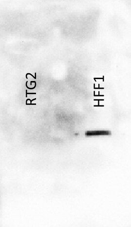
Application: Western BlotSample Tested: RTG-2 cells total lysate and HFF-1 cells total lysateSpecies: Rainbow trout, Oncorhynchus mykiss and HumanVerified Customer | Posted 05/02/201720ug of total cell lysate (RIPA) from RTG-2 and HFF-1 cells. Did not show any signal for rainbow trout cells.

There are no reviews that match your criteria.
Protocols
Find general support by application which include: protocols, troubleshooting, illustrated assays, videos and webinars.
- 7-Amino Actinomycin D (7-AAD) Cell Viability Flow Cytometry Protocol
- Appropriate Fixation of IHC/ICC Samples
- Cellular Response to Hypoxia Protocols
- ClariTSA™ Fluorophore Kits
- Detection & Visualization of Antibody Binding
- ELISA Sample Preparation & Collection Guide
- ELISA Troubleshooting Guide
- Extracellular Membrane Flow Cytometry Protocol
- Flow Cytometry Protocol for Cell Surface Markers
- Flow Cytometry Protocol for Staining Membrane Associated Proteins
- Flow Cytometry Staining Protocols
- Flow Cytometry Troubleshooting Guide
- How to Run an R&D Systems DuoSet ELISA
- How to Run an R&D Systems Quantikine ELISA
- How to Run an R&D Systems Quantikine™ QuicKit™ ELISA
- ICC Cell Smear Protocol for Suspension Cells
- ICC Immunocytochemistry Protocol Videos
- ICC for Adherent Cells
- Immunocytochemistry (ICC) Protocol
- Immunocytochemistry Troubleshooting
- Immunofluorescence of Organoids Embedded in Cultrex Basement Membrane Extract
- Immunohistochemistry (IHC) and Immunocytochemistry (ICC) Protocols
- Immunoprecipitation Protocol
- Intracellular Flow Cytometry Protocol Using Alcohol (Methanol)
- Intracellular Flow Cytometry Protocol Using Detergents
- Intracellular Nuclear Staining Flow Cytometry Protocol Using Detergents
- Intracellular Staining Flow Cytometry Protocol Using Alcohol Permeabilization
- Intracellular Staining Flow Cytometry Protocol Using Detergents to Permeabilize Cells
- Preparing Samples for IHC/ICC Experiments
- Preventing Non-Specific Staining (Non-Specific Binding)
- Primary Antibody Selection & Optimization
- Propidium Iodide Cell Viability Flow Cytometry Protocol
- Protocol for Liperfluo
- Protocol for VisUCyte™ HRP Polymer Detection Reagent
- Protocol for the Characterization of Human Th22 Cells
- Protocol for the Characterization of Human Th9 Cells
- Protocol for the Fluorescent ICC Staining of Cell Smears - Graphic
- Protocol for the Fluorescent ICC Staining of Cultured Cells on Coverslips - Graphic
- Protocol for the Preparation and Fluorescent ICC Staining of Cells on Coverslips
- Protocol for the Preparation and Fluorescent ICC Staining of Non-adherent Cells
- Protocol for the Preparation and Fluorescent ICC Staining of Stem Cells on Coverslips
- Protocol for the Preparation of a Cell Smear for Non-adherent Cell ICC - Graphic
- Protocol: Annexin V and PI Staining by Flow Cytometry
- Protocol: Annexin V and PI Staining for Apoptosis by Flow Cytometry
- Quantikine HS ELISA Kit Assay Principle, Alkaline Phosphatase
- Quantikine HS ELISA Kit Principle, Streptavidin-HRP Polymer
- R&D Systems Quality Control Western Blot Protocol
- Sandwich ELISA (Colorimetric) – Biotin/Streptavidin Detection Protocol
- Sandwich ELISA (Colorimetric) – Direct Detection Protocol
- TUNEL and Active Caspase-3 Detection by IHC/ICC Protocol
- The Importance of IHC/ICC Controls
- Troubleshooting Guide: ELISA
- Troubleshooting Guide: Fluorokine Flow Cytometry Kits
- Troubleshooting Guide: Western Blot Figures
- Western Blot Conditions
- Western Blot Protocol
- Western Blot Protocol for Cell Lysates
- Western Blot Troubleshooting
- Western Blot Troubleshooting Guide
- View all Protocols, Troubleshooting, Illustrated assays and Webinars
Loading...