Pancreatic Polypeptide/PP Antibody
Novus Biologicals | Catalog # NB100-1793
![Immunocytochemistry/ Immunofluorescence: Pancreatic Polypeptide/PP Antibody [NB100-1793]](https://resources.rndsystems.com/images/products/Pancreatic-Polypeptide-PP-Antibody-Immunocytochemistry-Immunofluorescence-NB100-1793-img0011.jpg "Immunocytochemistry/ Immunofluorescence: Pancreatic Polypeptide/PP Antibody [NB100-1793]")
Loading...
Key Product Details
Species Reactivity
Validated:
Human, Mouse, Rat, Monkey
Cited:
Human, Mouse
Predicted:
Canine (100%). Backed by our 100% Guarantee.
Applications
Validated:
Immunohistochemistry, Immunohistochemistry-Paraffin, Immunohistochemistry-Frozen, Immunoblotting, Peptide ELISA, Flow Cytometry, Immunocytochemistry/ Immunofluorescence
Cited:
Immunohistochemistry, Immunohistochemistry-Paraffin, Immunohistochemistry-Frozen, Immunoblotting, Immunocytochemistry/ Immunofluorescence, IF/IHC
Label
Unconjugated
Antibody Source
Polyclonal Goat IgG
Loading...
Product Specifications
Immunogen
Peptide with sequence C-TRPRYGKRHKEDT corresponding to internal region according to NP_002713.1.
Reactivity Notes
Monkey and Mouse reactivity reported from verified customer reviews.
Clonality
Polyclonal
Host
Goat
Isotype
IgG
Scientific Data Images for Pancreatic Polypeptide/PP Antibody
Immunocytochemistry/ Immunofluorescence: Pancreatic Polypeptide/PP Antibody [NB100-1793]
Pancreatic-Polypeptide-PP-Antibody-Immunocytochemistry-Immunofluorescence-NB100-1793-img0011.jpg![Immunohistochemistry-Paraffin: Pancreatic Polypeptide/PP Antibody [NB100-1793]](https://resources.rndsystems.com/images/products/Pancreatic-Polypeptide-PP-Antibody-Immunohistochemistry-Paraffin-NB100-1793-img0007.jpg "Immunohistochemistry-Paraffin: Pancreatic Polypeptide/PP Antibody [NB100-1793]")
Immunohistochemistry-Paraffin: Pancreatic Polypeptide/PP Antibody [NB100-1793]
Immunohistochemistry-Paraffin: Pancreatic Polypeptide/PP Antibody [NB100-1793] - Staining of paraffin embedded Human Pancreas with antibody at 3 ug/mL. Microwaved antigen retrieval with Tris/EDTA buffer pH 9, HRP-staining.![Flow Cytometry: Pancreatic Polypeptide/PP Antibody [NB100-1793]](https://resources.rndsystems.com/images/products/Pancreatic-Polypeptide-PP-Antibody-Flow-Cytometry-NB100-1793-img0010.jpg "Flow Cytometry: Pancreatic Polypeptide/PP Antibody [NB100-1793]")
Flow Cytometry: Pancreatic Polypeptide/PP Antibody [NB100-1793]
Flow Cytometry: Pancreatic Polypeptide/PP Antibody [NB100-1793] - Flow cytometric analysis of paraformaldehyde fixed U2OS cells (blue line), permeabilized with 0.5% Triton. Primary incubation 1hr (10 ug/mL) followed by Alexa Fluor 488 secondary antibody (1 ug/mL). IgG control: Unimmunized goat IgG (black line) followed by Alexa Fluor 488 secondary antibody.![Immunohistochemistry-Frozen: Pancreatic Polypeptide/PP Antibody [NB100-1793]](https://resources.rndsystems.com/images/products/Pancreatic-Polypeptide-PP-Antibody-Immunohistochemistry-Frozen-NB100-1793-img0004.jpg "Immunohistochemistry-Frozen: Pancreatic Polypeptide/PP Antibody [NB100-1793]")
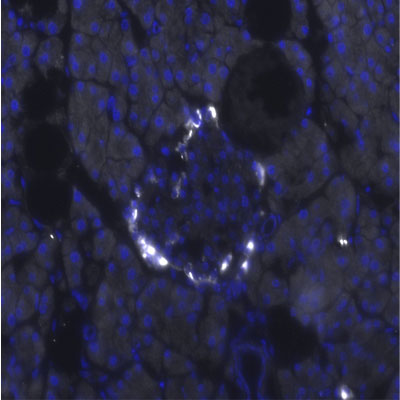
Immunohistochemistry-Frozen: Pancreatic Polypeptide/PP Antibody [NB100-1793]
Immunohistochemistry-Frozen: Pancreatic Polypeptide/PP Antibody [NB100-1793] - Analysis of Pancreatic Polypeptide in mouse adult pancreas tissue. IHC-Fr image submitted by a verified customer review.![Immunohistochemistry-Paraffin: Pancreatic Polypeptide/PP Antibody [NB100-1793]](https://resources.rndsystems.com/images/products/Pancreatic-Polypeptide-PP-Antibody-Immunohistochemistry-Paraffin-NB100-1793-img0006.jpg "Immunohistochemistry-Paraffin: Pancreatic Polypeptide/PP Antibody [NB100-1793]")
Immunohistochemistry-Paraffin: Pancreatic Polypeptide/PP Antibody [NB100-1793]
Immunohistochemistry-Paraffin: Pancreatic Polypeptide/PP Antibody [NB100-1793] - Adult macaque pancrease paraffin sections following standard protocols. IHC-P image submitted by a verified customer review.![Immunohistochemistry-Paraffin: Pancreatic Polypeptide/PP Antibody [NB100-1793]](https://resources.rndsystems.com/images/products/Pancreatic-Polypeptide-PP-Antibody-Immunohistochemistry-Paraffin-NB100-1793-img0008.jpg "Immunohistochemistry-Paraffin: Pancreatic Polypeptide/PP Antibody [NB100-1793]")
Immunohistochemistry-Paraffin: Pancreatic Polypeptide/PP Antibody [NB100-1793]
Immunohistochemistry-Paraffin: Pancreatic Polypeptide/PP Antibody [NB100-1793] - Staining of paraffin embedded Human Pancreas with antibody at 5 ug/mL. Steamed antigen retrieval with citrate buffer pH 6, AP-staining.![Immunohistochemistry-Paraffin: Pancreatic Polypeptide/PP Antibody [NB100-1793]](https://resources.rndsystems.com/images/products/Pancreatic-Polypeptide-PP-Antibody-Immunohistochemistry-Paraffin-NB100-1793-img0009.jpg "Immunohistochemistry-Paraffin: Pancreatic Polypeptide/PP Antibody [NB100-1793]")
Immunohistochemistry-Paraffin: Pancreatic Polypeptide/PP Antibody [NB100-1793]
Immunohistochemistry-Paraffin: Pancreatic Polypeptide/PP Antibody [NB100-1793] - Staining of paraffin embedded Human Intestine with antibody at 5 ug/mL. Steamed antigen retrieval with citrate buffer pH 6, AP-staining.
Immunohistochemistry: Pancreatic Polypeptide/PP Antibody [NB100-1793] -
Immunohistochemistry: Pancreatic Polypeptide/PP Antibody [NB100-1793] - Sorted CD133+ cells originate from pancreatic ducts.(A) Schematic diagram of the experimental procedure. Dissociated pancreatic cells were embedded & cultured as previously described (Lawson et al., 2007). Scale bar, 200 µm. (B) Confocal images of CD133 (green) & CPA1 (red) co-staining in adult human pancreas tissue. Scale bar, 20 µm. (C) CEL expression profiles of FACS-sorted human adult pancreatic cells & isolated islets (islet values normalized to 1). Data are presented as mean ± SEM (n=3). (D) Representative immunostaining pictures of sorted cells. Scale bar, 50 µm.DOI:http://dx.doi.org/10.7554/eLife.00940.005 Image collected & cropped by CiteAb from the following publication (https://pubmed.ncbi.nlm.nih.gov/24252877), licensed under a CC-BY license. Not internally tested by Novus Biologicals.
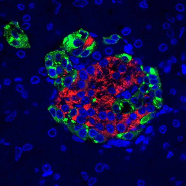
Immunocytochemistry/ Immunofluorescence: Pancreatic Polypeptide/PP Antibody [NB100-1793] -
Immunocytochemistry/ Immunofluorescence: Pancreatic Polypeptide/PP Antibody [NB100-1793] - Histologic comparison of wild type & Seriola dumerili mouse pancreata. Seriola dumerili Ins2 mice (bottom) have normal islet morphology & cyto-architecture compared to littermates with endogenous mouse Ins 1 & Ins 2 (top); insulin (red A–F), glucagon (green; A,D), pancreatic polypeptide (green; B,E), & somatostatin (green; C,F). G, H: Beta cell ultra-structure of NOD & Seriola dumerili Ins2 transgenic. Both NOD (G) & Seriola dumerili Ins 2 transgenic (H) islets contain insulin granules (yellow arrow), though Seriola dumerili Ins2 transgenic insulin granules are lighter in staining intensity compared to the NOD (n = 4; zoom in; inset). (I,J) Insulitis scoring pancreata from 12–15 weeks old NOD & Seriola dumerili Ins2 transgenics (n = 4 per group). Scale bar: 100 um. Image collected & cropped by CiteAb from the following publication (https://pubmed.ncbi.nlm.nih.gov/30899071), licensed under a CC-BY license. Not internally tested by Novus Biologicals.Applications for Pancreatic Polypeptide/PP Antibody
Application
Recommended Usage
Immunohistochemistry
5 ug/mL
Immunohistochemistry-Frozen
1:500
Immunohistochemistry-Paraffin
3 ug/ml
Peptide ELISA
Detection limit 1:32000
Application Notes
Use in Immunocytochemistry/immunofluorescence reported in scientific literature (PMID: 23221614). Use in immunoblotting reported in scientific literature (PMID: 27572106).
Reviewed Applications
Read 3 reviews rated 5 using NB100-1793 in the following applications:
Flow Cytometry Panel Builder
Bio-Techne Knows Flow Cytometry
Save time and reduce costly mistakes by quickly finding compatible reagents using the Panel Builder Tool.
Advanced Features
- Spectra Viewer - Custom analysis of spectra from multiple fluorochromes
- Spillover Popups - Visualize the spectra of individual fluorochromes
- Antigen Density Selector - Match fluorochrome brightness with antigen density

Formulation, Preparation, and Storage
Purification
Immunogen affinity purified
Formulation
Tris saline (20 mM Tris pH 7.3, 150 mM NaCl), 0.5% BSA
Preservative
0.02% Sodium Azide
Concentration
0.5 mg/ml
Shipping
The product is shipped with polar packs. Upon receipt, store it immediately at the temperature recommended below.
Stability & Storage
Store at -20C. Avoid freeze-thaw cycles.
Background: Pancreatic Polypeptide/PP
Additional Pancreatic Polypeptide/PP Products
Product Documents for Pancreatic Polypeptide/PP Antibody
Certificate of Analysis
To download a Certificate of Analysis, please enter a lot or batch number in the search box below.
Product Specific Notices for Pancreatic Polypeptide/PP Antibody
This product is for research use only and is not approved for use in humans or in clinical diagnosis. Primary Antibodies are guaranteed for 1 year from date of receipt.
Related Research Areas
Citations for Pancreatic Polypeptide/PP Antibody
 Powered by Bioz
Customer Reviews for Pancreatic Polypeptide/PP Antibody (3)
5 out of 5
3 Customer Ratings
Have you used Pancreatic Polypeptide/PP Antibody?
Submit a review and receive an Amazon gift card!
$25/€18/£15/$25CAN/¥2500 Yen for a review with an image
$10/€7/£6/$10CAN/¥1110 Yen for a review without an image
Submit a review
Customer Images
Showing
1
-
3 of
3 reviews
Showing All
Filter By:
-
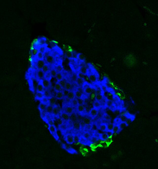
Application: Immunohistochemistry-ParaffinSample Tested: Mouse PancreasSpecies: MouseVerified Customer | Posted 04/28/2016Mouse pancreas stained for PP (green) and insulin (blue)

-
Application: Immunohistochemistry-ParaffinVerified Customer | Posted 12/18/2013Adult macaque pancreas paraffin sections following standard protocols
-
Application: Immunohistochemistry-FrozenSample Tested: Mouse Adult Pancreas TissueSpecies: MouseVerified Customer | Posted 08/21/2010
There are no reviews that match your criteria.
Protocols
Find general support by application which include: protocols, troubleshooting, illustrated assays, videos and webinars.
- 7-Amino Actinomycin D (7-AAD) Cell Viability Flow Cytometry Protocol
- Antigen Retrieval Protocol (PIER)
- Antigen Retrieval for Frozen Sections Protocol
- Appropriate Fixation of IHC/ICC Samples
- Cellular Response to Hypoxia Protocols
- Chromogenic IHC Staining of Formalin-Fixed Paraffin-Embedded (FFPE) Tissue Protocol
- Chromogenic Immunohistochemistry Staining of Frozen Tissue
- ClariTSA™ Fluorophore Kits
- Detection & Visualization of Antibody Binding
- ELISA Sample Preparation & Collection Guide
- ELISA Troubleshooting Guide
- Extracellular Membrane Flow Cytometry Protocol
- Flow Cytometry Protocol for Cell Surface Markers
- Flow Cytometry Protocol for Staining Membrane Associated Proteins
- Flow Cytometry Staining Protocols
- Flow Cytometry Troubleshooting Guide
- Fluorescent IHC Staining of Frozen Tissue Protocol
- Graphic Protocol for Heat-induced Epitope Retrieval
- Graphic Protocol for the Preparation and Fluorescent IHC Staining of Frozen Tissue Sections
- Graphic Protocol for the Preparation and Fluorescent IHC Staining of Paraffin-embedded Tissue Sections
- Graphic Protocol for the Preparation of Gelatin-coated Slides for Histological Tissue Sections
- How to Run an R&D Systems DuoSet ELISA
- How to Run an R&D Systems Quantikine ELISA
- How to Run an R&D Systems Quantikine™ QuicKit™ ELISA
- ICC Cell Smear Protocol for Suspension Cells
- ICC Immunocytochemistry Protocol Videos
- ICC for Adherent Cells
- IHC Sample Preparation (Frozen sections vs Paraffin)
- Immunocytochemistry (ICC) Protocol
- Immunocytochemistry Troubleshooting
- Immunofluorescence of Organoids Embedded in Cultrex Basement Membrane Extract
- Immunofluorescent IHC Staining of Formalin-Fixed Paraffin-Embedded (FFPE) Tissue Protocol
- Immunohistochemistry (IHC) and Immunocytochemistry (ICC) Protocols
- Immunohistochemistry Frozen Troubleshooting
- Immunohistochemistry Paraffin Troubleshooting
- Intracellular Flow Cytometry Protocol Using Alcohol (Methanol)
- Intracellular Flow Cytometry Protocol Using Detergents
- Intracellular Nuclear Staining Flow Cytometry Protocol Using Detergents
- Intracellular Staining Flow Cytometry Protocol Using Alcohol Permeabilization
- Intracellular Staining Flow Cytometry Protocol Using Detergents to Permeabilize Cells
- Preparing Samples for IHC/ICC Experiments
- Preventing Non-Specific Staining (Non-Specific Binding)
- Primary Antibody Selection & Optimization
- Propidium Iodide Cell Viability Flow Cytometry Protocol
- Protocol for Heat-Induced Epitope Retrieval (HIER)
- Protocol for Liperfluo
- Protocol for Making a 4% Formaldehyde Solution in PBS
- Protocol for VisUCyte™ HRP Polymer Detection Reagent
- Protocol for the Characterization of Human Th22 Cells
- Protocol for the Characterization of Human Th9 Cells
- Protocol for the Fluorescent ICC Staining of Cell Smears - Graphic
- Protocol for the Fluorescent ICC Staining of Cultured Cells on Coverslips - Graphic
- Protocol for the Preparation & Fixation of Cells on Coverslips
- Protocol for the Preparation and Chromogenic IHC Staining of Frozen Tissue Sections
- Protocol for the Preparation and Chromogenic IHC Staining of Frozen Tissue Sections - Graphic
- Protocol for the Preparation and Chromogenic IHC Staining of Paraffin-embedded Tissue Sections
- Protocol for the Preparation and Chromogenic IHC Staining of Paraffin-embedded Tissue Sections - Graphic
- Protocol for the Preparation and Fluorescent ICC Staining of Cells on Coverslips
- Protocol for the Preparation and Fluorescent ICC Staining of Non-adherent Cells
- Protocol for the Preparation and Fluorescent ICC Staining of Stem Cells on Coverslips
- Protocol for the Preparation and Fluorescent IHC Staining of Frozen Tissue Sections
- Protocol for the Preparation and Fluorescent IHC Staining of Paraffin-embedded Tissue Sections
- Protocol for the Preparation of Gelatin-coated Slides for Histological Tissue Sections
- Protocol for the Preparation of a Cell Smear for Non-adherent Cell ICC - Graphic
- Protocol: Annexin V and PI Staining by Flow Cytometry
- Protocol: Annexin V and PI Staining for Apoptosis by Flow Cytometry
- Quantikine HS ELISA Kit Assay Principle, Alkaline Phosphatase
- Quantikine HS ELISA Kit Principle, Streptavidin-HRP Polymer
- R&D Systems Quality Control Western Blot Protocol
- Sandwich ELISA (Colorimetric) – Biotin/Streptavidin Detection Protocol
- Sandwich ELISA (Colorimetric) – Direct Detection Protocol
- TUNEL and Active Caspase-3 Detection by IHC/ICC Protocol
- The Importance of IHC/ICC Controls
- Troubleshooting Guide: ELISA
- Troubleshooting Guide: Fluorokine Flow Cytometry Kits
- Troubleshooting Guide: Immunohistochemistry
- Troubleshooting Guide: Western Blot Figures
- Western Blot Conditions
- Western Blot Protocol
- Western Blot Protocol for Cell Lysates
- Western Blot Troubleshooting
- Western Blot Troubleshooting Guide
- View all Protocols, Troubleshooting, Illustrated assays and Webinars
Loading...