![Western Blot: Pax6 Antibody [NB300-750]](https://resources.rndsystems.com/images/products/Pax6-Antibody-Western-Blot-NB300-750-img0023.jpg "Western Blot: Pax6 Antibody [NB300-750]")
Key Product Details
Species Reactivity
Validated:
Cited:
Applications
Validated:
Cited:
Label
Antibody Source
Product Specifications
Immunogen
Reactivity Notes
Localization
Marker
Specificity
Clonality
Host
Isotype
Theoretical MW
Disclaimer note: The observed molecular weight of the protein may vary from the listed predicted molecular weight due to post translational modifications, post translation cleavages, relative charges, and other experimental factors.
Scientific Data Images for Pax6 Antibody
Western Blot: Pax6 Antibody [NB300-750]
Western Blot: Pax6 Antibody [NB300-750] - Analysis of sheep retinal extract![Immunocytochemistry/ Immunofluorescence: Pax6 Antibody [NB300-750]](https://resources.rndsystems.com/images/products/Pax6-Antibody-Immunocytochemistry-NB300-750-img0024.jpg "Immunocytochemistry/ Immunofluorescence: Pax6 Antibody [NB300-750]")
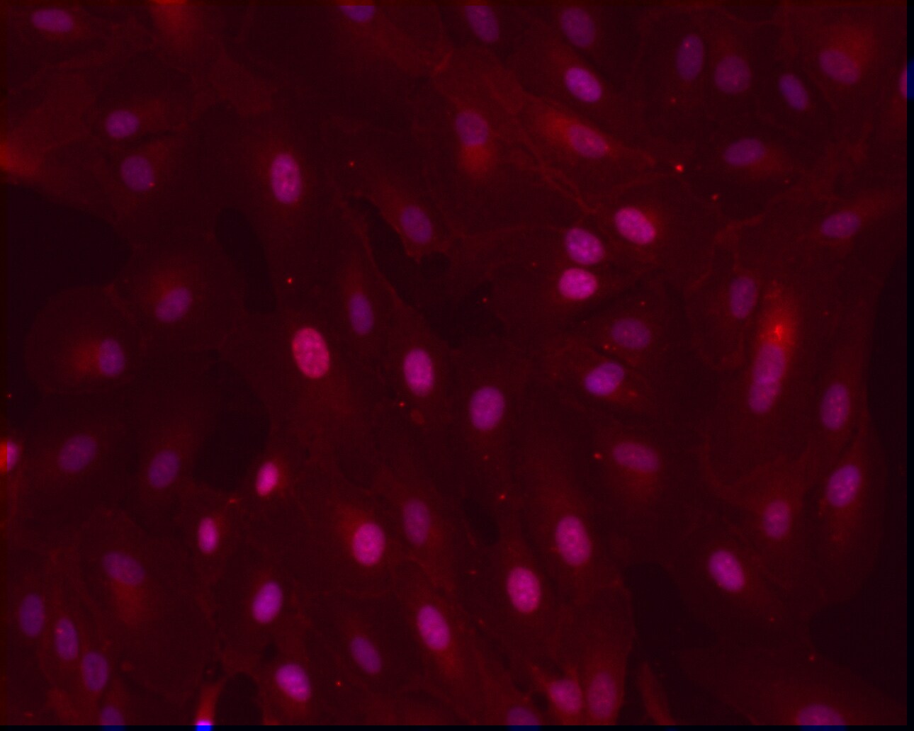
Immunocytochemistry/ Immunofluorescence: Pax6 Antibody [NB300-750]
Immunocytochemistry/Immunofluorescence: Pax6 Antibody [NB300-750] - ARPE-19 monolayer on glass substrate. Fixative Methanol Blocking Goat Serum. Dilution 1:100. Incubation 1h @ RT. This image was submitted via customer Review.![Western Blot: Pax6 Antibody [NB300-750]](https://resources.rndsystems.com/images/products/Pax6-Antibody-Western-Blot-NB300-750-img0017.jpg "Western Blot: Pax6 Antibody [NB300-750]")
Western Blot: Pax6 Antibody [NB300-750]
Western Blot: Pax6 Antibody [NB300-750] - Analysis of rat whole eye extract![Western Blot: Pax6 Antibody [NB300-750]](https://resources.rndsystems.com/images/products/Pax6-Antibody-Western-Blot-NB300-750-img0016.jpg "Western Blot: Pax6 Antibody [NB300-750]")
Western Blot: Pax6 Antibody [NB300-750]
Western Blot: Pax6 Antibody [NB300-750] - Analysis of various human, mouse and non-human primate whole cell lysates.![Western Blot: Pax6 Antibody [NB300-750]](https://resources.rndsystems.com/images/products/Pax6-Antibody-Western-Blot-NB300-750-img0019.jpg "Western Blot: Pax6 Antibody [NB300-750]")
Western Blot: Pax6 Antibody [NB300-750]
Western Blot: Pax6 Antibody [NB300-750] - Analysis of extracts from HeLa, U251 and mouse brain using PAX6 antibody (NB300-750, 1:100).![Western Blot: Pax6 Antibody [NB300-750]](https://resources.rndsystems.com/images/products/Pax6-Antibody-Western-Blot-NB300-750-img0022.jpg "Western Blot: Pax6 Antibody [NB300-750]")
Western Blot: Pax6 Antibody [NB300-750]
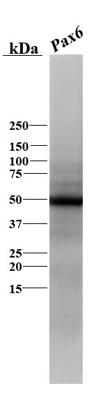
Western Blot: Pax6 Antibody [NB300-750] - Analysis of Pax6 in skate (Leucoraja erinacea) brain lysate using anti-Pax6 antibody. Image from verified customer review.![Immunocytochemistry/ Immunofluorescence: Pax6 Antibody [NB300-750]](https://resources.rndsystems.com/images/products/Pax6-Antibody-Immunocytochemistry-Immunofluorescence-NB300-750-img0011.jpg "Immunocytochemistry/ Immunofluorescence: Pax6 Antibody [NB300-750]")
Immunocytochemistry/ Immunofluorescence: Pax6 Antibody [NB300-750]
Immunocytochemistry/Immunofluorescence: Pax6 Antibody [NB300-750] - Formalin fixed cells were permeabilized with 0.1% Triton X-100 in TBS for 10 minutes at room temperature and blocked with 1% Blocker BSA for 15 minutes at room temperature. Cells were probed with a PAX6 polyclonal antibody at a dilution of 1:400 for 30 minutes at room temperature. F-Actin (red) was stained with DyLight 554 Phalloidin were stained with Hoechst 33342 dye.![Immunoprecipitation: Pax6 Antibody [NB300-750]](https://resources.rndsystems.com/images/products/Pax6-Antibody-Immunoprecipitation-NB300-750-img0020.jpg "Immunoprecipitation: Pax6 Antibody [NB300-750]")
Immunoprecipitation: Pax6 Antibody [NB300-750]
Immunoprecipitation: Pax6 Antibody [NB300-750] - Analysis of PAX6 was performed on 293T cells. Antigen: antibody complexes were formed by incubating 500ug whole cell lysate with 5ug of PAX6 polyclonal antibody overnight on a rocking platform at 4C. The immune complexes were captured on 50ul Protein A/G Plus Agarose, washed extensively, and eluted with 5X Lane Marker Reducing Sample Buffer. Samples were resolved on a 4-20% Tris-HCl polyacrylamide gel, transferred to a PVDF membrane, and blocked with 5% BSA/TBS-0.1%Tween for at least 1 hour. The membrane was probed with a PAX6 monoclonal antibody at a dilution of 1:1000 overnight rotating at 4C, washed in TBST, and probed with Clean-Blot IP Detection Reagent at a dilution of 1:1000 for at least one hour.
Immunocytochemistry/ Immunofluorescence: Pax6 Antibody [NB300-750] -
Immunocytochemistry/ Immunofluorescence: Pax6 Antibody [NB300-750] - Stepwise differentiation of human iPSCs towards renal progenitor cells (RPCs).(a) Schematic description of the two-stage protocol applied to iPSC-renal commitment. (b, c, d, e) Immunofluorescence of iPSCs (derived from retroviral transfected dermal fibroblasts) exposed to differentiating media. (b) The pluripotency markers SSEA4, TRA-1-81, Nanog, ME marker T(Bry) & IM marker Osr1. (c) IM & MM marker expression as Wt1, Pax8, Pax2, Six2 & Sall1. (d) Renal progenitor markers CD133, CD24, NCAM, glomerular epithelial marker Claudin1 & proximal tubular epithelial markers AQP1, GGT1. (e) Markers identifying endodermal AFP, ectodermal Pax6, & cardiac mesodermal Nkx2.5 derivation. Nuclei are stained with DAPI (blue). Scale bars: 20 μm (b, c, d, e). Image collected & cropped by CiteAb from the following publication (https://www.nature.com/articles/srep08826), licensed under a CC-BY license. Not internally tested by Novus Biologicals.Applications for Pax6 Antibody
Immunocytochemistry/ Immunofluorescence
Immunoprecipitation
Western Blot
Reviewed Applications
Read 4 reviews rated 4.5 using NB300-750 in the following applications:
Formulation, Preparation, and Storage
Purification
Formulation
Preservative
Concentration
Shipping
Stability & Storage
Background: Pax6
Long Name
Alternate Names
Gene Symbol
UniProt
Additional Pax6 Products
Product Documents for Pax6 Antibody
Certificate of Analysis
To download a Certificate of Analysis, please enter a lot or batch number in the search box below.
Product Specific Notices for Pax6 Antibody
This product is for research use only and is not approved for use in humans or in clinical diagnosis. Primary Antibodies are guaranteed for 1 year from date of receipt.
Citations for Pax6 Antibody
 Powered by Bioz
Customer Reviews for Pax6 Antibody (4)
Have you used Pax6 Antibody?
Submit a review and receive an Amazon gift card!
$25/€18/£15/$25CAN/¥2500 Yen for a review with an image
$10/€7/£6/$10CAN/¥1110 Yen for a review without an image
Submit a review
Customer Images
-(01-mg)_NB300-750_8281.jpg)
-
Application: ImmunocytochemistrySample Tested: ARPE-19 cellsSpecies: HumanVerified Customer | Posted 08/29/2017ARPE-19 monolayer on glass substrateFixative Methanol Blocking Goat Serum Dilution 1:100 Incubation 1h @ RT

-
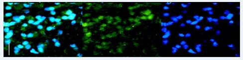
Application: ImmunofluorescenceSample Tested: HindbrainSpecies: OtherVerified Customer | Posted 04/20/2016Elasmobranch hindbrain- Pax6 in green
-
Application: Western BlotSample Tested: Skate brainSpecies: OtherVerified Customer | Posted 02/09/2016Pax6 in basal vertebrate
-
Application: Western BlotSample Tested:Species: HumanVerified Customer | Posted 06/13/2014Western blot analysis of extracts from HeLa, U251 and mouse brain using PAX6 antibody (NB300-750, 1:100).
There are no reviews that match your criteria.
Protocols
Find general support by application which include: protocols, troubleshooting, illustrated assays, videos and webinars.
- Antigen Retrieval Protocol (PIER)
- Antigen Retrieval for Frozen Sections Protocol
- Appropriate Fixation of IHC/ICC Samples
- Cellular Response to Hypoxia Protocols
- Chromogenic IHC Staining of Formalin-Fixed Paraffin-Embedded (FFPE) Tissue Protocol
- Chromogenic Immunohistochemistry Staining of Frozen Tissue
- ClariTSA™ Fluorophore Kits
- Detection & Visualization of Antibody Binding
- Fluorescent IHC Staining of Frozen Tissue Protocol
- Graphic Protocol for Heat-induced Epitope Retrieval
- Graphic Protocol for the Preparation and Fluorescent IHC Staining of Frozen Tissue Sections
- Graphic Protocol for the Preparation and Fluorescent IHC Staining of Paraffin-embedded Tissue Sections
- Graphic Protocol for the Preparation of Gelatin-coated Slides for Histological Tissue Sections
- ICC Cell Smear Protocol for Suspension Cells
- ICC Immunocytochemistry Protocol Videos
- ICC for Adherent Cells
- IHC Sample Preparation (Frozen sections vs Paraffin)
- Immunocytochemistry (ICC) Protocol
- Immunocytochemistry Troubleshooting
- Immunofluorescence of Organoids Embedded in Cultrex Basement Membrane Extract
- Immunofluorescent IHC Staining of Formalin-Fixed Paraffin-Embedded (FFPE) Tissue Protocol
- Immunohistochemistry (IHC) and Immunocytochemistry (ICC) Protocols
- Immunohistochemistry Frozen Troubleshooting
- Immunohistochemistry Paraffin Troubleshooting
- Immunoprecipitation Protocol
- Preparing Samples for IHC/ICC Experiments
- Preventing Non-Specific Staining (Non-Specific Binding)
- Primary Antibody Selection & Optimization
- Protocol for Heat-Induced Epitope Retrieval (HIER)
- Protocol for Making a 4% Formaldehyde Solution in PBS
- Protocol for VisUCyte™ HRP Polymer Detection Reagent
- Protocol for the Fluorescent ICC Staining of Cell Smears - Graphic
- Protocol for the Fluorescent ICC Staining of Cultured Cells on Coverslips - Graphic
- Protocol for the Preparation & Fixation of Cells on Coverslips
- Protocol for the Preparation and Chromogenic IHC Staining of Frozen Tissue Sections
- Protocol for the Preparation and Chromogenic IHC Staining of Frozen Tissue Sections - Graphic
- Protocol for the Preparation and Chromogenic IHC Staining of Paraffin-embedded Tissue Sections
- Protocol for the Preparation and Chromogenic IHC Staining of Paraffin-embedded Tissue Sections - Graphic
- Protocol for the Preparation and Fluorescent ICC Staining of Cells on Coverslips
- Protocol for the Preparation and Fluorescent ICC Staining of Non-adherent Cells
- Protocol for the Preparation and Fluorescent ICC Staining of Stem Cells on Coverslips
- Protocol for the Preparation and Fluorescent IHC Staining of Frozen Tissue Sections
- Protocol for the Preparation and Fluorescent IHC Staining of Paraffin-embedded Tissue Sections
- Protocol for the Preparation of Gelatin-coated Slides for Histological Tissue Sections
- Protocol for the Preparation of a Cell Smear for Non-adherent Cell ICC - Graphic
- R&D Systems Quality Control Western Blot Protocol
- TUNEL and Active Caspase-3 Detection by IHC/ICC Protocol
- The Importance of IHC/ICC Controls
- Troubleshooting Guide: Immunohistochemistry
- Troubleshooting Guide: Western Blot Figures
- Western Blot Conditions
- Western Blot Protocol
- Western Blot Protocol for Cell Lysates
- Western Blot Troubleshooting
- Western Blot Troubleshooting Guide
- View all Protocols, Troubleshooting, Illustrated assays and Webinars
FAQs for Pax6 Antibody
-
Q: I am just wondering if this specific epitope is conserved from rabbits to lampreys and skates
A: Yes, the immunogen is pretty conserved and is shared with the sequence available at Uniprot site so it may work in your mentioned species.
-
Q: What PAX6 antibody is recommend to use in ICC staining of human stem cells differentiated neural precursors?
A:
By performing a search on PAX6 then refining this to antibodies which have been validated with human samples and for ICC, I can see that we have three products to meet your requirements. You can see all three of these antibodies. These products are listed based on sales and citations, however all of our antibodies are covered by our 100% guarantee to work in the species and applications listed on our website and on our product datasheets. You can read more about our quality guarantee here.
-
Q: I am just wondering if this specific epitope is conserved from rabbits to lampreys and skates
A: Yes, the immunogen is pretty conserved and is shared with the sequence available at Uniprot site so it may work in your mentioned species.
-
Q: What PAX6 antibody is recommend to use in ICC staining of human stem cells differentiated neural precursors?
A:
By performing a search on PAX6 then refining this to antibodies which have been validated with human samples and for ICC, I can see that we have three products to meet your requirements. You can see all three of these antibodies. These products are listed based on sales and citations, however all of our antibodies are covered by our 100% guarantee to work in the species and applications listed on our website and on our product datasheets. You can read more about our quality guarantee here.

