Periostin/OSF-2 Antibody - C-terminus - Azide and BSA Free
Novus Biologicals | Catalog # NBP1-30042
![Western Blot: Periostin/OSF-2 AntibodyC-terminusAzide and BSA Free [NBP1-30042]](https://resources.rndsystems.com/images/products/Periostin-OSF-2-Antibody---C-terminus---Azide-and-BSA-Free-Western-Blot-NBP1-30042-img0008.jpg "Western Blot: Periostin/OSF-2 AntibodyC-terminusAzide and BSA Free [NBP1-30042]")
Loading...
Key Product Details
Validated by
Knockout/Knockdown
Species Reactivity
Validated:
Human, Mouse, Rat, Chicken
Cited:
Human, Mouse, Rat, Rabbit
Predicted:
Bovine (100%), Canine (100%), Equine (100%), Feline (100%), Finch (100%), Goat (100%), Guinea Pig (100%), Hamster (100%), Primate (%), Rabbit (100%), Sheep (100%), Vole (100%). Backed by our 100% Guarantee.
Applications
Validated:
Knockout Validated, Immunohistochemistry, Immunohistochemistry-Paraffin, Immunohistochemistry-Frozen, Western Blot, Immunocytochemistry/ Immunofluorescence
Cited:
Immunohistochemistry, Immunohistochemistry-Paraffin, Immunohistochemistry-Frozen, Western Blot, Immunocytochemistry/ Immunofluorescence, IF/IHC
Label
Unconjugated
Antibody Source
Polyclonal Rabbit IgG
Format
Azide and BSA Free
Loading...
Product Specifications
Immunogen
Bacterial fusion protein equivalent to a 188-amino acid polypeptide from the C-terminal region of mouse periostin which is comprised of six small alternatively-spliced exons. Accession # Q62009
Reactivity Notes
Expected reactivity to all mammalian and avian species. Based on customer review rabbit reacts.
Specificity
Specific for the ~93 kDa periostin doublet in mouse lung extract. Consistent with the fact that this antibody is made against an alternatively spliced region of periostin, this antibody recognizes only the largest two of the three forms of periostin recognized on Western blots by the Pan periostin antibody and also shows a distinctive staining pattern by immunohistochemistry. The antibody works well for immunohistochemistry on paraformaldehyde-fixed sections with a simple antigen-retrieval protocol (incubate slides for 20 minutes at 90 degrees C in 10 mM sodium citrate (pH 6.0)/ 0.1 % Tween-20).
Clonality
Polyclonal
Host
Rabbit
Isotype
IgG
Theoretical MW
93 kDa.
Disclaimer note: The observed molecular weight of the protein may vary from the listed predicted molecular weight due to post translational modifications, post translation cleavages, relative charges, and other experimental factors.
Disclaimer note: The observed molecular weight of the protein may vary from the listed predicted molecular weight due to post translational modifications, post translation cleavages, relative charges, and other experimental factors.
Description
Recommended that the undiluted antibody be aliquoted into smaller working volumes (10-30 uL/vial depending on usage).
Scientific Data Images for Periostin/OSF-2 Antibody - C-terminus - Azide and BSA Free
Western Blot: Periostin/OSF-2 AntibodyC-terminusAzide and BSA Free [NBP1-30042]
Periostin-OSF-2-Antibody---C-terminus---Azide-and-BSA-Free-Western-Blot-NBP1-30042-img0008.jpg![Immunocytochemistry/ Immunofluorescence: Periostin/OSF-2 Antibody - C-terminus - Azide and BSA Free [NBP1-30042]](https://resources.rndsystems.com/images/products/Periostin-OSF-2-Antibody---C-terminus---Azide-and-BSA-Free-Immunocytochemistry-Immunofluorescence-NBP1-30042-img0009.jpg "Immunocytochemistry/ Immunofluorescence: Periostin/OSF-2 Antibody - C-terminus - Azide and BSA Free [NBP1-30042]")
Immunocytochemistry/ Immunofluorescence: Periostin/OSF-2 Antibody - C-terminus - Azide and BSA Free [NBP1-30042]
Periostin-OSF-2-Antibody---C-terminus---Azide-and-BSA-Free-Immunocytochemistry-Immunofluorescence-NBP1-30042-img0009.jpg![Immunohistochemistry: Periostin/OSF-2 Antibody - C-terminus - Azide and BSA Free [NBP1-30042]](https://resources.rndsystems.com/images/products/Periostin-OSF-2-Antibody---C-terminus---Azide-and-BSA-Free-Immunohistochemistry-NBP1-30042-img0003.jpg "Immunohistochemistry: Periostin/OSF-2 Antibody - C-terminus - Azide and BSA Free [NBP1-30042]")
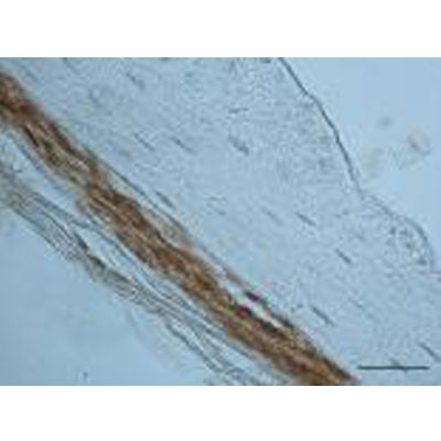
Immunohistochemistry: Periostin/OSF-2 Antibody - C-terminus - Azide and BSA Free [NBP1-30042]
Immunohistochemistry: Periostin/OSF-2 Antibody - C-terminus - Azide and BSA Free [NBP1-30042] - Analysis of periostin in rabbit periosteum. Image courtesy of product review submitted by Samer Zaky.![Western Blot: Periostin/OSF-2 AntibodyC-terminusAzide and BSA Free [NBP1-30042]](https://resources.rndsystems.com/images/products/Periostin-OSF-2-Antibody---C-terminus---Azide-and-BSA-Free-Western-Blot-NBP1-30042-img0004.jpg "Western Blot: Periostin/OSF-2 AntibodyC-terminusAzide and BSA Free [NBP1-30042]")
Western Blot: Periostin/OSF-2 AntibodyC-terminusAzide and BSA Free [NBP1-30042]
Western Blot: Periostin/OSF-2 Antibody - C-terminus - Azide and BSA Free [NBP1-30042] - Rat lung lysate showing specific immunolabeling of the ~ 93 kDa periostin protein doublet.![Western Blot: Periostin/OSF-2 AntibodyC-terminusAzide and BSA Free [NBP1-30042]](https://resources.rndsystems.com/images/products/Periostin-OSF-2-Antibody---C-terminus---Azide-and-BSA-Free-Western-Blot-NBP1-30042-img0007.jpg "Western Blot: Periostin/OSF-2 AntibodyC-terminusAzide and BSA Free [NBP1-30042]")
Western Blot: Periostin/OSF-2 AntibodyC-terminusAzide and BSA Free [NBP1-30042]
Periostin-OSF-2-Antibody---C-terminus---Azide-and-BSA-Free-Western-Blot-NBP1-30042-img0007.jpg![Immunocytochemistry/ Immunofluorescence: Periostin/OSF-2 Antibody - C-terminus - Azide and BSA Free [NBP1-30042]](https://resources.rndsystems.com/images/products/Periostin-OSF-2-Antibody---C-terminus---Azide-and-BSA-Free-Immunocytochemistry-Immunofluorescence-NBP1-30042-img0006.jpg "Immunocytochemistry/ Immunofluorescence: Periostin/OSF-2 Antibody - C-terminus - Azide and BSA Free [NBP1-30042]")
Immunocytochemistry/ Immunofluorescence: Periostin/OSF-2 Antibody - C-terminus - Azide and BSA Free [NBP1-30042]
Periostin-OSF-2-Antibody---C-terminus---Azide-and-BSA-Free-Immunocytochemistry-Immunofluorescence-NBP1-30042-img0006.jpg![Immunohistochemistry: Periostin/OSF-2 Antibody - C-terminus - Azide and BSA Free [NBP1-30042]](https://resources.rndsystems.com/images/products/Periostin-OSF-2%20Antibody%20-%20C-terminus%20-%20Azide%20and%20BSA%20Free-Immunohistochemistry-NBP1-30042-img0010.jpg "Immunohistochemistry: Periostin/OSF-2 Antibody - C-terminus - Azide and BSA Free [NBP1-30042]")
![Knockout Validated: Periostin/OSF-2 Antibody - C-terminus - Azide and BSA Free [NBP1-30042]](https://resources.rndsystems.com/images/products/Periostin-OSF-2-Antibody---C-terminus---Azide-and-BSA-Free-Knockout-Validated-NBP1-30042-img0005.jpg "Western Blot: Periostin/OSF-2 Antibody - C-terminus - Azide and BSA Free [NBP1-30042]")

Immunocytochemistry/ Immunofluorescence: Periostin/OSF-2 Antibody - C-terminus - Azide and BSA Free [NBP1-30042] -
Immunocytochemistry/ Immunofluorescence: Periostin/OSF-2 Antibody - C-terminus - Azide and BSA Free [NBP1-30042] - Cardiac muscle phenotypes of mice at 12 weeks of age.(A) Evaluation of cardiac function by transthoracic echocardiographic analyses. Left ventricular ejection fractions (LVEFs) are shown (n = 8–9). (B) H&E staining of cryosections from cardiac muscle. (C) Immunostaining of laminin ɑ2 (green) & periostin (red) with DAPI (blue) from cardiac muscle. (D) Western blot analyses of periostin. The graph shows the quantification of periostin levels normalized to GAPDH (n = 3). (E) qPCR analyses of Tgfb2 (TGF beta 2), Postn (periostin), & Fn1 (fibronectin) mRNA levels in cardiac muscle (n = 4). (F) qPCR analyses of Nppa (natriuretic peptide A) & Nppb (natriuretic peptide B) mRNA levels in cardiac muscle (n = 4). In qPCR analyses, data were normalized by Gapdh mRNA & expressed as fold increases from WT mice. ***P<0.001 compared with WT mice. Image collected & cropped by CiteAb from the following publication (https://pubmed.ncbi.nlm.nih.gov/31430335), licensed under a CC-BY license. Not internally tested by Novus Biologicals.
Western Blot: Periostin/OSF-2 Antibody - C-terminus - Azide and BSA Free [NBP1-30042] -
Western Blot: Periostin/OSF-2 Antibody - C-terminus - Azide and BSA Free [NBP1-30042] - TGFBI & periostin are induced in the heart after injury.(A) Western blot analysis of periostin & TGFBI in isolated adult cardiomyocytes & fibroblasts. Sarcomeric alpha -actin was used as a control for cardiomyocyte purity & GAPDH was used as a loading control. (B) Quantitative real time PCR for Postn & Tgfbi from 1 week sham or MI-operated hearts. mRNA levels were normalized to 18s ribosomal RNA. *p<0.05 for both genes in MI-operated animals compared to sham animals using an unpaired Student’s T-test. n = 3 animals. (C) Western blot analysis for periostin & TGFBI in the infarcted areas isolated from hearts 24 hours, 7 & 14 days after MI surgery. GAPDH was used as a loading control. Each lane corresponds to protein from one mouse. (D & E) Quantification of TGFBI (D) & periostin (E) protein levels from the conditions shown in panel C except that 3 hearts were analyzed in total each. (F) Immunohistochemistry for the indicated markers/proteins on sham or MI-operated hearts after 1 week. Images were taken at 400x magnification. Scale = 20 μm. Image collected & cropped by CiteAb from the following publication (https://dx.plos.org/10.1371/journal.pone.0181945), licensed under a CC-BY license. Not internally tested by Novus Biologicals.
Western Blot: Periostin/OSF-2 Antibody - C-terminus - Azide and BSA Free [NBP1-30042] -
Western Blot: Periostin/OSF-2 Antibody - C-terminus - Azide and BSA Free [NBP1-30042] - PostnMCM allele activity in vivo.(a) Schematic representation of the Postn genetic locus with a tamoxifen-regulated MerCreMer cDNA cassette inserted into exon 1 (E1), which was crossed with Rosa26 reporter mice (R26-eGFP) containing loxP sites flanking a stop cassette upstream of eGFP to allow for Cre-dependent lineage tracing. (b) Experimental scheme whereby PostnMCM/+; R26-eGFP mice were given tamoxifen for 8 weeks before harvesting at 16 weeks. (c) Representative histological section from the heart of mice described in a & b, which show exceptionally rare labelling of interstitial cells at baseline (arrow) with 8 weeks of tamoxifen. Nuclei are stained in blue. Inset shows alpha -actin stained cardiomyocytes (red) surrounding the one eGFP-labelled interstitial cell (green) (n=4 mice). (d) Experimental scheme whereby PostnMCM/+; R26-eGFP mice were MI injured or subjected to a sham procedure, then given tamoxifen for 1 week before harvesting. (e) Western blot analysis for periostin, eGFP, Cre (MerCreMer protein) & GAPDH as a control at day 0 before injury or day 7 after MI injury with 1 week of tamoxifen (n=3 mice per condition). (f) Representative histological sections showing eGFP-labelled interstitial cells in hearts of PostnMCM/+; R26-eGFP after MI injury with 7 days of tamoxifen labelling, but not with a sham procedure (n=6 mice for MI & n=3 for sham). (g) Whole-mount fluorescent images of hearts from PostnMCM/+; R26-eGFP mice for direct eGFP fluorescence over the given time course shown. A no tamoxifen control 7 days after MI is also shown (n=3 mice per time point & condition). Image collected & cropped by CiteAb from the following publication (https://pubmed.ncbi.nlm.nih.gov/27447449), licensed under a CC-BY license. Not internally tested by Novus Biologicals.Applications for Periostin/OSF-2 Antibody - C-terminus - Azide and BSA Free
Application
Recommended Usage
Immunocytochemistry/ Immunofluorescence
1:10-1:500
Immunohistochemistry
1:10-1:500
Immunohistochemistry-Paraffin
1:100
Western Blot
1:1000
Application Notes
For IHC-P antigen retrieval protocol (incubate slides for 20 minutes at 90 degrees C in 10 mM sodium citrate (pH 6.0)/ 0.1 % Tween-20). Immunocytochemistry/Immunofluorescence was reported in scientific literature. Use in Immunohistochemistry-Frozen reported in scientific literature (PMID: 31430335).
Reviewed Applications
Read 2 reviews rated 4 using NBP1-30042 in the following applications:
Formulation, Preparation, and Storage
Purification
Antigen Affinity-purified
Formulation
PBS
Format
Azide and BSA Free
Preservative
No Preservative
Concentration
Please see the vial label for concentration. If unlisted please contact technical services.
Shipping
The product is shipped with polar packs. Upon receipt, store it immediately at the temperature recommended below.
Stability & Storage
Store at -20C. Avoid freeze-thaw cycles.
Background: Periostin/OSF-2
Long Name
Osteoblast Specific Factor 2
Alternate Names
Fasciclin I-like, OSF-2, OSF2, POSTN, TRIF52
Gene Symbol
POSTN
UniProt
Additional Periostin/OSF-2 Products
Product Documents for Periostin/OSF-2 Antibody - C-terminus - Azide and BSA Free
Certificate of Analysis
To download a Certificate of Analysis, please enter a lot or batch number in the search box below.
Product Specific Notices for Periostin/OSF-2 Antibody - C-terminus - Azide and BSA Free
This product is for research use only and is not approved for use in humans or in clinical diagnosis. Primary Antibodies are guaranteed for 1 year from date of receipt.
Related Research Areas
Citations for Periostin/OSF-2 Antibody - C-terminus - Azide and BSA Free
 Powered by Bioz
Customer Reviews for Periostin/OSF-2 Antibody - C-terminus - Azide and BSA Free (2)
4 out of 5
2 Customer Ratings
Have you used Periostin/OSF-2 Antibody - C-terminus - Azide and BSA Free?
Submit a review and receive an Amazon gift card!
$25/€18/£15/$25CAN/¥2500 Yen for a review with an image
$10/€7/£6/$10CAN/¥1110 Yen for a review without an image
Submit a review
Customer Images
Showing
1
-
2 of
2 reviews
Showing All
Filter By:
-
Application: Immunohistochemistry-ParaffinSample Tested: See PMID 23574330Species: MouseVerified Customer | Posted 12/23/2014
-
Application: ImmunohistochemistrySample Tested: Rabbit periosteumSpecies: OtherVerified Customer | Posted 03/15/2011

There are no reviews that match your criteria.
Protocols
Find general support by application which include: protocols, troubleshooting, illustrated assays, videos and webinars.
- Antigen Retrieval Protocol (PIER)
- Antigen Retrieval for Frozen Sections Protocol
- Appropriate Fixation of IHC/ICC Samples
- Cellular Response to Hypoxia Protocols
- Chromogenic IHC Staining of Formalin-Fixed Paraffin-Embedded (FFPE) Tissue Protocol
- Chromogenic Immunohistochemistry Staining of Frozen Tissue
- ClariTSA™ Fluorophore Kits
- Detection & Visualization of Antibody Binding
- Fluorescent IHC Staining of Frozen Tissue Protocol
- Graphic Protocol for Heat-induced Epitope Retrieval
- Graphic Protocol for the Preparation and Fluorescent IHC Staining of Frozen Tissue Sections
- Graphic Protocol for the Preparation and Fluorescent IHC Staining of Paraffin-embedded Tissue Sections
- Graphic Protocol for the Preparation of Gelatin-coated Slides for Histological Tissue Sections
- ICC Cell Smear Protocol for Suspension Cells
- ICC Immunocytochemistry Protocol Videos
- ICC for Adherent Cells
- IHC Sample Preparation (Frozen sections vs Paraffin)
- Immunocytochemistry (ICC) Protocol
- Immunocytochemistry Troubleshooting
- Immunofluorescence of Organoids Embedded in Cultrex Basement Membrane Extract
- Immunofluorescent IHC Staining of Formalin-Fixed Paraffin-Embedded (FFPE) Tissue Protocol
- Immunohistochemistry (IHC) and Immunocytochemistry (ICC) Protocols
- Immunohistochemistry Frozen Troubleshooting
- Immunohistochemistry Paraffin Troubleshooting
- Preparing Samples for IHC/ICC Experiments
- Preventing Non-Specific Staining (Non-Specific Binding)
- Primary Antibody Selection & Optimization
- Protocol for Heat-Induced Epitope Retrieval (HIER)
- Protocol for Making a 4% Formaldehyde Solution in PBS
- Protocol for VisUCyte™ HRP Polymer Detection Reagent
- Protocol for the Fluorescent ICC Staining of Cell Smears - Graphic
- Protocol for the Fluorescent ICC Staining of Cultured Cells on Coverslips - Graphic
- Protocol for the Preparation & Fixation of Cells on Coverslips
- Protocol for the Preparation and Chromogenic IHC Staining of Frozen Tissue Sections
- Protocol for the Preparation and Chromogenic IHC Staining of Frozen Tissue Sections - Graphic
- Protocol for the Preparation and Chromogenic IHC Staining of Paraffin-embedded Tissue Sections
- Protocol for the Preparation and Chromogenic IHC Staining of Paraffin-embedded Tissue Sections - Graphic
- Protocol for the Preparation and Fluorescent ICC Staining of Cells on Coverslips
- Protocol for the Preparation and Fluorescent ICC Staining of Non-adherent Cells
- Protocol for the Preparation and Fluorescent ICC Staining of Stem Cells on Coverslips
- Protocol for the Preparation and Fluorescent IHC Staining of Frozen Tissue Sections
- Protocol for the Preparation and Fluorescent IHC Staining of Paraffin-embedded Tissue Sections
- Protocol for the Preparation of Gelatin-coated Slides for Histological Tissue Sections
- Protocol for the Preparation of a Cell Smear for Non-adherent Cell ICC - Graphic
- R&D Systems Quality Control Western Blot Protocol
- TUNEL and Active Caspase-3 Detection by IHC/ICC Protocol
- The Importance of IHC/ICC Controls
- Troubleshooting Guide: Immunohistochemistry
- Troubleshooting Guide: Western Blot Figures
- Western Blot Conditions
- Western Blot Protocol
- Western Blot Protocol for Cell Lysates
- Western Blot Troubleshooting
- Western Blot Troubleshooting Guide
- View all Protocols, Troubleshooting, Illustrated assays and Webinars
Loading...