![Western Blot: Rac1 Antibody [NB100-91266]](https://resources.rndsystems.com/images/products/Rac1-Antibody-Western-Blot-NB100-91266-img0011.jpg "Western Blot: Rac1 Antibody [NB100-91266]")
Loading...
Key Product Details
Species Reactivity
Validated:
Human, Mouse, Rat
Cited:
Mouse, Rat, Rabbit
Applications
Validated:
Immunohistochemistry, Immunohistochemistry-Paraffin, Immunohistochemistry-Frozen, Western Blot, Immunocytochemistry/ Immunofluorescence
Cited:
IF/IHC
Label
Unconjugated
Antibody Source
Polyclonal Rabbit IgG
Loading...
Product Specifications
Immunogen
This Rac1 antibody was developed against a synthetic peptide from amino acid region 100-150 as a part of human Rac1 conjugated to blue carrier protein.
Reactivity Notes
Predicted cross-reactivity based on antigen identity: Guinea pig, Bovine, Xenopus, Canine, Zebrafish, Chicken.
Specificity
Detects both RAC1 (Gene ID: 5879, UniProt: P63000) and RAC2 (Gene ID: 5880, UniProt: P15153).
Clonality
Polyclonal
Host
Rabbit
Isotype
IgG
Scientific Data Images for Rac1 Antibody
Western Blot: Rac1 Antibody [NB100-91266]
Western Blot: Rac1 Antibody [NB100-91266] - WB on tissue lysates. Blocking: 1% LFDM for 30 min at RT; primary antibody dilution: 1:1000 incubated overnight at 4C.![Immunocytochemistry/ Immunofluorescence: Rac1 Antibody [NB100-91266]](https://resources.rndsystems.com/images/products/Rac1-Antibody-Immunocytochemistry-Immunofluorescence-NB100-91266-img0009.jpg "Immunocytochemistry/ Immunofluorescence: Rac1 Antibody [NB100-91266]")
Immunocytochemistry/ Immunofluorescence: Rac1 Antibody [NB100-91266]
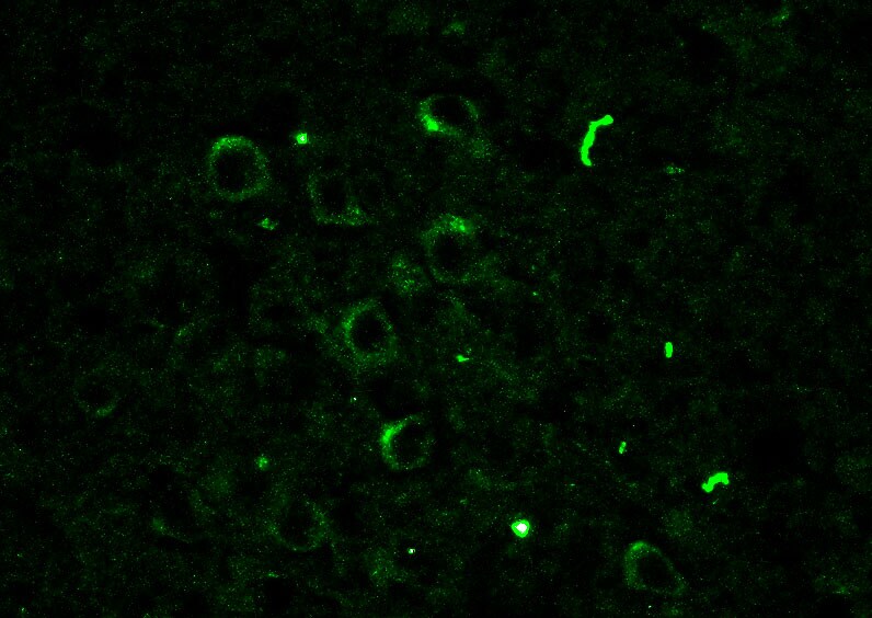
Immunocytochemistry/Immunofluorescence: Rac1 Antibody [NB100-91266] - Mouse Brain (cerebral cortex). ICC/IF image submitted by a verified customer review.![Immunohistochemistry: Rac1 Antibody [NB100-91266]](https://resources.rndsystems.com/images/products/Rac1-Antibody-Immunocytochemistry-NB100-91266-img0008.jpg "Immunohistochemistry: Rac1 Antibody [NB100-91266]")
Immunohistochemistry: Rac1 Antibody [NB100-91266]
Immunohistochemistry: Rac1 Antibody [NB100-91266] - Rat spinal cord using Rabbit antibody to RAC1, RAC2 (100-150) at 1:1000 dilution. Pre-absorption of the antibody with the immunising peptide completely abolishes the immunostaining (not shown).![Immunohistochemistry: Rac1 Antibody [NB100-91266]](https://resources.rndsystems.com/images/products/Rac1-Antibody-Immunohistochemistry-NB100-91266-img0005.jpg "Immunohistochemistry: Rac1 Antibody [NB100-91266]")
Immunohistochemistry: Rac1 Antibody [NB100-91266]
Immunohistochemistry: Rac1 Antibody [NB100-91266] - Rat spinal cord (ventral horn) using Rabbit antibody to RAC1, RAC2 whole serum at 1:1000 dilution. Pre-absorption of the antibody with the immunizing peptide completely abolishes the immunostaining (not shown).![Immunohistochemistry: Rac1 Antibody [NB100-91266]](https://resources.rndsystems.com/images/products/Rac1-Antibody-Immunohistochemistry-NB100-91266-img0004.jpg "Immunohistochemistry: Rac1 Antibody [NB100-91266]")
Immunohistochemistry: Rac1 Antibody [NB100-91266]
Immunohistochemistry: Rac1 Antibody [NB100-91266] - Rat ligated sciatic nerve using Rabbit antibody to RAC1, RAC2 whole serum at 1:1000 dilution. Pre-absorption of the antibody with the immunizing peptide completely abolishes the immunostaining (not shown).
Western Blot: Rac1 Antibody [NB100-91266] -
Melatonin injection restores renal cortical fibrosis in a CKD mouse model via increased expression of miR-4516. (A) Hematoxylin and eosin (H&E) staining was performed on kidney sections from a CKD mouse model following melatonin injection, or melatonin inhibition with miR-4516 inhibitor (scale bar = 1000 μm). (B,C) Expression of miR-4516 and ITGA9 was detected in the kidney cortex in each group by qPCR (n = 3). (D–F) Western blot analysis for ITGA9, Rac1, RhoA, CDC42, collagen type 1, and fibronectin expression using samples from the kidney cortex of each mouse model group (n = 3). Protein levels were determined by densitometry relative to alpha -tubulin. The values represent mean +/- SEM. * p < 0.05, ** p < 0.01 vs. healthy kidney, ##p < 0.01 vs. phosphate buffered saline (PBS), $$p < 0.01 vs. melatonin. Image collected and cropped by CiteAb from the following open publication (https://pubmed.ncbi.nlm.nih.gov/32727098), licensed under a CC-BY license. Not internally tested by Novus Biologicals.
Western Blot: Rac1 Antibody [NB100-91266] -
Cytoskeleton reorganization and ITGA9-Rho GTPase signaling pathways are activated due to decreased miR-4516 expression following P-cresol exposure. (A,B) Expression of miR-4516 and ITGA9 was detected in human proximal tubular epithelial (TH1) cells with P-cresol (0.1, 0.25, and 0.5 mM) or indoxyl sulfate (0.2, 0.4, and 0.8 mM) exposure for 72 h (n = 3). The values represent mean +/- SEM. * p < 0.05, ** p < 0.01 vs. control. (C,D) Western blot analysis for ITGA9, Rac1, RhoA, and CDC42 in TH1 cells after exposure to various doses of P-cresol (0, 0.1, 0.25, and 0.5 mM) for 72 h (n = 3). Protein expression was determined by densitometry relative to beta -actin. The values represent mean +/- SEM. ** p < 0.01 vs. control. Image collected and cropped by CiteAb from the following open publication (https://pubmed.ncbi.nlm.nih.gov/32727098), licensed under a CC-BY license. Not internally tested by Novus Biologicals.Applications for Rac1 Antibody
Application
Recommended Usage
Immunocytochemistry/ Immunofluorescence
1:10 - 1:500
Immunohistochemistry
1:1000 - 1:2000
Immunohistochemistry-Frozen
1:1000 - 1:2000
Immunohistochemistry-Paraffin
1:1000 - 1:2000
Western Blot
1:1000-1:2000
Application Notes
This Rac1 antibody is validated for ICC/IF from a verified customer review.
Reviewed Applications
Read 1 review rated 4 using NB100-91266 in the following applications:
Formulation, Preparation, and Storage
Purification
Unpurified
Reconstitution
Reconstitute in 0.1 ml of sterile water. Centrifuge to remove any insoluble material. Glycerol may be added (1:1) for additional stability. Please note the sample size is provided in reconstituted format.
Formulation
Lyophilized from whole antisera
Preservative
No Preservative
Concentration
This product is unpurified. The exact concentration of antibody is not quantifiable.
Shipping
The product is shipped at ambient temperature. Upon receipt, store it immediately at the temperature recommended below.
Stability & Storage
Store at 4C short term. Aliquot and store at -20C long term. Avoid freeze-thaw cycles.
Calculators
Background: Rac1
Long Name
Ras-related C3 Botulinum Toxin Substrate 1 (Rho Family, small GTP binding Protein Rac1)
Alternate Names
p21-Rac1, TC-25
Gene Symbol
RAC1
UniProt
Additional Rac1 Products
Product Documents for Rac1 Antibody
Certificate of Analysis
To download a Certificate of Analysis, please enter a lot or batch number in the search box below.
Product Specific Notices for Rac1 Antibody
This product is for research use only and is not approved for use in humans or in clinical diagnosis. Primary Antibodies are guaranteed for 1 year from date of receipt.
Related Research Areas
Citations for Rac1 Antibody
 Powered by Bioz
Customer Reviews for Rac1 Antibody (1)
4 out of 5
1 Customer Rating
Have you used Rac1 Antibody?
Submit a review and receive an Amazon gift card!
$25/€18/£15/$25CAN/¥2500 Yen for a review with an image
$10/€7/£6/$10CAN/¥1110 Yen for a review without an image
Submit a review
Customer Images
Showing
1
-
1 of
1 review
Showing All
Filter By:
-
Application: ImmunohistochemistrySample Tested: Mouse brain and Brain (cerebral cortex)Species: MouseVerified Customer | Posted 05/11/2017

There are no reviews that match your criteria.
Protocols
Find general support by application which include: protocols, troubleshooting, illustrated assays, videos and webinars.
- Antigen Retrieval Protocol (PIER)
- Antigen Retrieval for Frozen Sections Protocol
- Appropriate Fixation of IHC/ICC Samples
- Cellular Response to Hypoxia Protocols
- Chromogenic IHC Staining of Formalin-Fixed Paraffin-Embedded (FFPE) Tissue Protocol
- Chromogenic Immunohistochemistry Staining of Frozen Tissue
- ClariTSA™ Fluorophore Kits
- Detection & Visualization of Antibody Binding
- Fluorescent IHC Staining of Frozen Tissue Protocol
- Graphic Protocol for Heat-induced Epitope Retrieval
- Graphic Protocol for the Preparation and Fluorescent IHC Staining of Frozen Tissue Sections
- Graphic Protocol for the Preparation and Fluorescent IHC Staining of Paraffin-embedded Tissue Sections
- Graphic Protocol for the Preparation of Gelatin-coated Slides for Histological Tissue Sections
- ICC Cell Smear Protocol for Suspension Cells
- ICC Immunocytochemistry Protocol Videos
- ICC for Adherent Cells
- IHC Sample Preparation (Frozen sections vs Paraffin)
- Immunocytochemistry (ICC) Protocol
- Immunocytochemistry Troubleshooting
- Immunofluorescence of Organoids Embedded in Cultrex Basement Membrane Extract
- Immunofluorescent IHC Staining of Formalin-Fixed Paraffin-Embedded (FFPE) Tissue Protocol
- Immunohistochemistry (IHC) and Immunocytochemistry (ICC) Protocols
- Immunohistochemistry Frozen Troubleshooting
- Immunohistochemistry Paraffin Troubleshooting
- Preparing Samples for IHC/ICC Experiments
- Preventing Non-Specific Staining (Non-Specific Binding)
- Primary Antibody Selection & Optimization
- Protocol for Heat-Induced Epitope Retrieval (HIER)
- Protocol for Making a 4% Formaldehyde Solution in PBS
- Protocol for VisUCyte™ HRP Polymer Detection Reagent
- Protocol for the Fluorescent ICC Staining of Cell Smears - Graphic
- Protocol for the Fluorescent ICC Staining of Cultured Cells on Coverslips - Graphic
- Protocol for the Preparation & Fixation of Cells on Coverslips
- Protocol for the Preparation and Chromogenic IHC Staining of Frozen Tissue Sections
- Protocol for the Preparation and Chromogenic IHC Staining of Frozen Tissue Sections - Graphic
- Protocol for the Preparation and Chromogenic IHC Staining of Paraffin-embedded Tissue Sections
- Protocol for the Preparation and Chromogenic IHC Staining of Paraffin-embedded Tissue Sections - Graphic
- Protocol for the Preparation and Fluorescent ICC Staining of Cells on Coverslips
- Protocol for the Preparation and Fluorescent ICC Staining of Non-adherent Cells
- Protocol for the Preparation and Fluorescent ICC Staining of Stem Cells on Coverslips
- Protocol for the Preparation and Fluorescent IHC Staining of Frozen Tissue Sections
- Protocol for the Preparation and Fluorescent IHC Staining of Paraffin-embedded Tissue Sections
- Protocol for the Preparation of Gelatin-coated Slides for Histological Tissue Sections
- Protocol for the Preparation of a Cell Smear for Non-adherent Cell ICC - Graphic
- R&D Systems Quality Control Western Blot Protocol
- TUNEL and Active Caspase-3 Detection by IHC/ICC Protocol
- The Importance of IHC/ICC Controls
- Troubleshooting Guide: Immunohistochemistry
- Troubleshooting Guide: Western Blot Figures
- Western Blot Conditions
- Western Blot Protocol
- Western Blot Protocol for Cell Lysates
- Western Blot Troubleshooting
- Western Blot Troubleshooting Guide
- View all Protocols, Troubleshooting, Illustrated assays and Webinars


