CX3CL1, also named neurotactin, is a member of the delta chemokine subfamily that contains a novel C-X3-C motif. Unlike other known chemokines, CX3CL1 is a type 1 membrane protein containing a chemokine domain tethered on a long mucin-like stalk. Rat CX3CL1 cDNA encodes a 393 amino acid (aa) residue precursor protein with two alternative (21 aa or 24 aa residue) putative signal peptides, a 74 aa or 76 aa residue globular chemokine domain, a 238 aa residue stalk region rich in Gly, Pro, Ser and Thr and containing degenerate mucin-like repeats, a 19 aa residue transmembrane segment and a 36 aa residue cytoplasmic domain. The extracellular domain of CX3CL1 can potentially be released as a soluble protein by proteolysis at the conserved dibasic motif proximal to the transmembrane region. With the exception of the stalk region, rat CX3CL1 shares a high degree of amino acid sequence homology (83% sequence identity) with human and mouse CX3CL1. CX3CL1 is expressed in various tissues including heart, brain, lung, kidney, skeletal muscle, and testis. In rat brain, CX3CL1 expression was found to be localized principally to neurons. The expression of CX3CL1 was also reported to be up-regulated on activated endothelial cells. Membrane-bound CX3CL1 has been shown to promote adhesion of leukocytes. The soluble chemokine domain of human CX3CL1 was reported to be chemotactic for T cells and monocytes while the soluble chemokine domain of mouse CX3CL1 was reported to chemoattract neutrophils and T‑lymphocytes but not monocytes. CX3CR1, previously named V28 or chemokine beta receptor‑like 1, has been found to be a specific receptor for CX3CL1. In addition, US28, a 7TM receptor encoded by human cytomegalovirus that binds multiple CC chemokines, has also been shown to bind CX3CL1 with high-affinity.
Rat CX3CL1/Fractalkine Chemokine Domain Antibody
R&D Systems | Catalog # AF537


Key Product Details
Species Reactivity
Validated:
Rat
Cited:
Mouse, Rat, Transgenic Mouse
Applications
Validated:
Immunohistochemistry, Western Blot, ELISA Capture (Matched Antibody Pair), Neutralization
Cited:
Immunohistochemistry, Immunohistochemistry-Paraffin, Western Blot, Neutralization, Immunocytochemistry, In vivo assay
Label
Unconjugated
Antibody Source
Polyclonal Goat IgG
Loading...
Product Specifications
Immunogen
E. coli-derived recombinant rat CX3CL1/Fractalkine
Gln25-Gly100
Accession # O55145
Gln25-Gly100
Accession # O55145
Specificity
Detects rat CX3CL1/Fractalkine in ELISAs and Western blots. Neutralizes the biological activity of recombinant human CX3CL1 and recombinant mouse CX3CL1 with similar effectiveness.
Clonality
Polyclonal
Host
Goat
Isotype
IgG
Endotoxin Level
<0.10 EU per 1 μg of the antibody by the LAL method.
Scientific Data Images for Rat CX3CL1/Fractalkine Chemokine Domain Antibody
Chemotaxis Induced by CX3CL1/Fractalkine and Neutralization by Rat CX3CL1/Fractalkine Antibody.
Recombinant Rat CX3CL1/Fractalkine (Catalog # 536-FR) chemoattracts the BaF3 mouse pro-B cell line transfected with human CX3CR1 in a dose-dependent manner (orange line). The amount of cells that migrated through to the lower chemotaxis chamber was measured by Resazurin (Catalog # AR002). Chemotaxis elicited by Recombinant Rat CX3CL1/Fractalkine (40 ng/mL) is neutralized (green line) by increasing concentrations of Goat Anti-Rat CX3CL1/Fractalkine Chemokine Domain Antigen Affinity-purified Polyclonal Antibody (Catalog # AF537). The ND50 is typically 0.3-1.2 µg/mL.
CX3CL1/Fractalkine in Mouse Brain.
CX3CL1/Fractalkine was detected in perfusion fixed frozen sections of mouse brain (cingulate cortex) using 1.7 µg/mL Goat Anti-Rat CX3CL1/Fractalkine Chemokine Domain Antigen Affinity-purified Polyclonal Antibody (Catalog # AF537) overnight at 4 °C. Tissue was stained with the Anti-Goat HRP-DAB Cell & Tissue Staining Kit (brown; Catalog # CTS008) and counterstained with hematoxylin (blue). View our protocol for Chromogenic IHC Staining of Frozen Tissue Sections.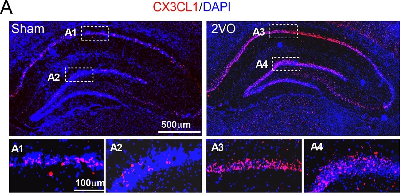
Detection of Rat CX3CL1/Fractalkine by Immunohistochemistry
Upregulation of the expression of CX3CL1 and CX3CR1 in the hippocampus of 2VO and miR-195 loss-of-function rats. a, b Expression of CX3CL1 (red) and CX3CR1 (green) in the hippocampus of sham and 2VO rats was shown by immunofluorescence staining. The scale bar is 500 um. c Expression of CX3CL1 and CX3CR1 in the hippocampus of sham and 2VO rats detected by western blot technique. Bars represent the mean ± SD, n = 6, *P < 0.05 vs sham group, Student’s t test. d Lenti-pre-AMO-miR-195 upregulated the expression of CX3CL1 in rat hippocampus. Mean ± SD, n = 6, *P < 0.05 vs sham; #P < 0.05 vs lenti-pre-AMO-miR-195. Data were analyzed using one-way ANOVA followed by Tukey test. e Lenti-pre-AMO-miR-195 upregulated the expression of CX3CR1 in the rat hippocampus. Mean ± SD, n = 6, *P < 0.05 vs sham; #P < 0.05 vs lenti-pre-AMO-miR-195. Data were analyzed using one-way ANOVA followed by Tukey test Image collected and cropped by CiteAb from the following open publication (https://pubmed.ncbi.nlm.nih.gov/32819407), licensed under a CC-BY license. Not internally tested by R&D Systems.
Rat CX3CL1 / Fractalkine ELISA Standard Curve
Recombinant Rat CX3CL1/Fractalkine (Catalog # 536-FR) was serially diluted and captured by Goat Anti-Rat CX3CL1/Fractalkine Chemokine Domain Antigen Affinity-purified Polyclonal Antibody (Catalog # AF537) coated on a Clear Polystyrene Microplate (Catalog # DY990). Goat Anti-Rat CX3CL1/Fractalkine Chemokine Domain Antigen Affinity-purified Polyclonal Antibody (Catalog # AF537) was biotinylated and incubated with the protein captured on the plate. Detection of the standard curve was achieved by incubating Streptavidin-HRP (Catalog # DY998)
Rat CX3CL1 / Fractalkine ELISA Standard Curve
Recombinant Rat CX3CL1/Fractalkine (Catalog # 536-FR) was serially diluted and captured by Goat Anti-Rat CX3CL1/Fractalkine Chemokine Domain Antigen Affinity-purified Polyclonal Antibody (Catalog # AF537) coated on a Clear Polystyrene Microplate (Catalog # DY990). Goat Anti-Rat CX3CL1/Fractalkine Chemokine Domain Antigen Affinity-purified Polyclonal Antibody (Catalog # AF537) was biotinylated and incubated with the protein captured on the plate. Detection of the standard curve was achieved by incubating Streptavidin-HRP (Catalog # DY998)Applications for Rat CX3CL1/Fractalkine Chemokine Domain Antibody
Application
Recommended Usage
Immunohistochemistry
5-15 µg/mL
Sample: Perfusion fixed frozen sections of mouse brain (cingulate cortex)
Sample: Perfusion fixed frozen sections of mouse brain (cingulate cortex)
Western Blot
0.1 µg/mL
Sample: Recombinant Rat CX3CL1/Fractalkine (Catalog # 536-FR)
Sample: Recombinant Rat CX3CL1/Fractalkine (Catalog # 536-FR)
Neutralization
Measured by its ability to neutralize CX3CL1/Fractalkine-induced chemotaxis in the BaF3 mouse pro‑B cell line transfected with human CX3CR1. The Neutralization Dose (ND50) is typically 0.3-1.2 µg/mL in the presence of 40 ng/mL Recombinant Rat CX3CL1/Fractalkine.
Rat CX3CL1/Fractalkine Sandwich Immunoassay
Please Note: Optimal dilutions of this antibody should be experimentally determined.
Reviewed Applications
Read 2 reviews rated 4 using AF537 in the following applications:
Formulation, Preparation, and Storage
Purification
Antigen Affinity-purified
Reconstitution
Reconstitute at 0.2 mg/mL in sterile PBS. For liquid material, refer to CoA for concentration.
Loading...
Formulation
Lyophilized from a 0.2 μm filtered solution in PBS with Trehalose. *Small pack size (SP) is supplied either lyophilized or as a 0.2 µm filtered solution in PBS.
Shipping
Lyophilized product is shipped at ambient temperature. Liquid small pack size (-SP) is shipped with polar packs. Upon receipt, store immediately at the temperature recommended below.
Stability & Storage
Use a manual defrost freezer and avoid repeated freeze-thaw cycles.
- 12 months from date of receipt, -20 to -70 °C as supplied.
- 1 month, 2 to 8 °C under sterile conditions after reconstitution.
- 6 months, -20 to -70 °C under sterile conditions after reconstitution.
Calculators
Background: CX3CL1/Fractalkine
References
- Kledal, T.N. et al. (1998) FEBS Lett. 441:209.
- Combadiere, C. et al. (1998) J. Biol. Chem. 273:23799.
- Harrison, J.L. et al. (1998) Proc. Natl. Acad. Sci. USA 95:10896.
- Rossi, D.L. et al. (1998) Genomics 47:163.
Alternate Names
FKN, Fractalkine, Neurotactin
Gene Symbol
CX3CL1
UniProt
Additional CX3CL1/Fractalkine Products
Product Documents for Rat CX3CL1/Fractalkine Chemokine Domain Antibody
Certificate of Analysis
To download a Certificate of Analysis, please enter a lot or batch number in the search box below.
Note: Certificate of Analysis not available for kit components.
Product Specific Notices for Rat CX3CL1/Fractalkine Chemokine Domain Antibody
For research use only
Related Research Areas
Citations for Rat CX3CL1/Fractalkine Chemokine Domain Antibody
 Powered by Bioz
Customer Reviews for Rat CX3CL1/Fractalkine Chemokine Domain Antibody (2)
4 out of 5
2 Customer Ratings
Have you used Rat CX3CL1/Fractalkine Chemokine Domain Antibody?
Submit a review and receive an Amazon gift card!
$25/€18/£15/$25CAN/¥2500 for a review with an image
$10/€7/£6/$10CAN/¥1110 for a review without an image
Submit a review
Customer Images
Showing
1
-
2 of
2 reviews
Showing All
Filter By:
-
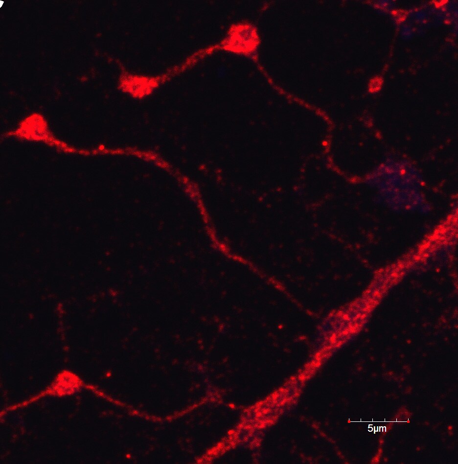
Application: Immunocytochemistry/ImmunofluorescenceSample Tested: Cortical neuronsSpecies: RatVerified Customer | Posted 12/19/2016Results published in: Article title: Noradrenaline induces CX3CL1 production and release by neurons Article reference: NP6524 Journal title: Neuropharmacology Final version published online: 08-Dec-2016 DOI information: 10.1016/j.neuropharm.2016.12.001

-
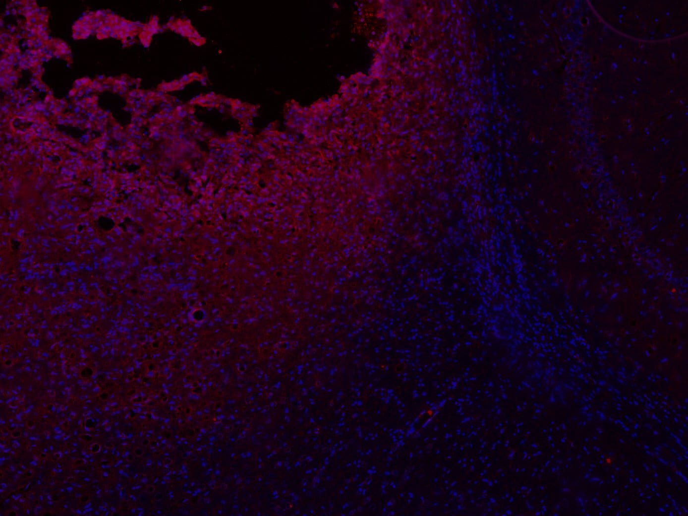
Application: ImmunohistochemistrySample Tested: Brain (cortex) tissueSpecies: RatVerified Customer | Posted 12/01/2016Traumatic brain injury site in rat brain Fractalkine antibody (red) and DAPI (blue)
There are no reviews that match your criteria.
Protocols
Find general support by application which include: protocols, troubleshooting, illustrated assays, videos and webinars.
- Antigen Retrieval Protocol (PIER)
- Antigen Retrieval for Frozen Sections Protocol
- Appropriate Fixation of IHC/ICC Samples
- Cellular Response to Hypoxia Protocols
- Chromogenic IHC Staining of Formalin-Fixed Paraffin-Embedded (FFPE) Tissue Protocol
- Chromogenic Immunohistochemistry Staining of Frozen Tissue
- ClariTSA™ Fluorophore Kits
- Detection & Visualization of Antibody Binding
- Fluorescent IHC Staining of Frozen Tissue Protocol
- Graphic Protocol for Heat-induced Epitope Retrieval
- Graphic Protocol for the Preparation and Fluorescent IHC Staining of Frozen Tissue Sections
- Graphic Protocol for the Preparation and Fluorescent IHC Staining of Paraffin-embedded Tissue Sections
- Graphic Protocol for the Preparation of Gelatin-coated Slides for Histological Tissue Sections
- IHC Sample Preparation (Frozen sections vs Paraffin)
- Immunofluorescent IHC Staining of Formalin-Fixed Paraffin-Embedded (FFPE) Tissue Protocol
- Immunohistochemistry (IHC) and Immunocytochemistry (ICC) Protocols
- Immunohistochemistry Frozen Troubleshooting
- Immunohistochemistry Paraffin Troubleshooting
- Preparing Samples for IHC/ICC Experiments
- Preventing Non-Specific Staining (Non-Specific Binding)
- Primary Antibody Selection & Optimization
- Protocol for Heat-Induced Epitope Retrieval (HIER)
- Protocol for Making a 4% Formaldehyde Solution in PBS
- Protocol for VisUCyte™ HRP Polymer Detection Reagent
- Protocol for the Preparation & Fixation of Cells on Coverslips
- Protocol for the Preparation and Chromogenic IHC Staining of Frozen Tissue Sections
- Protocol for the Preparation and Chromogenic IHC Staining of Frozen Tissue Sections - Graphic
- Protocol for the Preparation and Chromogenic IHC Staining of Paraffin-embedded Tissue Sections
- Protocol for the Preparation and Chromogenic IHC Staining of Paraffin-embedded Tissue Sections - Graphic
- Protocol for the Preparation and Fluorescent IHC Staining of Frozen Tissue Sections
- Protocol for the Preparation and Fluorescent IHC Staining of Paraffin-embedded Tissue Sections
- Protocol for the Preparation of Gelatin-coated Slides for Histological Tissue Sections
- R&D Systems Quality Control Western Blot Protocol
- TUNEL and Active Caspase-3 Detection by IHC/ICC Protocol
- The Importance of IHC/ICC Controls
- Troubleshooting Guide: Immunohistochemistry
- Troubleshooting Guide: Western Blot Figures
- Western Blot Conditions
- Western Blot Protocol
- Western Blot Protocol for Cell Lysates
- Western Blot Troubleshooting
- Western Blot Troubleshooting Guide
- View all Protocols, Troubleshooting, Illustrated assays and Webinars
Loading...
Associated Pathways