RIPK3/RIP3 Antibody - BSA Free
Novus Biologicals | Catalog # NBP1-77299
![Western Blot: RIPK3/RIP3 AntibodyBSA Free [NBP1-77299]](https://resources.rndsystems.com/images/products/RIPK3-RIP3-Antibody-Western-Blot-NBP1-77299-img0014.jpg "Western Blot: RIPK3/RIP3 AntibodyBSA Free [NBP1-77299]")
Key Product Details
Validated by
Knockout/Knockdown
Species Reactivity
Validated:
Human, Mouse, Rat
Cited:
Human, Mouse, Rat
Applications
Validated:
Knockout Validated, Immunohistochemistry, Immunohistochemistry-Paraffin, Western Blot, ELISA, Immunocytochemistry/ Immunofluorescence, Simple Western, Immunoprecipitation, Gel Supershift Assay, Knockdown Validated
Cited:
Knockout Validated, Immunohistochemistry, Immunohistochemistry-Paraffin, Western Blot, Immunocytochemistry/ Immunofluorescence, Immunoprecipitation, Proximity Ligation Assay, Gel Supershift Assay, IF/IHC, Knockdown Validated
Label
Unconjugated
Antibody Source
Polyclonal Rabbit IgG
Format
BSA Free
Loading...
Product Specifications
Immunogen
RIPK3/RIP3 Antibody was made to a 14 amino acid peptide near the carboxy terminus of murine RIP3. The immunogen is located within the last 50 amino acids of RIP3.
Reactivity Notes
Human, mouse and rat reactivity reported in multiple pieces of scientific literature.
Specificity
Mouse RIPK3/RIP3 Antibody has one isoform (486aa, 53 kDa). Human RIP3 has 3 isoforms, including isoform 1 (518aa, 57 kDa), isoform 2 (252aa, 28 kDa) and isoform 3 (231aa, 25 kDa). Rat RIP3 also has one isoform (478aa, 52 kDa). NBP1-77299 can detect can detect isoforms of mouse and rat as well as human isoform 1.
Clonality
Polyclonal
Host
Rabbit
Isotype
IgG
Theoretical MW
53 kDa.
Disclaimer note: The observed molecular weight of the protein may vary from the listed predicted molecular weight due to post translational modifications, post translation cleavages, relative charges, and other experimental factors.
Disclaimer note: The observed molecular weight of the protein may vary from the listed predicted molecular weight due to post translational modifications, post translation cleavages, relative charges, and other experimental factors.
Scientific Data Images for RIPK3/RIP3 Antibody - BSA Free
Western Blot: RIPK3/RIP3 AntibodyBSA Free [NBP1-77299]
Western Blot: RIPK3/RIP3 Antibody [NBP1-77299] - Analysis of RIP3 in HeLa cell lysate with RIPK3/RIP3 antibody [NBP1-77299] at (A) 1 and (B) 2 ug/mL. Observed molecular weight ~60 kDa. Theoretical molecular weight 53 kDa.![Western Blot: RIPK3/RIP3 AntibodyBSA Free [NBP1-77299]](https://resources.rndsystems.com/images/products/RIPK3-RIP3-Antibody-Western-Blot-NBP1-77299-img0022.jpg "Western Blot: RIPK3/RIP3 AntibodyBSA Free [NBP1-77299]")
Western Blot: RIPK3/RIP3 AntibodyBSA Free [NBP1-77299]
Western Blot: RIPK3/RIP3 Antibody [NBP1-77299] - Mouse Cell lines. 15 ug of lysates per lane. Antibodies: [NBP1-77299], (0.5 ug/mL), 1h incubation at RT in 5% NFDM/TBST. Secondary: Goat anti-rabbit IgG HRP conjugate at 1:10000 dilution. Observed molecular weight ~50 kDa. Theoretical molecular weight 53 kDa.![Western Blot: RIPK3/RIP3 AntibodyBSA Free [NBP1-77299]](https://resources.rndsystems.com/images/products/RIPK3-RIP3-Antibody-Western-Blot-NBP1-77299-img0024.jpg "Western Blot: RIPK3/RIP3 AntibodyBSA Free [NBP1-77299]")
Western Blot: RIPK3/RIP3 AntibodyBSA Free [NBP1-77299]
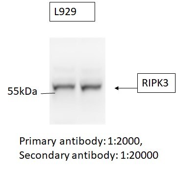
Western Blot: RIPK3/RIP3 Antibody [NBP1-77299] - Analysis using RIPK3/RIP3 antibody [NBP1-77299]. L929 cell line (mouse). 20 ug whole cell lysate. Primary antibody: 1:2000. Secondary antibody: 1:20000. Western blot image submitted by a verified customer review. Observed molecular weight ~57 kDa. Theoretical molecular weight 53 kDa.![Western Blot: RIPK3/RIP3 AntibodyBSA Free [NBP1-77299]](https://resources.rndsystems.com/images/products/RIPK3-RIP3-Antibody-Western-Blot-NBP1-77299-img0021.jpg "Western Blot: RIPK3/RIP3 AntibodyBSA Free [NBP1-77299]")
Western Blot: RIPK3/RIP3 AntibodyBSA Free [NBP1-77299]
Western Blot: RIPK3/RIP3 Antibody [NBP1-77299] - C2C12 Cells. 15 ug of lysates per lane. Antibodies: RIPK3/RIP3 antibody [NBP1-77299], 1h incubation at RT in 5% NFDM/TBST. Secondary: Goat anti-rabbit IgG HRP conjugate at 1:10000 dilution. Lane 1: NBP1-77299, 0.1 ug/mL in the presence of peptide blocking. Lane 2: NBP1-77299, 0.1 ug/mL Lane 3: NBP1-77299, 0.2 ug/mL Lane 4: NBP1-77299, 0.5 ug/mL. Observed molecular weight ~55 kDa. Theoretical molecular weight 53 kDa.![Knockout Validated: RIPK3/RIP3 Antibody - BSA Free [NBP1-77299]](https://resources.rndsystems.com/images/products/RIPK3-RIP3-Antibody-Knockout-Validated-NBP1-77299-img0025.jpg "Western Blot: RIPK3/RIP3 Antibody - BSA Free [NBP1-77299]")
![Knockdown Validated: RIPK3/RIP3 Antibody - BSA Free [NBP1-77299]](https://resources.rndsystems.com/images/products/RIPK3-RIP3-Antibody-Knockdown-Validated-NBP1-77299-img0026.jpg "Knockdown Validated: RIPK3/RIP3 Antibody - BSA Free [NBP1-77299]")

Western Blot: RIPK3/RIP3 Antibody - BSA Free [NBP1-77299] -
Western Blot: RIPK3/RIP3 Antibody - BSA Free [NBP1-77299] - Bone-marrow-derived macrophages exhibit significant reduction in RIPK3 levels in Ripk3 delta Global, Ripk3 delta MΦ-Cre & Ripk3 delta EC-Cre mice. (A,B) BMDMs were isolated, differentiated on chamber slides for 7 days, immunostained for macrophage markers CD68 (red; A) & CD11b (pink; B), & co-stained for nuclei (Hoechst; blue). All cells from seven separate BMDM isolations were positive for these markers (n=7). (C) A phagocytosis assay was performed on BMDMs with fluorescent polystyrene beads. Phagocytic cells display TRITC+/Hoechst+ signal, & 85% of cells from two separate BMDM isolations were double positive (n=2). (D-F) Protein or RNA was collected from control, Ripk3 delta Global, Ripk3 delta MΦ-Cre & Ripk3 delta EC-Cre BMDMs. RNA was converted to cDNA & analyzed by qPCR for Ripk3 levels (D). Protein lysates were immunoblotted to identify CD11b, RIPK3 & beta -actin (loading control) (E) & quantified (F). (G) RNA from control & Ripk3 delta Global BMDMs was converted to cDNA & analyzed by qPCR for Ripk3-Exon 10 levels. For panels D,F,G, each dot represents a BMDM isolation from an individual animal. Statistics for panels D & F were calculated using one-way ANOVA with Dunnett's multiple comparisons test. Overall ANOVA P-values (prior to the post hoc tests) are 0.0002 (D) & <0.0001 (F). Statistics for panel G were calculated using an unpaired t-test with Welch's correction. *P<0.05, **P<0.01, ***P<0.001, ****P<0.0001. Data are mean±s.d. Scale bars: 25 µm. Image collected & cropped by CiteAb from the following publication (https://pubmed.ncbi.nlm.nih.gov/31953345), licensed under a CC-BY license. Not internally tested by Novus Biologicals.
Western Blot: RIPK3/RIP3 Antibody - BSA Free [NBP1-77299] -
Western Blot: RIPK3/RIP3 Antibody - BSA Free [NBP1-77299] - p-MLKL levels are nearly undetectable in advanced plaques. (A-C) After 3 months on a Western diet, protein was collected from control (n=7), Ripk3 delta MΦ-Cre (n=5), Ripk3 delta EC-Cre (n=3) & Ripk3 delta SMC-Cre (n=3) aortas. Protein lysates were immunoblotted to identify p-MLKL (Abcam; #ab196436), MLKL & beta -actin (loading control) (A) & quantified (B,C). A faint positive p-MLKL signal can be seen in the representative blot in the control arch region; however, we could only detect p-MLKL in two out of the 18 aortas analyzed (B). Note that the same transfer membrane used for detecting CD11b (in Fig. 4S) was reprobed for p-MLKL & MLKL in A; the beta -actin control blots are therefore the same in both figures. (D,E) Protein lysates were immunoblotted to identify RIPK3 & GAPDH (loading control) (D) & quantified (E). For panels B,C,E, each dot represents an individual animal. Statistics were calculated using two-way ANOVA. Overall ANOVA P-values are 0.76 (B), 0.07 (C) & 0.46 (E). Data are mean±s.d. Image collected & cropped by CiteAb from the following publication (https://pubmed.ncbi.nlm.nih.gov/31953345), licensed under a CC-BY license. Not internally tested by Novus Biologicals.
Immunocytochemistry/ Immunofluorescence: RIPK3/RIP3 Antibody - BSA Free [NBP1-77299] -
Immunocytochemistry/ Immunofluorescence: RIPK3/RIP3 Antibody - BSA Free [NBP1-77299] - Validation of RIPK3/RIP3 in Rat Kidney Tissue. Immunofluorescent analysis of 4% paraformaldehyde-fixed Rat Kidney tissue labeling RIPK3/RIP3 with at 20 ug/mL, followed by goat anti-rabbit IgG secondary antibody at 1/500 dilution (red).
Immunohistochemistry: RIPK3/RIP3 Antibody - BSA Free [NBP1-77299] -
Immunohistochemistry: RIPK3/RIP3 Antibody - BSA Free [NBP1-77299] - Validation of RIPK3/RIP3 in Mouse Kidney Tissue. Immunohistochemical analysis of paraffin-embedded mouse kidney tissue using anti-RIPK3/RIP3 antibody at 2.5 ug/ml. Tissue was fixed with formaldehyde and blocked with 10% serum for 1 h at RT; antigen retrieval was by heat mediation with a citrate buffer (pH6). Samples were incubated with primary antibody overnight at 4C. A goat anti-rabbit IgG H&L (HRP) at 1/250 was used as secondary. Counter stained with Hematoxylin.
Immunohistochemistry: RIPK3/RIP3 Antibody - BSA Free [NBP1-77299] -
Immunohistochemistry: RIPK3/RIP3 Antibody - BSA Free [NBP1-77299] - Validation of RIPK3/RIP3 in Rat Kidney Tissue.Immunohistochemical analysis of paraffin-embedded rat kidney tissue using anti-RIPK3/RIP3 antibody at 5 ug/ml. Tissue was fixed with formaldehyde and blocked with 10% serum for 1 h at RT; antigen retrieval was by heat mediation with a citrate buffer (pH6). Samples were incubated with primary antibody overnight at 4C. A goat anti-rabbit IgG H&L (HRP) at 1/250 was used as secondary. Counter stained with Hematoxylin.
Western Blot: RIPK3/RIP3 Antibody - BSA Free [NBP1-77299] -
Western Blot: RIPK3/RIP3 Antibody - BSA Free [NBP1-77299] - Validation in Human Cell Lines. Loading: 15 ug of lysates per lane.Antibodies: RIPK3/RIP3, (0.5 ug/mL), 1h incubation at RT in 5% NFDM/TBST.Secondary: Goat anti-rabbit IgG HRP conjugate at 1:10000 dilution.
Knockout Validated: RIPK3/RIP3 Antibody - BSA Free [NBP1-77299] -
RIP3−/− mice exhibit a decrease in kidney injury after renal I/R. (A) Genotype used to confirm the RIP3+/− and RIP3−/− mice versus the littermate WT mouse. Note that the RIP3+/− heterozygote mouse displayed both alleles (knockout and WT bands). (B) Immunoblot confirming the knockdown of RIP3 in RIP3−/− versus littermate WT mice. (C) Left: H&E staining images showing an increase in kidney injury in WT mice after renal I/R compared to the sham-operated animals. However, in RIP3−/− mice, the tubular injury was reduced when compared to littermate WT mice after renal I/R. Right: Graph showing an increase in kidney injury score in WT mice after renal I/R compared to the sham-operated animals. However, in RIP3−/− mice, the tubular injury was reduced when compared to littermate WT mice after renal I/R. Note that there was no difference in kidney injury between WT and RIP3−/− sham mice. In addition, the cell necrosis, loss of the brush border, cast formation, and tubular dilatation were defined as: 0, none; 1, ≤10%; 2, 11%–25%; 3, 26%–45%; 4, 46%–75%; 5, >76%. Values are expressed as means +/- SEM; * p < 0.05 versus WT group; #p < 0.05 versus WT-I/R group, (n = 6/group). (D) Graph showing increases in the levels of serum creatine in WT mice after renal I/R compared to sham-operated animals. However, in RIP3−/− mice, the level of serum creatine was reduced when compared to littermate WT mice after renal I/R. Note that there was no difference in the levels of serum creatine between WT and RIP3−/− sham mice. Values are expressed as means +/- SEM; * p < 0.05 versus WT-Sham group, respectively; #p < 0.05 versus WT-I/R group (n = 6/group). Image collected and cropped by CiteAb from the following open publication (https://pubmed.ncbi.nlm.nih.gov/35741025), licensed under a CC-BY license. Not internally tested by Novus Biologicals.
Knockout Validated: RIPK3/RIP3 Antibody - BSA Free [NBP1-77299] -
RIP3−/− mice exhibit a decrease in kidney injury after renal I/R. (A) Genotype used to confirm the RIP3+/− and RIP3−/− mice versus the littermate WT mouse. Note that the RIP3+/− heterozygote mouse displayed both alleles (knockout and WT bands). (B) Immunoblot confirming the knockdown of RIP3 in RIP3−/− versus littermate WT mice. (C) Left: H&E staining images showing an increase in kidney injury in WT mice after renal I/R compared to the sham-operated animals. However, in RIP3−/− mice, the tubular injury was reduced when compared to littermate WT mice after renal I/R. Right: Graph showing an increase in kidney injury score in WT mice after renal I/R compared to the sham-operated animals. However, in RIP3−/− mice, the tubular injury was reduced when compared to littermate WT mice after renal I/R. Note that there was no difference in kidney injury between WT and RIP3−/− sham mice. In addition, the cell necrosis, loss of the brush border, cast formation, and tubular dilatation were defined as: 0, none; 1, ≤10%; 2, 11%–25%; 3, 26%–45%; 4, 46%–75%; 5, >76%. Values are expressed as means +/- SEM; * p < 0.05 versus WT group; #p < 0.05 versus WT-I/R group, (n = 6/group). (D) Graph showing increases in the levels of serum creatine in WT mice after renal I/R compared to sham-operated animals. However, in RIP3−/− mice, the level of serum creatine was reduced when compared to littermate WT mice after renal I/R. Note that there was no difference in the levels of serum creatine between WT and RIP3−/− sham mice. Values are expressed as means +/- SEM; * p < 0.05 versus WT-Sham group, respectively; #p < 0.05 versus WT-I/R group (n = 6/group). Image collected and cropped by CiteAb from the following open publication (https://pubmed.ncbi.nlm.nih.gov/35741025), licensed under a CC-BY license. Not internally tested by Novus Biologicals.Applications for RIPK3/RIP3 Antibody - BSA Free
Application
Recommended Usage
ELISA
1:100 - 1:2000
Immunocytochemistry/ Immunofluorescence
20 ug/ml
Immunohistochemistry
5 ug/ml
Immunohistochemistry-Paraffin
5 ug/ml
Immunoprecipitation
20 ug/mL
Simple Western
1:25
Western Blot
0.1-0.5 ug/ml
Application Notes
Gel supershift assay reported in scientific literature [PMID: 27721066]. Immunoprecipitation reported in scientific literature [PMID: 27861127]. RIPK3/RIP3 antibody validated for WB from a verified customer review. Knockout validation reported in scientific literature [PMID: 32246911]. Knockdown validation reported in scientific literature [PMID: 31655343]. ICC/IF, IHC, and WB reported in multiple pieces of scientific literature.
See Simple Western Antibody Database for Simple Western validation: Tested in Mouse, rat heart tissue 0.25 mg/mL, separated by Size, antibody dilution of 1:25, apparent MW was 70 kDa
See Simple Western Antibody Database for Simple Western validation: Tested in Mouse, rat heart tissue 0.25 mg/mL, separated by Size, antibody dilution of 1:25, apparent MW was 70 kDa
Reviewed Applications
Read 1 review rated 5 using NBP1-77299 in the following applications:
Formulation, Preparation, and Storage
Purification
Peptide affinity purified
Formulation
PBS
Format
BSA Free
Preservative
0.02% Sodium Azide
Concentration
1 mg/ml
Shipping
The product is shipped with polar packs. Upon receipt, store it immediately at the temperature recommended below.
Stability & Storage
Store at 4C.
Background: RIPK3/RIP3
RIPK3 has several phosphorylation sites that are required for its role in necroptosis. For example, serine204 in mouse, which is conserved in the human (serine199), is necessary for necroptosis while serine residues 232 and 227 are both required for RIPK3s interaction with MLKL (2). The core necroptosis proteins RIPK1/RIPK3 and MLKL are implicated in several disease states such as neurodegeneration, cardiovascular, hepatic and pulmonary disease (3).
References
1. Orozco, S., & Oberst, A. (2017). RIPK3 in cell death and inflammation: the good, the bad, and the ugly. Immunological Reviews. https://doi.org/10.1111/imr.12536
2. Dhuriya, Y. K., & Sharma, D. (2018). Necroptosis: A regulated inflammatory mode of cell death. Journal of Neuroinflammation. https://doi.org/10.1186/s12974-018-1235-0
3. Choi, M. E., Price, D. R., Ryter, S. W., & Choi, A. M. K. (2019). Necroptosis: A crucial pathogenic mediator of human disease. JCI Insight. https://doi.org/10.1172/jci.insight.128834
4. Lee, K.-H., & Kang, T.-B. (2019). The Molecular Links between Cell Death and Inflammasome. Cells. https://doi.org/10.3390/cells8091057
Long Name
Receptor (TNFRSF)-Interacting Serine-Threonine Kinase 3
Alternate Names
RIP3
Entrez Gene IDs
6063101 (Mouse)
Gene Symbol
RIPK3
UniProt
Additional RIPK3/RIP3 Products
Product Documents for RIPK3/RIP3 Antibody - BSA Free
Certificate of Analysis
To download a Certificate of Analysis, please enter a lot or batch number in the search box below.
Product Specific Notices for RIPK3/RIP3 Antibody - BSA Free
This product is for research use only and is not approved for use in humans or in clinical diagnosis. Primary Antibodies are guaranteed for 1 year from date of receipt.
Related Research Areas
Citations for RIPK3/RIP3 Antibody - BSA Free
 Powered by Bioz
Customer Reviews for RIPK3/RIP3 Antibody - BSA Free (1)
5 out of 5
1 Customer Rating
Have you used RIPK3/RIP3 Antibody - BSA Free?
Submit a review and receive an Amazon gift card!
$25/€18/£15/$25CAN/¥2500 Yen for a review with an image
$10/€7/£6/$10CAN/¥1110 Yen for a review without an image
Submit a review
Customer Images
Showing
1
-
1 of
1 review
Showing All
Filter By:
-
Application: Western BlotSample Tested: 20 ug whole cell lysateSpecies: L929 cellsVerified Customer | Posted 05/04/2020L929 cell line Primary antibody: 1:2000, Secondary antibody: 1:20000L929 cell line Primary antibody: 1:2000, Secondary antibody: 1:20000

There are no reviews that match your criteria.
Protocols
Find general support by application which include: protocols, troubleshooting, illustrated assays, videos and webinars.
- Antigen Retrieval Protocol (PIER)
- Antigen Retrieval for Frozen Sections Protocol
- Appropriate Fixation of IHC/ICC Samples
- Cellular Response to Hypoxia Protocols
- Chromogenic IHC Staining of Formalin-Fixed Paraffin-Embedded (FFPE) Tissue Protocol
- Chromogenic Immunohistochemistry Staining of Frozen Tissue
- ClariTSA™ Fluorophore Kits
- Detection & Visualization of Antibody Binding
- ELISA Sample Preparation & Collection Guide
- ELISA Troubleshooting Guide
- Fluorescent IHC Staining of Frozen Tissue Protocol
- Graphic Protocol for Heat-induced Epitope Retrieval
- Graphic Protocol for the Preparation and Fluorescent IHC Staining of Frozen Tissue Sections
- Graphic Protocol for the Preparation and Fluorescent IHC Staining of Paraffin-embedded Tissue Sections
- Graphic Protocol for the Preparation of Gelatin-coated Slides for Histological Tissue Sections
- How to Run an R&D Systems DuoSet ELISA
- How to Run an R&D Systems Quantikine ELISA
- How to Run an R&D Systems Quantikine™ QuicKit™ ELISA
- ICC Cell Smear Protocol for Suspension Cells
- ICC Immunocytochemistry Protocol Videos
- ICC for Adherent Cells
- IHC Sample Preparation (Frozen sections vs Paraffin)
- Immunocytochemistry (ICC) Protocol
- Immunocytochemistry Troubleshooting
- Immunofluorescence of Organoids Embedded in Cultrex Basement Membrane Extract
- Immunofluorescent IHC Staining of Formalin-Fixed Paraffin-Embedded (FFPE) Tissue Protocol
- Immunohistochemistry (IHC) and Immunocytochemistry (ICC) Protocols
- Immunohistochemistry Frozen Troubleshooting
- Immunohistochemistry Paraffin Troubleshooting
- Immunoprecipitation Protocol
- Preparing Samples for IHC/ICC Experiments
- Preventing Non-Specific Staining (Non-Specific Binding)
- Primary Antibody Selection & Optimization
- Protocol for Heat-Induced Epitope Retrieval (HIER)
- Protocol for Making a 4% Formaldehyde Solution in PBS
- Protocol for VisUCyte™ HRP Polymer Detection Reagent
- Protocol for the Fluorescent ICC Staining of Cell Smears - Graphic
- Protocol for the Fluorescent ICC Staining of Cultured Cells on Coverslips - Graphic
- Protocol for the Preparation & Fixation of Cells on Coverslips
- Protocol for the Preparation and Chromogenic IHC Staining of Frozen Tissue Sections
- Protocol for the Preparation and Chromogenic IHC Staining of Frozen Tissue Sections - Graphic
- Protocol for the Preparation and Chromogenic IHC Staining of Paraffin-embedded Tissue Sections
- Protocol for the Preparation and Chromogenic IHC Staining of Paraffin-embedded Tissue Sections - Graphic
- Protocol for the Preparation and Fluorescent ICC Staining of Cells on Coverslips
- Protocol for the Preparation and Fluorescent ICC Staining of Non-adherent Cells
- Protocol for the Preparation and Fluorescent ICC Staining of Stem Cells on Coverslips
- Protocol for the Preparation and Fluorescent IHC Staining of Frozen Tissue Sections
- Protocol for the Preparation and Fluorescent IHC Staining of Paraffin-embedded Tissue Sections
- Protocol for the Preparation of Gelatin-coated Slides for Histological Tissue Sections
- Protocol for the Preparation of a Cell Smear for Non-adherent Cell ICC - Graphic
- Quantikine HS ELISA Kit Assay Principle, Alkaline Phosphatase
- Quantikine HS ELISA Kit Principle, Streptavidin-HRP Polymer
- R&D Systems Quality Control Western Blot Protocol
- Sandwich ELISA (Colorimetric) – Biotin/Streptavidin Detection Protocol
- Sandwich ELISA (Colorimetric) – Direct Detection Protocol
- TUNEL and Active Caspase-3 Detection by IHC/ICC Protocol
- The Importance of IHC/ICC Controls
- Troubleshooting Guide: ELISA
- Troubleshooting Guide: Immunohistochemistry
- Troubleshooting Guide: Western Blot Figures
- Western Blot Conditions
- Western Blot Protocol
- Western Blot Protocol for Cell Lysates
- Western Blot Troubleshooting
- Western Blot Troubleshooting Guide
- View all Protocols, Troubleshooting, Illustrated assays and Webinars
FAQs for RIPK3/RIP3 Antibody - BSA Free
Showing
1
-
5 of
5 FAQs
Showing All
-
Q: I am currently looking for a good antibody working as an ELISA capture antibody for mouse RIP3. Would you let me know if this application was tested during the development of your NovusBio (NBP1-77299) antibody.
A: Our product NBP1-77299 has been validated for use in ELISA. We tested this product in a direct ELISA format; it has not yet been tested in a sandwich ELISA format. The immunogen used to make this product falls between amino acids 473 to 486 of the mouse protein. If your detection antibody is outside of this range, it is possible that this combination will yield positive results.
-
Q: If this product is used in an application or species as a part of a customer review, will that validate this product in the application/species?
A: If any of our primary antibodes are used in an untested application or species and it is shown to work through images from customer reviews or through publications, this validates the application/species for this product. Please check out our Innovator's Reward Program if you decide to test an antibody with a species or application that is not currently listed.
-
Q: What human isoforms can be detected by this antibody?
A: RIPK3/RIP3 Antibody [NBP1-77299] can detect human isoform 1. This antibody can also detect isoforms of mouse and rat.
-
Q: What is the theoretical molecular weight for your RIPK3/RIP3 antibodies?
A: This depends on the immunogen. We have one RIPK3/RIP3 antibody [NBP1-77299] that is raised against murine RIP3. The theoretical molecular weight for this product is 53 kDa. The rest of our RIPK3/RIP3 antibodies are developed against human RIP3 and have a theoretical molecular weight of 56.7 kDa.
-
Q: What research areas can this product be used in?
A: All RIPK3/RIP3 products can be used in: Cancer, Cell Biology, Immunology, Necroptosis, Protein Kinase, Signal Transduction.
-
Q: I am currently looking for a good antibody working as an ELISA capture antibody for mouse RIP3. Would you let me know if this application was tested during the development of your NovusBio (NBP1-77299) antibody.
A: Our product NBP1-77299 has been validated for use in ELISA. We tested this product in a direct ELISA format; it has not yet been tested in a sandwich ELISA format. The immunogen used to make this product falls between amino acids 473 to 486 of the mouse protein. If your detection antibody is outside of this range, it is possible that this combination will yield positive results.
-
Q: If this product is used in an application or species as a part of a customer review, will that validate this product in the application/species?
A: If any of our primary antibodes are used in an untested application or species and it is shown to work through images from customer reviews or through publications, this validates the application/species for this product. Please check out our Innovator's Reward Program if you decide to test an antibody with a species or application that is not currently listed.
-
Q: What human isoforms can be detected by this antibody?
A: RIPK3/RIP3 Antibody [NBP1-77299] can detect human isoform 1. This antibody can also detect isoforms of mouse and rat.
-
Q: What is the theoretical molecular weight for your RIPK3/RIP3 antibodies?
A: This depends on the immunogen. We have one RIPK3/RIP3 antibody [NBP1-77299] that is raised against murine RIP3. The theoretical molecular weight for this product is 53 kDa. The rest of our RIPK3/RIP3 antibodies are developed against human RIP3 and have a theoretical molecular weight of 56.7 kDa.
-
Q: What research areas can this product be used in?
A: All RIPK3/RIP3 products can be used in: Cancer, Cell Biology, Immunology, Necroptosis, Protein Kinase, Signal Transduction.
Loading...