Key Product Details
Species Reactivity
Validated:
Human, Mouse, Rat, Bovine
Cited:
Human, Mouse, Rat
Applications
Validated:
Immunohistochemistry, Immunohistochemistry-Paraffin, Western Blot, Flow Cytometry, Immunocytochemistry/ Immunofluorescence
Cited:
Immunohistochemistry-Paraffin, Western Blot, IF/IHC
Label
Unconjugated
Antibody Source
Monoclonal Mouse IgG2a Kappa Clone # 4C4.9
Loading...
Product Specifications
Immunogen
Purified bovine brain S100B protein (Uniprot: P04271)
Localization
Cytoplasmic and Nuclear
Marker
Astrocyte and Melanoma Marker
Specificity
S100 belongs to the family of calcium binding proteins. S100A and S100B proteins are two members of the S100 family. S100A is composed of an alpha and a beta chain whereas S100B is composed of two beta chains. This antibody is specific against an epitope located on the beta-chain (i.e. in S-100A and S-100B) but not on the alpha-chain of S-100 (i.e. in S-100A and S100A0). This antibody can be used to localize S-100A and S-100B in various tissue sections. S-100 protein has been found in normal melanocytes, Langerhans cells, histiocytes, chondrocytes, lipocytes, skeletal and cardiac muscle, Schwann cells, epithelial and myoepithelial cells of the breast, salivary and sweat glands, as well as in glial cells. Neoplasms derived from these cells also express S-100 protein, albeit non-uniformly. A large number of well-differentiated tumors of the salivary gland, adipose and cartilaginous tissue, and Schwann cell-derived tumors express S-100 protein. Almost all malignant melanomas and cases of histiocytosis X are positive for S-100 protein.
Clonality
Monoclonal
Host
Mouse
Isotype
IgG2a Kappa
Description
200ug/ml of antibody purified from Bioreactor Concentrate by Protein A or G. Prepared in 10 mM PBS with 0.05% BSA & 0.05% azide. Also available WITHOUT BSA & azide at 1.0 mg/ml. (NBP2-33168)
Antibody with azide - store at 2 to 8C. Antibody without azide - store at -20 to -80C.
Antibody with azide - store at 2 to 8C. Antibody without azide - store at -20 to -80C.
Scientific Data Images for S100B Antibody (4C4.9)
![Western Blot: S100B Antibody (4C4.9) [NBP2-29403]](https://resources.rndsystems.com/images/products/S100B-Antibody-4C4-9-Western-Blot-NBP2-29403-img0001.jpg "Western Blot: S100B Antibody (4C4.9) [NBP2-29403]")
Western Blot: S100B Antibody (4C4.9) [NBP2-29403]
Western Blot: S100B Antibody (4C4.9) [NBP2-29403] - Analysis of human brain lysate using S100 antibody at 1 ug/ml.![Immunocytochemistry/ Immunofluorescence: S100B Antibody (4C4.9) [NBP2-29403]](https://resources.rndsystems.com/images/products/S100B-Antibody-4C4-9-Immunocytochemistry-Immunofluorescence-NBP2-29403-img0004.jpg "Immunocytochemistry/ Immunofluorescence: S100B Antibody (4C4.9) [NBP2-29403]")
Immunocytochemistry/ Immunofluorescence: S100B Antibody (4C4.9) [NBP2-29403]
Immunocytochemistry/Immunofluorescence: S100B Antibody (4C4.9) [NBP2-29403] - Confocal Immunofluorescent analysis of A2058 cells using AF488-labeled S100B Monoclonal Antibody (4C4.9) (Green). F-actin filaments were labeled with DyLight 554 Phalloidin (red). DAPI was used to stain the cell nuclei (blue).![Immunohistochemistry-Paraffin: S100B Antibody (4C4.9) [NBP2-29403]](https://resources.rndsystems.com/images/products/S100B-Antibody-4C4-9-Immunohistochemistry-Paraffin-NBP2-29403-img0009.jpg "Immunohistochemistry-Paraffin: S100B Antibody (4C4.9) [NBP2-29403]")
Immunohistochemistry-Paraffin: S100B Antibody (4C4.9) [NBP2-29403]
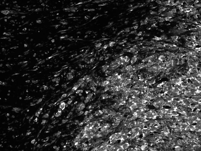
Immunohistochemistry-Paraffin: S100B Antibody (4C4.9) [NBP2-29403] - CODEX multiplexing image of S100b in primary melanoma of the skin. Image from verified customer review.![Immunohistochemistry-Paraffin: S100B Antibody (4C4.9) [NBP2-29403]](https://resources.rndsystems.com/images/products/S100B-Antibody-4C4-9-Immunohistochemistry-Paraffin-NBP2-29403-img0003.jpg "Immunohistochemistry-Paraffin: S100B Antibody (4C4.9) [NBP2-29403]")
Immunohistochemistry-Paraffin: S100B Antibody (4C4.9) [NBP2-29403]
Immunohistochemistry-Paraffin: S100B Antibody (4C4.9) [NBP2-29403] - Formalin-fixed, paraffin-embedded human melanoma stained with S100B antibody(4C4.9).![Immunohistochemistry-Paraffin: S100B Antibody (4C4.9) [NBP2-29403]](https://resources.rndsystems.com/images/products/S100B-Antibody-4C4-9-Immunohistochemistry-Paraffin-NBP2-29403-img0007.jpg "Immunohistochemistry-Paraffin: S100B Antibody (4C4.9) [NBP2-29403]")
Immunohistochemistry-Paraffin: S100B Antibody (4C4.9) [NBP2-29403]
Immunohistochemistry-Paraffin: S100B Antibody (4C4.9) [NBP2-29403] - Confocal Immunofluorescent analysis of A2058 cells using AF488-labeled S100B Monoclonal antibody (4C4.9) (Green). F-actin filaments were labeled with DyLight 554 Phalloidin (red). DAPI was used to stain the cell nuclei (blue).![Immunohistochemistry-Paraffin: S100B Antibody (4C4.9) [NBP2-29403]](https://resources.rndsystems.com/images/products/S100B-Antibody-4C4-9-Immunohistochemistry-Paraffin-NBP2-29403-img0008.jpg "Immunohistochemistry-Paraffin: S100B Antibody (4C4.9) [NBP2-29403]")
Immunohistochemistry-Paraffin: S100B Antibody (4C4.9) [NBP2-29403]
Immunohistochemistry-Paraffin: S100B Antibody (4C4.9) [NBP2-29403] - Formalin-fixed, paraffin-embedded human Melanoma stained with S100B Antibody (4C4.9) [NBP2-29403] -")
Western Blot: S100B Antibody (4C4.9) [NBP2-29403] -
Injection of H-sEVs increases tumor ECM1 levels, cancer growth and metastasis in CD-fed 4T1-bearing mouse model.a Location of the injected 4T1 cells in the mammary fat pad of the female BALB/c mice (upper panel indicated by an arrow) and accumulation of the tail vein injected DIR-labeled sEVs in the mammary pad of the mice (lower panel). b A schematic diagram showing the sEVs treatment protocol. c Protein expressions of ECM1, MMP3 and S100A/B in tumors of CD-fed 4T1-bearing BALB/c mice after C-sEVs or H-sEVs treatments. d Tumors, e tumor size, f tumor weight and g lung metastasis of the CD-fed 4T1-bearing BALB/c mice after the sEVs treatments. Mouse tumor sizes are presented as the mean +/- SEM, other data are presented as mean +/- SD; two-sided unpaired t-test for (c, f, g); n = 7 mice in each group; p values are indicated in graphs. C-sEVs, circulating sEVs in CD-fed BALB/c mice; H-sEVs, circulating sEVs in HFD-fed BALB/c mice; ECM1 extracellular matrix protein 1; MMP3 matrix metallopeptidase 3. Source data are provided as a Source Data file. Image collected and cropped by CiteAb from the following open publication (https://www.nature.com/articles/s41467-024-45995-5), licensed under a CC-BY license. Not internally tested by Novus Biologicals. [NBP2-29403] -")
Western Blot: S100B Antibody (4C4.9) [NBP2-29403] -
ECM1 in the sEVs plays a role in enhancing BC growth and metastasis.a A schematic diagram showing the sEVs treatment protocol. b Protein expressions of ECM1, MMP3 and S100A/B in the tumors of E0771-bearing B6/J-Rab27a-Cas9-KO mice after C-sEVs, D-sEVs or ECM-sEVs treatments. c Tumors, d tumor size, e tumor weight and f lung metastasis of the E0771-bearing B6/J-Rab27a-Cas9-KO mice after sEVs treatments. Mouse tumor sizes are presented as the mean +/- SEM, other data are presented as mean +/- SD; one-way ANOVA with Tukey’s multiple comparison test for (b, d, e); n = 7 mice in each group, p values are indicated in graphs. Rab27aKO, B6/J-Rab27a-Cas9-KO mice; C-sEVs, circulating sEVs in CD-fed C57BL/6 mice; D-sEVs, circulating sEVs in high-fat-diet induced obesity C57BL/6 mice; ECM-sEVs, ECM1 construct-loaded C-sEVs; ECM1, extracellular matrix protein 1; MMP3, matrix metallopeptidase 3. Source data are provided as a Source Data file. Image collected and cropped by CiteAb from the following open publication (https://www.nature.com/articles/s41467-024-45995-5), licensed under a CC-BY license. Not internally tested by Novus Biologicals.Applications for S100B Antibody (4C4.9)
Application
Recommended Usage
Flow Cytometry
1-2 ug/million cells
Immunocytochemistry/ Immunofluorescence
1-2 ug/ml
Immunohistochemistry-Paraffin
0.25-0.5 ug/ml
Western Blot
1-2 ug/ml
Application Notes
Immunohistochemistry (Formalin-fixed): 0.25-0.5ug/ml for 30 minutes at RT. Staining of formalin-fixed tissues requires heating tissue sections in 10mM Tris with 1mM EDTA, pH 9.0, for 45 min at 95C followed by cooling at RT for 20 minutes.
Optimal dilution for a specific application should be determined.
Optimal dilution for a specific application should be determined.
Reviewed Applications
Read 1 review rated 5 using NBP2-29403 in the following applications:
Flow Cytometry Panel Builder
Bio-Techne Knows Flow Cytometry
Save time and reduce costly mistakes by quickly finding compatible reagents using the Panel Builder Tool.
Advanced Features
- Spectra Viewer - Custom analysis of spectra from multiple fluorochromes
- Spillover Popups - Visualize the spectra of individual fluorochromes
- Antigen Density Selector - Match fluorochrome brightness with antigen density

Formulation, Preparation, and Storage
Purification
Protein A or G purified
Formulation
10 mM PBS with 0.05% BSA
Preservative
0.05% Sodium Azide
Concentration
0.2 mg/ml
Shipping
The product is shipped with polar packs. Upon receipt, store it immediately at the temperature recommended below.
Stability & Storage
Store at 4C.
Background: S100B
References
1. Yardan, T., Erenler, A. K., Baydin, A., Aydin, K., & Cokluk, C. (2011). Usefulness of S100B protein in neurological disorders. JPMA. The Journal of the Pakistan Medical Association, 61(3), 276-281.
2. Langeh, U., & Singh, S. (2021). Targeting S100B Protein as a Surrogate Biomarker and its Role in Various Neurological Disorders. Current neuropharmacology, 19(2), 265-277. https://doi.org/10.2174/1570159X18666200729100427
3. Thelin, E. P., Nelson, D. W., & Bellander, B. M. (2017). A review of the clinical utility of serum S100B protein levels in the assessment of traumatic brain injury. Acta neurochirurgica, 159(2), 209-225. https://doi.org/10.1007/s00701-016-3046-3
4. Wang, K. K., Yang, Z., Zhu, T., Shi, Y., Rubenstein, R., Tyndall, J. A., & Manley, G. T. (2018). An update on diagnostic and prognostic biomarkers for traumatic brain injury. Expert review of molecular diagnostics, 18(2), 165-180. https://doi.org/10.1080/14737159.2018.1428089
Long Name
S100 Calcium Binding Protein B
Alternate Names
beta (neural), NEF, S100, S100 beta, S100 calcium binding protein B, S100 calcium-binding protein B, S100 calcium-binding protein, beta (neural), S-100 calcium-binding protein, beta chain, 10protein S100-B, S-100 protein beta chain, S-100 protein subunit beta, S100beta
Gene Symbol
S100B
Additional S100B Products
Product Documents for S100B Antibody (4C4.9)
Certificate of Analysis
To download a Certificate of Analysis, please enter a lot or batch number in the search box below.
Product Specific Notices for S100B Antibody (4C4.9)
This product is for research use only and is not approved for use in humans or in clinical diagnosis. Primary Antibodies are guaranteed for 1 year from date of receipt.
Citations for S100B Antibody (4C4.9)
 Powered by Bioz
Customer Reviews for S100B Antibody (4C4.9) (1)
5 out of 5
1 Customer Rating
Have you used S100B Antibody (4C4.9)?
Submit a review and receive an Amazon gift card!
$25/€18/£15/$25CAN/¥2500 Yen for a review with an image
$10/€7/£6/$10CAN/¥1110 Yen for a review without an image
Submit a review
Customer Images
Showing
1
-
1 of
1 review
Showing All
Filter By:
-
Application: Immunohistochemistry-ParaffinSample Tested: human melanomaSpecies: HumanVerified Customer | Posted 11/02/2022CODEX multiplexing image of S100b in primary melanoma of the skin

There are no reviews that match your criteria.
Protocols
Find general support by application which include: protocols, troubleshooting, illustrated assays, videos and webinars.
- 7-Amino Actinomycin D (7-AAD) Cell Viability Flow Cytometry Protocol
- Antigen Retrieval Protocol (PIER)
- Antigen Retrieval for Frozen Sections Protocol
- Appropriate Fixation of IHC/ICC Samples
- Cellular Response to Hypoxia Protocols
- Chromogenic IHC Staining of Formalin-Fixed Paraffin-Embedded (FFPE) Tissue Protocol
- Chromogenic Immunohistochemistry Staining of Frozen Tissue
- ClariTSA™ Fluorophore Kits
- Detection & Visualization of Antibody Binding
- Extracellular Membrane Flow Cytometry Protocol
- Flow Cytometry Protocol for Cell Surface Markers
- Flow Cytometry Protocol for Staining Membrane Associated Proteins
- Flow Cytometry Staining Protocols
- Flow Cytometry Troubleshooting Guide
- Fluorescent IHC Staining of Frozen Tissue Protocol
- Graphic Protocol for Heat-induced Epitope Retrieval
- Graphic Protocol for the Preparation and Fluorescent IHC Staining of Frozen Tissue Sections
- Graphic Protocol for the Preparation and Fluorescent IHC Staining of Paraffin-embedded Tissue Sections
- Graphic Protocol for the Preparation of Gelatin-coated Slides for Histological Tissue Sections
- ICC Cell Smear Protocol for Suspension Cells
- ICC Immunocytochemistry Protocol Videos
- ICC for Adherent Cells
- IHC Sample Preparation (Frozen sections vs Paraffin)
- Immunocytochemistry (ICC) Protocol
- Immunocytochemistry Troubleshooting
- Immunofluorescence of Organoids Embedded in Cultrex Basement Membrane Extract
- Immunofluorescent IHC Staining of Formalin-Fixed Paraffin-Embedded (FFPE) Tissue Protocol
- Immunohistochemistry (IHC) and Immunocytochemistry (ICC) Protocols
- Immunohistochemistry Frozen Troubleshooting
- Immunohistochemistry Paraffin Troubleshooting
- Intracellular Flow Cytometry Protocol Using Alcohol (Methanol)
- Intracellular Flow Cytometry Protocol Using Detergents
- Intracellular Nuclear Staining Flow Cytometry Protocol Using Detergents
- Intracellular Staining Flow Cytometry Protocol Using Alcohol Permeabilization
- Intracellular Staining Flow Cytometry Protocol Using Detergents to Permeabilize Cells
- Preparing Samples for IHC/ICC Experiments
- Preventing Non-Specific Staining (Non-Specific Binding)
- Primary Antibody Selection & Optimization
- Propidium Iodide Cell Viability Flow Cytometry Protocol
- Protocol for Heat-Induced Epitope Retrieval (HIER)
- Protocol for Liperfluo
- Protocol for Making a 4% Formaldehyde Solution in PBS
- Protocol for VisUCyte™ HRP Polymer Detection Reagent
- Protocol for the Characterization of Human Th22 Cells
- Protocol for the Characterization of Human Th9 Cells
- Protocol for the Fluorescent ICC Staining of Cell Smears - Graphic
- Protocol for the Fluorescent ICC Staining of Cultured Cells on Coverslips - Graphic
- Protocol for the Preparation & Fixation of Cells on Coverslips
- Protocol for the Preparation and Chromogenic IHC Staining of Frozen Tissue Sections
- Protocol for the Preparation and Chromogenic IHC Staining of Frozen Tissue Sections - Graphic
- Protocol for the Preparation and Chromogenic IHC Staining of Paraffin-embedded Tissue Sections
- Protocol for the Preparation and Chromogenic IHC Staining of Paraffin-embedded Tissue Sections - Graphic
- Protocol for the Preparation and Fluorescent ICC Staining of Cells on Coverslips
- Protocol for the Preparation and Fluorescent ICC Staining of Non-adherent Cells
- Protocol for the Preparation and Fluorescent ICC Staining of Stem Cells on Coverslips
- Protocol for the Preparation and Fluorescent IHC Staining of Frozen Tissue Sections
- Protocol for the Preparation and Fluorescent IHC Staining of Paraffin-embedded Tissue Sections
- Protocol for the Preparation of Gelatin-coated Slides for Histological Tissue Sections
- Protocol for the Preparation of a Cell Smear for Non-adherent Cell ICC - Graphic
- Protocol: Annexin V and PI Staining by Flow Cytometry
- Protocol: Annexin V and PI Staining for Apoptosis by Flow Cytometry
- R&D Systems Quality Control Western Blot Protocol
- TUNEL and Active Caspase-3 Detection by IHC/ICC Protocol
- The Importance of IHC/ICC Controls
- Troubleshooting Guide: Fluorokine Flow Cytometry Kits
- Troubleshooting Guide: Immunohistochemistry
- Troubleshooting Guide: Western Blot Figures
- Western Blot Conditions
- Western Blot Protocol
- Western Blot Protocol for Cell Lysates
- Western Blot Troubleshooting
- Western Blot Troubleshooting Guide
- View all Protocols, Troubleshooting, Illustrated assays and Webinars
Loading...
Associated Pathways
