SARS Nucleocapsid Protein Antibody - BSA Free
Novus Biologicals | Catalog # NB100-56683
![Simple Western: SARS Nucleocapsid Protein Antibody [NB100-56683]](https://resources.rndsystems.com/images/products/SARS-Nucleocapsid-Protein-Antibody-Simple-Western-NB100-56683-img0004.jpg "Simple Western: SARS Nucleocapsid Protein Antibody [NB100-56683]")
Loading...
Key Product Details
Validated by
Orthogonal Validation
Species Reactivity
Validated:
SARS-CoV, SARS-CoV-2
Cited:
Human, Hamster, Virus - SARS-CoV-2
Applications
Validated:
Immunohistochemistry, Immunohistochemistry-Paraffin, Western Blot, ELISA, Dual RNAscope ISH-IHC, Immunocytochemistry/ Immunofluorescence, Simple Western
Cited:
Immunohistochemistry-Paraffin, Western Blot, Immunocytochemistry/ Immunofluorescence, IF/IHC
Label
Unconjugated
Antibody Source
Polyclonal Rabbit IgG
Format
BSA Free
Loading...
Product Specifications
Immunogen
The antibody was developed by immunizing rabbits with a synthetic peptide corresponding to amino acids 354-370 (NKHIDAYKTFPPTEPKK-C) from the N (SARS Nucleocapsid) for the Human SARS coronavirus (Genbank accession no. YP_009724397.2)
Specificity
The was tested on a human cell line transfected with full-length SARS Nucleocapsid cDNA with a predicted molecular weight of 46 kDa.
Clonality
Polyclonal
Host
Rabbit
Isotype
IgG
Scientific Data Images for SARS Nucleocapsid Protein Antibody - BSA Free
Simple Western: SARS Nucleocapsid Protein Antibody [NB100-56683]
Simple Western: SARS Nucleocapsid Protein Antibody [NB100-56683] - Simple Western lane view shows recombinant SARS-CoV-2 Nucleocapsid Protein (Catalog # 10474-CV), loaded at 20 ng/mL. A specific band was detected for SARS-CoV-2 Nucleocapsid Protein at approximately 60 kDa (as indicated) using a serial dilution of Rabbit Anti-SARS-CoV Nucleocapsid Protein Polyclonal Antibody (Catalog # NB100-56683) followed by incubation with HRP-conjugated Anti-Goat IgG Secondary Antibody. This experiment was conducted under reducing conditions and using the 12-230 kDa separation system.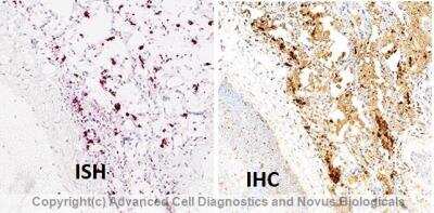
Dual RNAscope ISH-IHC: SARS Nucleocapsid Protein Antibody [NB100-56683] - Formalin-fixed paraffin-embedded tissue sections of SARS-CoV-2 infected human lung tissue were probed for SARS-CoV-2 viral RNA (ACD anti-sense specific probe v-nCoV2019-S [848561]); Fast Red chromogen, ACD [322360]). Adjacent tissue section was processed for immunohistochemistry using rabbit polyclonal anti-SARS Nucleocapsid Antibody [NB100-56683] at 15ug/mL with 1 hr incubation at 25 degrees Celsius followed by incubation with anti-rabbit IgG VisUCyte HRP Polymer Antibody [VC003] and DAB chromogen (yellow-brown). Tissue was counterstained with hematoxylin (blue). Specific staining was localized to SARS-CoV-2 infected cells.
![Simple Western: SARS Nucleocapsid Protein Antibody [NB100-56683]](https://resources.rndsystems.com/images/products/SARS-Nucleocapsid-Protein-Antibody-Simple-Western-NB100-56683-img0003.jpg "Simple Western: SARS Nucleocapsid Protein Antibody [NB100-56683]")
Simple Western: SARS Nucleocapsid Protein Antibody [NB100-56683]
Simple Western: SARS Nucleocapsid Protein Antibody [NB100-56683] - Simple Western lane view shows lysates of SARS-CoV-2 (1:50), MERS (1:100), OC43 (1:100), and 229E (1:100). A specific band was detected for SARS-CoV-2 Nucleocapsid Protein at approximately 60 kDa (as indicated) only in the SARS-CoV-2 lysate using 25 ug/mL of Rabbit Anti-SARS-CoV Nucleocapsid Protein Polyclonal Antibody (Catalog # NB100-56683) followed by incubation with HRP-conjugated Anti-Goat IgG Secondary Antibody. This experiment was conducted under reducing conditions and using the 12-230 kDa separation system. Note: some reactivity observed with FL Std 230. SARS-CoV-2 lysate courtesy of University of Maryland.![Western Blot: SARS Nucleocapsid Protein Antibody [NB100-56683]](https://resources.rndsystems.com/images/products/SARS-Nucleocapsid-Protein-Antibody-Western-Blot-NB100-56683-img0002.jpg "Western Blot: SARS Nucleocapsid Protein Antibody [NB100-56683]")
Western Blot: SARS Nucleocapsid Protein Antibody [NB100-56683]
Western Blot: SARS Nucleocapsid Protein Antibody [NB100-56683] - Analysis of SARS Nucleocapsid in (A) untransfected mouse melanoma cell lysate and (B) transfected cell lysate using this antibody at a 1:2000 dilution.Applications for SARS Nucleocapsid Protein Antibody - BSA Free
Application
Recommended Usage
ELISA
1:100-1:2000
Immunocytochemistry/ Immunofluorescence
reported in scientific literature (PMID 16014910)
Simple Western
1:50
Western Blot
1:100-1:2000
Formulation, Preparation, and Storage
Purification
Protein G purified
Formulation
PBS
Format
BSA Free
Preservative
0.05% Sodium Azide
Concentration
1 mg/ml
Shipping
The product is shipped with polar packs. Upon receipt, store it immediately at the temperature recommended below.
Stability & Storage
Store at 4C short term. Aliquot and store at -20C long term. Avoid freeze-thaw cycles.
Background: SARS Nucleocapsid Protein
Alternate Names
N, N protein, N structural protein, NC, Nucleocapsid protein, Nucleoprotein, Protein N, SARS coronavirus N protein, SARS coronavirus nucleocapsid protein, SARS CoV, SARS CoV N protein, SARS CoV nucleocapsid protein, SARS N protein, SARS Nucleoprotein, SARSCoV, SARSCoV N protein, SARSCoV nucleocapsid protein, Severe acute respiratory syndrome, COVID-19 nucleocapsid protein, SARSCOV2 N protein, SARS-COV-2 nucleocapsid protein, SARS-COV-2 Nucleoprotein
Gene Symbol
N
UniProt
Additional SARS Nucleocapsid Protein Products
Product Documents for SARS Nucleocapsid Protein Antibody - BSA Free
Certificate of Analysis
To download a Certificate of Analysis, please enter a lot or batch number in the search box below.
Product Specific Notices for SARS Nucleocapsid Protein Antibody - BSA Free
This product is for research use only and is not approved for use in humans or in clinical diagnosis. Primary Antibodies are guaranteed for 1 year from date of receipt.
Citations for SARS Nucleocapsid Protein Antibody - BSA Free
 Powered by Bioz
Customer Reviews for SARS Nucleocapsid Protein Antibody - BSA Free
There are currently no reviews for this product. Be the first to review SARS Nucleocapsid Protein Antibody - BSA Free and earn rewards!
Have you used SARS Nucleocapsid Protein Antibody - BSA Free?
Submit a review and receive an Amazon gift card!
$25/€18/£15/$25CAN/¥2500 Yen for a review with an image
$10/€7/£6/$10CAN/¥1110 Yen for a review without an image
Submit a review
Protocols
Find general support by application which include: protocols, troubleshooting, illustrated assays, videos and webinars.
- Antigen Retrieval Protocol (PIER)
- Antigen Retrieval for Frozen Sections Protocol
- Appropriate Fixation of IHC/ICC Samples
- Cellular Response to Hypoxia Protocols
- Chromogenic IHC Staining of Formalin-Fixed Paraffin-Embedded (FFPE) Tissue Protocol
- Chromogenic Immunohistochemistry Staining of Frozen Tissue
- ClariTSA™ Fluorophore Kits
- Detection & Visualization of Antibody Binding
- ELISA Sample Preparation & Collection Guide
- ELISA Troubleshooting Guide
- Fluorescent IHC Staining of Frozen Tissue Protocol
- Graphic Protocol for Heat-induced Epitope Retrieval
- Graphic Protocol for the Preparation and Fluorescent IHC Staining of Frozen Tissue Sections
- Graphic Protocol for the Preparation and Fluorescent IHC Staining of Paraffin-embedded Tissue Sections
- Graphic Protocol for the Preparation of Gelatin-coated Slides for Histological Tissue Sections
- How to Run an R&D Systems DuoSet ELISA
- How to Run an R&D Systems Quantikine ELISA
- How to Run an R&D Systems Quantikine™ QuicKit™ ELISA
- ICC Cell Smear Protocol for Suspension Cells
- ICC Immunocytochemistry Protocol Videos
- ICC for Adherent Cells
- IHC Sample Preparation (Frozen sections vs Paraffin)
- ISH-IHC Protocol for Chromogenic Detection on Formalin Fixed Paraffin Embedded (FFPE) Tissue
- Immunocytochemistry (ICC) Protocol
- Immunocytochemistry Troubleshooting
- Immunofluorescence of Organoids Embedded in Cultrex Basement Membrane Extract
- Immunofluorescent IHC Staining of Formalin-Fixed Paraffin-Embedded (FFPE) Tissue Protocol
- Immunohistochemistry (IHC) and Immunocytochemistry (ICC) Protocols
- Immunohistochemistry Frozen Troubleshooting
- Immunohistochemistry Paraffin Troubleshooting
- Preparing Samples for IHC/ICC Experiments
- Preventing Non-Specific Staining (Non-Specific Binding)
- Primary Antibody Selection & Optimization
- Protocol for Heat-Induced Epitope Retrieval (HIER)
- Protocol for Making a 4% Formaldehyde Solution in PBS
- Protocol for VisUCyte™ HRP Polymer Detection Reagent
- Protocol for the Fluorescent ICC Staining of Cell Smears - Graphic
- Protocol for the Fluorescent ICC Staining of Cultured Cells on Coverslips - Graphic
- Protocol for the Preparation & Fixation of Cells on Coverslips
- Protocol for the Preparation and Chromogenic IHC Staining of Frozen Tissue Sections
- Protocol for the Preparation and Chromogenic IHC Staining of Frozen Tissue Sections - Graphic
- Protocol for the Preparation and Chromogenic IHC Staining of Paraffin-embedded Tissue Sections
- Protocol for the Preparation and Chromogenic IHC Staining of Paraffin-embedded Tissue Sections - Graphic
- Protocol for the Preparation and Fluorescent ICC Staining of Cells on Coverslips
- Protocol for the Preparation and Fluorescent ICC Staining of Non-adherent Cells
- Protocol for the Preparation and Fluorescent ICC Staining of Stem Cells on Coverslips
- Protocol for the Preparation and Fluorescent IHC Staining of Frozen Tissue Sections
- Protocol for the Preparation and Fluorescent IHC Staining of Paraffin-embedded Tissue Sections
- Protocol for the Preparation of Gelatin-coated Slides for Histological Tissue Sections
- Protocol for the Preparation of a Cell Smear for Non-adherent Cell ICC - Graphic
- Quantikine HS ELISA Kit Assay Principle, Alkaline Phosphatase
- Quantikine HS ELISA Kit Principle, Streptavidin-HRP Polymer
- R&D Systems Quality Control Western Blot Protocol
- Sandwich ELISA (Colorimetric) – Biotin/Streptavidin Detection Protocol
- Sandwich ELISA (Colorimetric) – Direct Detection Protocol
- TUNEL and Active Caspase-3 Detection by IHC/ICC Protocol
- The Importance of IHC/ICC Controls
- Troubleshooting Guide: ELISA
- Troubleshooting Guide: Immunohistochemistry
- Troubleshooting Guide: Western Blot Figures
- Western Blot Conditions
- Western Blot Protocol
- Western Blot Protocol for Cell Lysates
- Western Blot Troubleshooting
- Western Blot Troubleshooting Guide
- View all Protocols, Troubleshooting, Illustrated assays and Webinars
Loading...