SIN3A Antibody
Novus Biologicals | Catalog # NB600-1263
![Western Blot: SIN3A Antibody [NB600-1263]](https://resources.rndsystems.com/images/products/SIN3A-Antibody-Western-Blot-NB600-1263-img0003.jpg "Western Blot: SIN3A Antibody [NB600-1263]")
Loading...
Key Product Details
Validated by
Biological Validation
Species Reactivity
Validated:
Human, Mouse, Rat
Cited:
Human, Mouse
Applications
Validated:
Immunohistochemistry, Immunohistochemistry-Paraffin, Western Blot, Flow Cytometry, Immunocytochemistry/ Immunofluorescence, Immunoprecipitation, Chromatin Immunoprecipitation (ChIP)
Cited:
Flow Cytometry, Chemotaxis
Label
Unconjugated
Antibody Source
Polyclonal Rabbit IgG
Loading...
Product Specifications
Immunogen
Synthetic peptide corresponding to residues M(1) K R R L D D Q E S D V Y A A Q Q R R(19) of mouse mSin3A.
Reactivity Notes
Rat reactivity reported in scientific literature (PMID: 23280436).
Specificity
mSin 3A
Clonality
Polyclonal
Host
Rabbit
Isotype
IgG
Theoretical MW
145 kDa.
Disclaimer note: The observed molecular weight of the protein may vary from the listed predicted molecular weight due to post translational modifications, post translation cleavages, relative charges, and other experimental factors.
Disclaimer note: The observed molecular weight of the protein may vary from the listed predicted molecular weight due to post translational modifications, post translation cleavages, relative charges, and other experimental factors.
Scientific Data Images for SIN3A Antibody
Western Blot: SIN3A Antibody [NB600-1263]
Western Blot: SIN3A Antibody [NB600-1263] - Analysis of K562 cell extract.![Immunocytochemistry/ Immunofluorescence: SIN3A Antibody [NB600-1263]](https://resources.rndsystems.com/images/products/SIN3A-Antibody-Immunocytochemistry-Immunofluorescence-NB600-1263-img0008.jpg "Immunocytochemistry/ Immunofluorescence: SIN3A Antibody [NB600-1263]")
Immunocytochemistry/ Immunofluorescence: SIN3A Antibody [NB600-1263]
Immunocytochemistry/Immunofluorescence: SIN3A Antibody [NB600-1263] - Analysis of Sin3A (green) showing staining in the nucleus of A431 cells (right) compared to a negative control without primary antibody (left).![Immunohistochemistry-Paraffin: SIN3A Antibody [NB600-1263]](https://resources.rndsystems.com/images/products/SIN3A-Antibody-Immunohistochemistry-Paraffin-NB600-1263-img0009.jpg "Immunohistochemistry-Paraffin: SIN3A Antibody [NB600-1263]")
Immunohistochemistry-Paraffin: SIN3A Antibody [NB600-1263]
Immunohistochemistry-Paraffin: SIN3A Antibody [NB600-1263] - Analysis showing staining in the nucleus of human ovary carcinoma (right) compared to a negative control without primary antibody (left).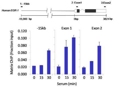
Chromatin Immunoprecipitation: SIN3A Antibody [NB600-1263] - Analysis of Sin3A performed using cross-linked chromatin from colon carcinoma cells treated with serum. Immunoprecipitation performed using a multiplex microplate Matrix ChIP assay with Sin3A polyclonal antibody. Chromatin aliquots from cells were used per ChIP pull-down. Quantitative PCR data done in quadruplicate using 1ul of eluted DNA in 2ul SYBR real-time PCR reactions containing primers to amplify -15kb upstream of the Egr1 gene or exon-1 or exon-2 of Egr1. Quantitation of immunoprecipitated chromatin is presented as signal relative to the total amount of input chromatin. A representation of the Egr-1 locus is shown; boxes represent exons (black boxes = translated, white boxes = untranslated), the zigzag line represents an intron, and the straight line represents upstream sequence. Regions amplified by Egr-1 primers are represented by black bars. Data courtesy of the Innovators Program.
![Immunocytochemistry/ Immunofluorescence: SIN3A Antibody [NB600-1263]](https://resources.rndsystems.com/images/products/SIN3A-Antibody-Immunocytochemistry-Immunofluorescence-NB600-1263-img0006.jpg "Immunocytochemistry/ Immunofluorescence: SIN3A Antibody [NB600-1263]")
Immunocytochemistry/ Immunofluorescence: SIN3A Antibody [NB600-1263]
Immunocytochemistry/Immunofluorescence: SIN3A Antibody [NB600-1263] - Analysis of Sin3A (green) showing staining in the nucleus of NIH-3T3 cells (right) compared to a negative control without primary antibody (left).![Immunocytochemistry/ Immunofluorescence: SIN3A Antibody [NB600-1263]](https://resources.rndsystems.com/images/products/SIN3A-Antibody-Immunocytochemistry-Immunofluorescence-NB600-1263-img0007.jpg "Immunocytochemistry/ Immunofluorescence: SIN3A Antibody [NB600-1263]")
Immunocytochemistry/ Immunofluorescence: SIN3A Antibody [NB600-1263]
Immunocytochemistry/Immunofluorescence: SIN3A Antibody [NB600-1263] - Analysis of Sin3A (green) showing staining in the nucleus of Hela cells (right) compared to a negative control without primary antibody (left).Applications for SIN3A Antibody
Application
Recommended Usage
Chromatin Immunoprecipitation (ChIP)
1-3 ul
Immunocytochemistry/ Immunofluorescence
1:50 - 1:500
Immunohistochemistry
1:10 - 1:500
Immunohistochemistry-Paraffin
1:50 - 1:500
Immunoprecipitation
1:10 - 1:500
Western Blot
0.5 ug/ml
Application Notes
Use in FLOW reported in scientific literature (PMID 28260693).
Flow Cytometry Panel Builder
Bio-Techne Knows Flow Cytometry
Save time and reduce costly mistakes by quickly finding compatible reagents using the Panel Builder Tool.
Advanced Features
- Spectra Viewer - Custom analysis of spectra from multiple fluorochromes
- Spillover Popups - Visualize the spectra of individual fluorochromes
- Antigen Density Selector - Match fluorochrome brightness with antigen density

Formulation, Preparation, and Storage
Purification
Immunogen affinity purified
Formulation
PBS with 1 mg/ml BSA
Preservative
0.05% Sodium Azide
Concentration
1 mg/ml
Shipping
The product is shipped with polar packs. Upon receipt, store it immediately at the temperature recommended below.
Stability & Storage
Store at -20C. Avoid freeze-thaw cycles.
Background: SIN3A
Long Name
SIN3 Homolog A
Alternate Names
DKFZp434K2235, FLJ90319, Histone deacetylase complex subunit Sin3a, KIAA0700, paired amphipathic helix protein Sin3a, SIN3 homolog A, transcription regulator (yeast), SIN3 homolog A, transcriptional regulator (yeast), Transcriptional corepressor Sin3a, transcriptional co-repressor Sin3A, transcriptional regulator, SIN3A
Gene Symbol
SIN3A
UniProt
Additional SIN3A Products
Product Documents for SIN3A Antibody
Certificate of Analysis
To download a Certificate of Analysis, please enter a lot or batch number in the search box below.
Product Specific Notices for SIN3A Antibody
This product is for research use only and is not approved for use in humans or in clinical diagnosis. Primary Antibodies are guaranteed for 1 year from date of receipt.
Related Research Areas
Citations for SIN3A Antibody
 Powered by Bioz
Customer Reviews for SIN3A Antibody
There are currently no reviews for this product. Be the first to review SIN3A Antibody and earn rewards!
Have you used SIN3A Antibody?
Submit a review and receive an Amazon gift card!
$25/€18/£15/$25CAN/¥2500 Yen for a review with an image
$10/€7/£6/$10CAN/¥1110 Yen for a review without an image
Submit a review
Protocols
Find general support by application which include: protocols, troubleshooting, illustrated assays, videos and webinars.
- 7-Amino Actinomycin D (7-AAD) Cell Viability Flow Cytometry Protocol
- Antigen Retrieval Protocol (PIER)
- Antigen Retrieval for Frozen Sections Protocol
- Appropriate Fixation of IHC/ICC Samples
- Cellular Response to Hypoxia Protocols
- ChIP Protocol Video
- Chromatin Immunoprecipitation (ChIP) Protocol
- Chromatin Immunoprecipitation Protocol
- Chromogenic IHC Staining of Formalin-Fixed Paraffin-Embedded (FFPE) Tissue Protocol
- Chromogenic Immunohistochemistry Staining of Frozen Tissue
- ClariTSA™ Fluorophore Kits
- Detection & Visualization of Antibody Binding
- Extracellular Membrane Flow Cytometry Protocol
- Flow Cytometry Protocol for Cell Surface Markers
- Flow Cytometry Protocol for Staining Membrane Associated Proteins
- Flow Cytometry Staining Protocols
- Flow Cytometry Troubleshooting Guide
- Fluorescent IHC Staining of Frozen Tissue Protocol
- Graphic Protocol for Heat-induced Epitope Retrieval
- Graphic Protocol for the Preparation and Fluorescent IHC Staining of Frozen Tissue Sections
- Graphic Protocol for the Preparation and Fluorescent IHC Staining of Paraffin-embedded Tissue Sections
- Graphic Protocol for the Preparation of Gelatin-coated Slides for Histological Tissue Sections
- ICC Cell Smear Protocol for Suspension Cells
- ICC Immunocytochemistry Protocol Videos
- ICC for Adherent Cells
- IHC Sample Preparation (Frozen sections vs Paraffin)
- Immunocytochemistry (ICC) Protocol
- Immunocytochemistry Troubleshooting
- Immunofluorescence of Organoids Embedded in Cultrex Basement Membrane Extract
- Immunofluorescent IHC Staining of Formalin-Fixed Paraffin-Embedded (FFPE) Tissue Protocol
- Immunohistochemistry (IHC) and Immunocytochemistry (ICC) Protocols
- Immunohistochemistry Frozen Troubleshooting
- Immunohistochemistry Paraffin Troubleshooting
- Immunoprecipitation Protocol
- Intracellular Flow Cytometry Protocol Using Alcohol (Methanol)
- Intracellular Flow Cytometry Protocol Using Detergents
- Intracellular Nuclear Staining Flow Cytometry Protocol Using Detergents
- Intracellular Staining Flow Cytometry Protocol Using Alcohol Permeabilization
- Intracellular Staining Flow Cytometry Protocol Using Detergents to Permeabilize Cells
- Preparing Samples for IHC/ICC Experiments
- Preventing Non-Specific Staining (Non-Specific Binding)
- Primary Antibody Selection & Optimization
- Propidium Iodide Cell Viability Flow Cytometry Protocol
- Protocol for Heat-Induced Epitope Retrieval (HIER)
- Protocol for Liperfluo
- Protocol for Making a 4% Formaldehyde Solution in PBS
- Protocol for VisUCyte™ HRP Polymer Detection Reagent
- Protocol for the Characterization of Human Th22 Cells
- Protocol for the Characterization of Human Th9 Cells
- Protocol for the Fluorescent ICC Staining of Cell Smears - Graphic
- Protocol for the Fluorescent ICC Staining of Cultured Cells on Coverslips - Graphic
- Protocol for the Preparation & Fixation of Cells on Coverslips
- Protocol for the Preparation and Chromogenic IHC Staining of Frozen Tissue Sections
- Protocol for the Preparation and Chromogenic IHC Staining of Frozen Tissue Sections - Graphic
- Protocol for the Preparation and Chromogenic IHC Staining of Paraffin-embedded Tissue Sections
- Protocol for the Preparation and Chromogenic IHC Staining of Paraffin-embedded Tissue Sections - Graphic
- Protocol for the Preparation and Fluorescent ICC Staining of Cells on Coverslips
- Protocol for the Preparation and Fluorescent ICC Staining of Non-adherent Cells
- Protocol for the Preparation and Fluorescent ICC Staining of Stem Cells on Coverslips
- Protocol for the Preparation and Fluorescent IHC Staining of Frozen Tissue Sections
- Protocol for the Preparation and Fluorescent IHC Staining of Paraffin-embedded Tissue Sections
- Protocol for the Preparation of Gelatin-coated Slides for Histological Tissue Sections
- Protocol for the Preparation of a Cell Smear for Non-adherent Cell ICC - Graphic
- Protocol: Annexin V and PI Staining by Flow Cytometry
- Protocol: Annexin V and PI Staining for Apoptosis by Flow Cytometry
- R&D Systems Quality Control Western Blot Protocol
- TUNEL and Active Caspase-3 Detection by IHC/ICC Protocol
- The Importance of IHC/ICC Controls
- Troubleshooting Guide: Fluorokine Flow Cytometry Kits
- Troubleshooting Guide: Immunohistochemistry
- Troubleshooting Guide: Western Blot Figures
- Western Blot Conditions
- Western Blot Protocol
- Western Blot Protocol for Cell Lysates
- Western Blot Troubleshooting
- Western Blot Troubleshooting Guide
- View all Protocols, Troubleshooting, Illustrated assays and Webinars
Loading...