Smad3 Antibody - BSA Free
Novus Biologicals | Catalog # NB100-56479
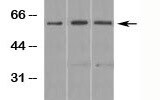
![Western Blot: Smad3 Antibody [NB100-56479]](https://resources.rndsystems.com/images/products/Smad3-Antibody-Western-Blot-NB100-56479-img0008.jpg "Western Blot: Smad3 Antibody [NB100-56479]")
Key Product Details
Validated by
Species Reactivity
Validated:
Cited:
Predicted:
Applications
Validated:
Cited:
Label
Antibody Source
Format
Product Specifications
Immunogen
Clonality
Host
Isotype
Scientific Data Images for Smad3 Antibody - BSA Free
![Immunocytochemistry/ Immunofluorescence: Smad3 Antibody [NB100-56479]](https://resources.rndsystems.com/images/products/Smad3-Antibody-Immunocytochemistry-Immunofluorescence-NB100-56479-img0006.jpg "Immunocytochemistry/ Immunofluorescence: Smad3 Antibody [NB100-56479]")
Immunocytochemistry/ Immunofluorescence: Smad3 Antibody [NB100-56479]
Immunocytochemistry/Immunofluorescence: Smad3 Antibody [NB100-56479] - HeLa cells were fixed for 10 minutes using 10% formalin and then permeabilized for 5 minutes using 1X PBS + 0.5% Triton X-100. The cells were incubated with anti-Smad3 at 10 ug/mL overnight at 4C and detected with an anti-rabbit DyLight 488 (Green) at a 1:500 dilution. Nuclei were counterstained with DAPI (Blue). Cells were imaged using a 40X objective.![Immunohistochemistry-Paraffin: Smad3 Antibody [NB100-56479]](https://resources.rndsystems.com/images/products/Smad3-Antibody-Immunohistochemistry-Paraffin-NB100-56479-img0004.jpg "Immunohistochemistry-Paraffin: Smad3 Antibody [NB100-56479]")
Immunohistochemistry-Paraffin: Smad3 Antibody [NB100-56479]
Immunohistochemistry-Paraffin: Smad3 Antibody [NB100-56479] - Human liver tissue probed with SMAD3 antibody (bottom left, right) and isotype control antibody (top left) at 10 ug/mL.![Western Blot: Smad3 Antibody [NB100-56479]](https://resources.rndsystems.com/images/products/Smad3-Antibody-Western-Blot-NB100-56479-img0005.jpg "Western Blot: Smad3 Antibody [NB100-56479]")
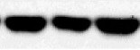
Western Blot: Smad3 Antibody [NB100-56479]
Western Blot: Smad3 Antibody [NB100-56479] - Analysis using SMAD3 antibody. Lysate from human liver in the 1) absence and 2) presence of immunizing peptide, 3) mouse liver and 4) rat liver probed with SMAD3 antibody at 0.5 ug/mL. I goat anti-rabbit Ig HRP secondary antibody and PicoTect ECL substrate solution were used for this test.
Western Blot: Smad3 Antibody - BSA Free [NB100-56479] -
Effects of Sr and SB-505124 on the transforming growth factor beta (TGF beta )/SMAD family member (SMAD) signaling pathway and downstream gene transcription in bovine chondrocytes. Chondrocytes were treated with different doses of Sr (0, 0.1, 1, 10 μg/ml) with or without activin receptor-like kinase 5 (ALK5) kinase inhibitor (10 μM SB-505124). (A) Western blot analysis and relative protein expression levels of SMAD3, pSMAD3, SMAD1/5/9, pSMAD1/5/9, TGF beta, bone morphogenetic protein 2 (BMP2), and runt-related transcription factor 2 (RUNX2). (B) mRNA expression levels of TGF beta 1, serpin family E member 1 (SERPINE1), ALK5, ALK1, and inhibitor of DNA binding 1 (ID1). All experiments were repeated at least thrice. Data are means +/- SEM (n = 3 in each group) *p < 0.05; **p < 0.01. Image collected and cropped by CiteAb from the following open publication (https://pubmed.ncbi.nlm.nih.gov/35712700), licensed under a CC-BY license. Not internally tested by Novus Biologicals.Applications for Smad3 Antibody - BSA Free
Immunohistochemistry-Paraffin
Simple Western
Western Blot
Reviewed Applications
Read 5 reviews rated 3.2 using NB100-56479 in the following applications:
Formulation, Preparation, and Storage
Purification
Formulation
Format
Preservative
Concentration
Shipping
Stability & Storage
Background: Smad3
Long Name
Alternate Names
Gene Symbol
UniProt
Additional Smad3 Products
Product Documents for Smad3 Antibody - BSA Free
Certificate of Analysis
To download a Certificate of Analysis, please enter a lot or batch number in the search box below.
Product Specific Notices for Smad3 Antibody - BSA Free
This product is for research use only and is not approved for use in humans or in clinical diagnosis. Primary Antibodies are guaranteed for 1 year from date of receipt.
Related Research Areas
Citations for Smad3 Antibody - BSA Free
 Powered by Bioz
Customer Reviews for Smad3 Antibody - BSA Free (5)
Have you used Smad3 Antibody - BSA Free?
Submit a review and receive an Amazon gift card!
$25/€18/£15/$25CAN/¥2500 Yen for a review with an image
$10/€7/£6/$10CAN/¥1110 Yen for a review without an image
Submit a review
Customer Images
-
Application: Western BlotSample Tested: Lung tissueSpecies: MouseVerified Customer | Posted 08/02/2021Analysis of smad3 in lung tissue.

-
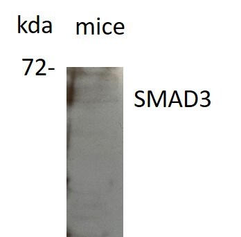
Application: Western BlotSample Tested: eye tissueSpecies: miceVerified Customer | Posted 05/26/2021poor qualityused at 1:100 overnight.
Bio-Techne ResponseThank you for reviewing our product. We are sorry to that that this product did not perform as expected. We have been in touch with the customer to resolve this issue according to our Product Guarantee and to the customer’s satisfaction.
-
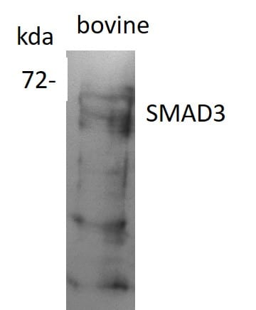
Application: Western BlotSample Tested: eye tissueSpecies: BovineVerified Customer | Posted 05/26/2021works just fineUsed at 1:1000- for overnight.
Bio-Techne ResponseThank you for reviewing our product. We are sorry to that that this product did not perform as expected. We have been in touch with the customer to resolve this issue according to our Product Guarantee and to the customer’s satisfaction.
-
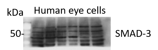
Application: Western BlotSample Tested: ocular tissueSpecies: HumanVerified Customer | Posted 05/24/2021Gives multiple bandsused at 1:1000 and incubated at 4degrees overnight
-
Application: Western BlotSample Tested: Human fibroblast and HeLa whole cell lysateSpecies: MouseVerified Customer | Posted 06/17/2019untreated whole cell lysate band observed around 55 Kda1:1000 dilution 1 hour blocking in milk 5%
There are no reviews that match your criteria.
Protocols
Find general support by application which include: protocols, troubleshooting, illustrated assays, videos and webinars.
- Antigen Retrieval Protocol (PIER)
- Antigen Retrieval for Frozen Sections Protocol
- Appropriate Fixation of IHC/ICC Samples
- Cellular Response to Hypoxia Protocols
- Chromogenic IHC Staining of Formalin-Fixed Paraffin-Embedded (FFPE) Tissue Protocol
- Chromogenic Immunohistochemistry Staining of Frozen Tissue
- ClariTSA™ Fluorophore Kits
- Detection & Visualization of Antibody Binding
- Fluorescent IHC Staining of Frozen Tissue Protocol
- Graphic Protocol for Heat-induced Epitope Retrieval
- Graphic Protocol for the Preparation and Fluorescent IHC Staining of Frozen Tissue Sections
- Graphic Protocol for the Preparation and Fluorescent IHC Staining of Paraffin-embedded Tissue Sections
- Graphic Protocol for the Preparation of Gelatin-coated Slides for Histological Tissue Sections
- ICC Cell Smear Protocol for Suspension Cells
- ICC Immunocytochemistry Protocol Videos
- ICC for Adherent Cells
- IHC Sample Preparation (Frozen sections vs Paraffin)
- Immunocytochemistry (ICC) Protocol
- Immunocytochemistry Troubleshooting
- Immunofluorescence of Organoids Embedded in Cultrex Basement Membrane Extract
- Immunofluorescent IHC Staining of Formalin-Fixed Paraffin-Embedded (FFPE) Tissue Protocol
- Immunohistochemistry (IHC) and Immunocytochemistry (ICC) Protocols
- Immunohistochemistry Frozen Troubleshooting
- Immunohistochemistry Paraffin Troubleshooting
- Preparing Samples for IHC/ICC Experiments
- Preventing Non-Specific Staining (Non-Specific Binding)
- Primary Antibody Selection & Optimization
- Protocol for Heat-Induced Epitope Retrieval (HIER)
- Protocol for Making a 4% Formaldehyde Solution in PBS
- Protocol for VisUCyte™ HRP Polymer Detection Reagent
- Protocol for the Fluorescent ICC Staining of Cell Smears - Graphic
- Protocol for the Fluorescent ICC Staining of Cultured Cells on Coverslips - Graphic
- Protocol for the Preparation & Fixation of Cells on Coverslips
- Protocol for the Preparation and Chromogenic IHC Staining of Frozen Tissue Sections
- Protocol for the Preparation and Chromogenic IHC Staining of Frozen Tissue Sections - Graphic
- Protocol for the Preparation and Chromogenic IHC Staining of Paraffin-embedded Tissue Sections
- Protocol for the Preparation and Chromogenic IHC Staining of Paraffin-embedded Tissue Sections - Graphic
- Protocol for the Preparation and Fluorescent ICC Staining of Cells on Coverslips
- Protocol for the Preparation and Fluorescent ICC Staining of Non-adherent Cells
- Protocol for the Preparation and Fluorescent ICC Staining of Stem Cells on Coverslips
- Protocol for the Preparation and Fluorescent IHC Staining of Frozen Tissue Sections
- Protocol for the Preparation and Fluorescent IHC Staining of Paraffin-embedded Tissue Sections
- Protocol for the Preparation of Gelatin-coated Slides for Histological Tissue Sections
- Protocol for the Preparation of a Cell Smear for Non-adherent Cell ICC - Graphic
- R&D Systems Quality Control Western Blot Protocol
- TUNEL and Active Caspase-3 Detection by IHC/ICC Protocol
- The Importance of IHC/ICC Controls
- Troubleshooting Guide: Immunohistochemistry
- Troubleshooting Guide: Western Blot Figures
- Western Blot Conditions
- Western Blot Protocol
- Western Blot Protocol for Cell Lysates
- Western Blot Troubleshooting
- Western Blot Troubleshooting Guide
- View all Protocols, Troubleshooting, Illustrated assays and Webinars
FAQs for Smad3 Antibody - BSA Free
-
Q: I was wondering if you can recommend any primary polyclonal Smad3 antibody (reactivity: human) for gel supershifts?
A: We do provide a wide range of Smad3 antibodies, but unfortunately none of them have been tested for gel shift assay, so we can't guarantee they will work for an application never tested before.
Associated Pathways


