SMN Antibody (2B1) [Alexa Fluor® 647]
Novus Biologicals | Catalog # NB100-1936AF647
Key Product Details
Species Reactivity
Validated:
Cited:
Applications
Validated:
Cited:
Label
Antibody Source
Product Specifications
Immunogen
Reactivity Notes
Localization
Clonality
Host
Isotype
Scientific Data Images for SMN Antibody (2B1) [Alexa Fluor® 647]
![SMN Antibody (2B1) [Alexa Fluor® 647]](https://resources.rndsystems.com/images/products/nb100-1936af647_mouse-monoclonal-smn-antibody-2b1-alexa-fluor-647-immunocytochemistry-immunofluorescence-1432024145552..jpg "SMN (2B1) in U-2 OS Human Cell Line -")
SMN (2B1) in U-2 OS Human Cell Line -
SMN (2B1) was detected in immersion fixed U-2 OS human osteosarcoma cell line using Mouse anti-SMN (2B1) Protein G Purified Monoclonal Antibody conjugated to Alexa Fluor® 647 (Catalog # NB100-1936AF647) (light blue) at 5 µg/mL overnight at 4C. Cells were counterstained with DAPI (dark blue). Cells were imaged using a 100X objective and digitally deconvolved.![SMN Antibody (2B1) [Alexa Fluor® 647]](https://resources.rndsystems.com/images/products/nb100-1936af647_mouse-monoclonal-smn-antibody-2b1-alexa-fluor-647-flow-intracellular--3092024154753..jpg "Detection of SMN (2B1) in A431 Human Cell Line by Flow Cytometry.")
Detection of SMN (2B1) in A431 Human Cell Line by Flow Cytometry.
An intracellular stain was performed on A431 human skin carcinoma cell line using Mouse anti- SMN (2B1) Protein-G purified Monoclonal Antibody conjugated to Alexa Fluor® 647 (Catalog # NB100-1936AF647, blue histogram) or matched control antibody (orange histogram) at 2.5 µg/mL for 30 minutes at RT.![SMN Antibody (2B1) [Alexa Fluor® 647]](https://resources.rndsystems.com/images/products/nb100-1936af647_mouse-monoclonal-smn-antibody-2b1-alexa-fluor-647-310202415532044.jpg "Immunocytochemistry/ Immunofluorescence: SMN Antibody (2B1) [Alexa Fluor® 647] [NB100-1936AF647] -")
Immunocytochemistry/ Immunofluorescence: SMN Antibody (2B1) [Alexa Fluor® 647] [NB100-1936AF647] -
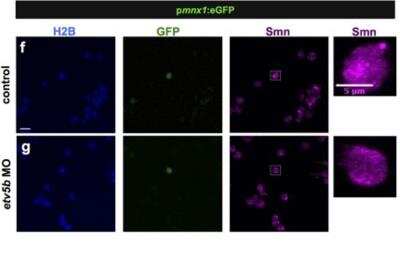
Immunocytochemistry/ Immunofluorescence: SMN Antibody (2B1) [Alexa Fluor® 647] [NB100-1936AF647] - Etv5b activates the smn promoter leading to increased Smn levels in motoneurons.(a–d) Whole-mount in situ hybridization of etv5b expression in 24 hpf embryos. Expression pattern in the entire embryo (a), in magnified lateral (b) & dorsal (c) views as well as in a transverse section (d) of the trunk. In the latter case, yellow arrowhead denote signal in the motoneuron region. Scale bars: 100 μm (a) & 50 μm (b–d). nc, notochord. (e) RT-PCR in control (C - only psmn:mCherry) versus etv5b overexpressing (OE - etv5b mRNA & psmn:mCherry) embryos at early gastrula stage. (f,g) scIF of control (f) & etv5b MO (g) cells from pmnx1:eGFP embryos. H2B, GFP & Smn signals are shown in Z-projected confocal sections. GFP+ cells marked by white rectangles are magnified on the right. Scale bars: 10 μm for low & 5 μm for high magnification. (h) Relative Smn signal in control & in etv5b MO cells from one representative experiment. Exact values are (mean ± SD): 0.99 ± 0.25 (control) & 0.7 ± 0.14 (etvb5 MO), p = 0.003 with Wilcoxon Sum Rank Test. (i) Average relative Smn levels in control & in etv5b MO motoneurons. The exact value of decrease is 0.75 ± 0.04 (mean ± SD). N = 3 experiments, n = number of analyzed cells. Image collected & cropped by CiteAb from the following publication (https://www.nature.com/articles/srep27470), licensed under a CC-BY license. Not internally tested by Novus Biologicals.![SMN Antibody (2B1) [Alexa Fluor® 647]](https://resources.rndsystems.com/images/products/nb100-1936af647_mouse-monoclonal-smn-antibody-2b1-alexa-fluor-647-310202415523912.jpg "Immunocytochemistry/ Immunofluorescence: SMN Antibody (2B1) [Alexa Fluor® 647] [NB100-1936AF647] -")
Immunocytochemistry/ Immunofluorescence: SMN Antibody (2B1) [Alexa Fluor® 647] [NB100-1936AF647] -
Immunocytochemistry/ Immunofluorescence: SMN Antibody (2B1) [Alexa Fluor® 647] [NB100-1936AF647] - Motoneurons exhibit elevated Smn levels.(a–c) Single-cell immunofluorescent (scIF) experiments. 24 hpf pmnx1:eGFP embryos were dissociated & immunostained for Smn ((a) see also Methods). DIC image of cells following 3-hour (b) & 24-hour (c) incubation are shown with GFP in green. Note that the GFP signal in the cell body is oversaturated so that the weaker signal in the axon becomes visible. In (c) DNA is in blue & the motoneuron marker Znp1 (synaptotagmin) is in red; the axon growth cone is magnified in the corner of the images. Scale bars are 100 μm (a), 10 μm (low magnification in (b,c)) & 2 μm (high magnification in (c)). (d) scIF on pmnx1:eGFP embryos. Histone 2B (H2B), GFP & Smn signals are shown in Z-projected confocal sections. Cells marked by white rectangles are magnified on the right. Scale bars: 10 μm for low & 5 μm for high magnification. (e) To account for potential variability in the immunostaining, Smn levels were quantified relative to H2B. Diamonds denote GFP negative (GFP−) & GFP positive (GFP+) cells from one representative experiment. Blue bars indicate mean ± SD with significance values of *p < 0.05 & **p < 0.01. Exact values are (mean ± SD) 0.51 ± 0.14 (GFP−) & 0.87 ± 0.17 (GFP+), p = 0.001 with Wilcoxon Sum Rank Test. For more details, see Materials & Methods & Supplementary Table S1. (f) Average increase of Smn levels in motoneurons versus control cells. The exact value of enrichment is (mean ± SD): 1.67 ± 0.14. N = 3 experiments, n = number of analyzed cells. Image collected & cropped by CiteAb from the following publication (https://www.nature.com/articles/srep27470), licensed under a CC-BY license. Not internally tested by Novus Biologicals.Applications for SMN Antibody (2B1) [Alexa Fluor® 647]
ELISA
Flow Cytometry
Immunocytochemistry/ Immunofluorescence
Immunohistochemistry
Immunohistochemistry-Paraffin
Immunoprecipitation
Western Blot
Reviewed Applications
Read 1 review rated 3 using NB100-1936AF647 in the following applications:
Spectra Viewer
Plan Your Experiments
Use our spectra viewer to interactively plan your experiments, assessing multiplexing options. View the excitation and emission spectra for our fluorescent dye range and other commonly used dyes.
Spectra Viewer
Flow Cytometry Panel Builder
Bio-Techne Knows Flow Cytometry
Save time and reduce costly mistakes by quickly finding compatible reagents using the Panel Builder Tool.
Advanced Features
- Spectra Viewer - Custom analysis of spectra from multiple fluorochromes
- Spillover Popups - Visualize the spectra of individual fluorochromes
- Antigen Density Selector - Match fluorochrome brightness with antigen density

Formulation, Preparation, and Storage
Purification
Formulation
Preservative
Concentration
Shipping
Stability & Storage
Background: SMN
Alternate Names
Gene Symbol
Additional SMN Products
Product Documents for SMN Antibody (2B1) [Alexa Fluor® 647]
Certificate of Analysis
To download a Certificate of Analysis, please enter a lot or batch number in the search box below.
Product Specific Notices for SMN Antibody (2B1) [Alexa Fluor® 647]
Alexa Fluor (R) products are provided under an intellectual property license from Life Technologies Corporation. The purchase of this product conveys to the buyer the non-transferable right to use the purchased product and components of the product only in research conducted by the buyer (whether the buyer is an academic or for-profit entity). The sale of this product is expressly conditioned on the buyer not using the product or its components, or any materials made using the product or its components, in any activity to generate revenue, which may include, but is not limited to use of the product or its components: (i) in manufacturing; (ii) to provide a service, information, or data in return for payment; (iii) for therapeutic, diagnostic or prophylactic purposes; or (iv) for resale, regardless of whether they are resold for use in research. For information on purchasing a license to this product for purposes other than as described above, contact Life Technologies Corporation, 5791 Van Allen Way, Carlsbad, CA 92008 USA or outlicensing@lifetech.com. This conjugate is made on demand. Actual recovery may vary from the stated volume of this product. The volume will be greater than or equal to the unit size stated on the datasheet.
This product is for research use only and is not approved for use in humans or in clinical diagnosis. Primary Antibodies are guaranteed for 1 year from date of receipt.
Citations for SMN Antibody (2B1) [Alexa Fluor® 647]
 Powered by Bioz
Customer Reviews for SMN Antibody (2B1) [Alexa Fluor® 647] (1)
Have you used SMN Antibody (2B1) [Alexa Fluor® 647]?
Submit a review and receive an Amazon gift card!
$25/€18/£15/$25CAN/¥2500 Yen for a review with an image
$10/€7/£6/$10CAN/¥1110 Yen for a review without an image
Submit a review
Customer Images
![SMN Antibody (2B1) [Alexa Fluor® 647] NB100-1936AF647](https://resources.rndsystems.com/images/reviews/Western-Blot__NB100-1936AF647_19681.jpg)
-
Application: Western BlotSample Tested: Zebrafish embryos, HEK, HUVEC cellsSpecies: OtherVerified Customer | Posted 05/20/2015SMN expression in zebrafish embyros
![SMN Antibody (2B1) [Alexa Fluor® 647] NB100-1936AF647](data:image/png;base64,R0lGODlhAQABAAD/ACwAAAAAAQABAAACADs=)
There are no reviews that match your criteria.
Protocols
Find general support by application which include: protocols, troubleshooting, illustrated assays, videos and webinars.
- 7-Amino Actinomycin D (7-AAD) Cell Viability Flow Cytometry Protocol
- Antigen Retrieval Protocol (PIER)
- Antigen Retrieval for Frozen Sections Protocol
- Appropriate Fixation of IHC/ICC Samples
- Cellular Response to Hypoxia Protocols
- Chromogenic IHC Staining of Formalin-Fixed Paraffin-Embedded (FFPE) Tissue Protocol
- Chromogenic Immunohistochemistry Staining of Frozen Tissue
- ClariTSA™ Fluorophore Kits
- Detection & Visualization of Antibody Binding
- ELISA Sample Preparation & Collection Guide
- ELISA Troubleshooting Guide
- Extracellular Membrane Flow Cytometry Protocol
- Flow Cytometry Protocol for Cell Surface Markers
- Flow Cytometry Protocol for Staining Membrane Associated Proteins
- Flow Cytometry Staining Protocols
- Flow Cytometry Troubleshooting Guide
- Fluorescent IHC Staining of Frozen Tissue Protocol
- Graphic Protocol for Heat-induced Epitope Retrieval
- Graphic Protocol for the Preparation and Fluorescent IHC Staining of Frozen Tissue Sections
- Graphic Protocol for the Preparation and Fluorescent IHC Staining of Paraffin-embedded Tissue Sections
- Graphic Protocol for the Preparation of Gelatin-coated Slides for Histological Tissue Sections
- How to Run an R&D Systems DuoSet ELISA
- How to Run an R&D Systems Quantikine ELISA
- How to Run an R&D Systems Quantikine™ QuicKit™ ELISA
- ICC Cell Smear Protocol for Suspension Cells
- ICC Immunocytochemistry Protocol Videos
- ICC for Adherent Cells
- IHC Sample Preparation (Frozen sections vs Paraffin)
- Immunocytochemistry (ICC) Protocol
- Immunocytochemistry Troubleshooting
- Immunofluorescence of Organoids Embedded in Cultrex Basement Membrane Extract
- Immunofluorescent IHC Staining of Formalin-Fixed Paraffin-Embedded (FFPE) Tissue Protocol
- Immunohistochemistry (IHC) and Immunocytochemistry (ICC) Protocols
- Immunohistochemistry Frozen Troubleshooting
- Immunohistochemistry Paraffin Troubleshooting
- Immunoprecipitation Protocol
- Intracellular Flow Cytometry Protocol Using Alcohol (Methanol)
- Intracellular Flow Cytometry Protocol Using Detergents
- Intracellular Nuclear Staining Flow Cytometry Protocol Using Detergents
- Intracellular Staining Flow Cytometry Protocol Using Alcohol Permeabilization
- Intracellular Staining Flow Cytometry Protocol Using Detergents to Permeabilize Cells
- Preparing Samples for IHC/ICC Experiments
- Preventing Non-Specific Staining (Non-Specific Binding)
- Primary Antibody Selection & Optimization
- Propidium Iodide Cell Viability Flow Cytometry Protocol
- Protocol for Heat-Induced Epitope Retrieval (HIER)
- Protocol for Liperfluo
- Protocol for Making a 4% Formaldehyde Solution in PBS
- Protocol for VisUCyte™ HRP Polymer Detection Reagent
- Protocol for the Characterization of Human Th22 Cells
- Protocol for the Characterization of Human Th9 Cells
- Protocol for the Fluorescent ICC Staining of Cell Smears - Graphic
- Protocol for the Fluorescent ICC Staining of Cultured Cells on Coverslips - Graphic
- Protocol for the Preparation & Fixation of Cells on Coverslips
- Protocol for the Preparation and Chromogenic IHC Staining of Frozen Tissue Sections
- Protocol for the Preparation and Chromogenic IHC Staining of Frozen Tissue Sections - Graphic
- Protocol for the Preparation and Chromogenic IHC Staining of Paraffin-embedded Tissue Sections
- Protocol for the Preparation and Chromogenic IHC Staining of Paraffin-embedded Tissue Sections - Graphic
- Protocol for the Preparation and Fluorescent ICC Staining of Cells on Coverslips
- Protocol for the Preparation and Fluorescent ICC Staining of Non-adherent Cells
- Protocol for the Preparation and Fluorescent ICC Staining of Stem Cells on Coverslips
- Protocol for the Preparation and Fluorescent IHC Staining of Frozen Tissue Sections
- Protocol for the Preparation and Fluorescent IHC Staining of Paraffin-embedded Tissue Sections
- Protocol for the Preparation of Gelatin-coated Slides for Histological Tissue Sections
- Protocol for the Preparation of a Cell Smear for Non-adherent Cell ICC - Graphic
- Protocol: Annexin V and PI Staining by Flow Cytometry
- Protocol: Annexin V and PI Staining for Apoptosis by Flow Cytometry
- Quantikine HS ELISA Kit Assay Principle, Alkaline Phosphatase
- Quantikine HS ELISA Kit Principle, Streptavidin-HRP Polymer
- R&D Systems Quality Control Western Blot Protocol
- Sandwich ELISA (Colorimetric) – Biotin/Streptavidin Detection Protocol
- Sandwich ELISA (Colorimetric) – Direct Detection Protocol
- TUNEL and Active Caspase-3 Detection by IHC/ICC Protocol
- The Importance of IHC/ICC Controls
- Troubleshooting Guide: ELISA
- Troubleshooting Guide: Fluorokine Flow Cytometry Kits
- Troubleshooting Guide: Immunohistochemistry
- Troubleshooting Guide: Western Blot Figures
- Western Blot Conditions
- Western Blot Protocol
- Western Blot Protocol for Cell Lysates
- Western Blot Troubleshooting
- Western Blot Troubleshooting Guide
- View all Protocols, Troubleshooting, Illustrated assays and Webinars
FAQs for SMN Antibody (2B1) [Alexa Fluor® 647]
-
Q: Could you recommond the Human specific SMN antibody? It will only recognize human SMN, without cross reaction with mouse smn.
A: We do not have an SMN antibody that has been validated NOT to work in mouse samples. When an antibody is found to not react with a certain species, we mark the data sheet with a (-) next to the species' name. Unfortunately, the SMN antibodies that do not list mouse have not been tested in mouse, and may react.
-
Q: I was wondering if the Alexa 647 conjugated anti-SMN antibody (NB100-1936AF647) can be used in Western Blot, and if so, the recommended working concentration is 2ug/ml?
A: Our SMN antibody with catalogue number NB100-1936AF647 is covered by our 100% quality guarantee for the Western blotting application. The recommended starting dilution for the unconjugated antibody, NB100-1936, is 2ug/ml. With an unconjugated antibody however the signal will be amplified through the use of a conjugated secondary for detection. Since NB100-1936AF647 is directly conjugated, and no secondary antibody is required for detection, it is possible that you might need to use a higher concentration of the antibody to obtain a satisfactory signal. We do always recommend that our customers optimise their staining conditions to suit their particular sample type, and you might wish to try a number of different dilutions when using this antibody initially.
-
Q: Could you recommond the Human specific SMN antibody? It will only recognize human SMN, without cross reaction with mouse smn.
A: We do not have an SMN antibody that has been validated NOT to work in mouse samples. When an antibody is found to not react with a certain species, we mark the data sheet with a (-) next to the species' name. Unfortunately, the SMN antibodies that do not list mouse have not been tested in mouse, and may react.
-
Q: I was wondering if the Alexa 647 conjugated anti-SMN antibody (NB100-1936AF647) can be used in Western Blot, and if so, the recommended working concentration is 2ug/ml?
A: Our SMN antibody with catalogue number NB100-1936AF647 is covered by our 100% quality guarantee for the Western blotting application. The recommended starting dilution for the unconjugated antibody, NB100-1936, is 2ug/ml. With an unconjugated antibody however the signal will be amplified through the use of a conjugated secondary for detection. Since NB100-1936AF647 is directly conjugated, and no secondary antibody is required for detection, it is possible that you might need to use a higher concentration of the antibody to obtain a satisfactory signal. We do always recommend that our customers optimise their staining conditions to suit their particular sample type, and you might wish to try a number of different dilutions when using this antibody initially.