SOD2/Mn-SOD Antibody - BSA Free
Novus Biologicals | Catalog # NB100-1992
![Immunohistochemistry: SOD2/Mn-SOD Antibody [NB100-1992]](https://resources.rndsystems.com/images/products/SOD2-Mn-SOD-Antibody-Immunohistochemistry-NB100-1992-img0013.jpg "Immunohistochemistry: SOD2/Mn-SOD Antibody [NB100-1992]")
Key Product Details
Validated by
Species Reactivity
Validated:
Cited:
Applications
Validated:
Cited:
Label
Antibody Source
Format
Product Specifications
Immunogen
Reactivity Notes
Localization
Specificity
Clonality
Host
Isotype
Scientific Data Images for SOD2/Mn-SOD Antibody - BSA Free
![Immunocytochemistry/ Immunofluorescence: SOD2/Mn-SOD Antibody [NB100-1992]](https://resources.rndsystems.com/images/products/SOD2-Mn-SOD-Antibody-Immunocytochemistry-Immunofluorescence-NB100-1992-img0010.jpg "Immunocytochemistry/ Immunofluorescence: SOD2/Mn-SOD Antibody [NB100-1992]")
Immunocytochemistry/ Immunofluorescence: SOD2/Mn-SOD Antibody [NB100-1992]
Immunocytochemistry/Immunofluorescence: SOD2/Mn-SOD Antibody [NB100-1992] - Analysis using Rabbit Anti-SOD (Mn) Polyclonal Antibody. Tissue: HeLa Cells. Species: Human. Fixation: 2% Formaldehyde for 20 min at RT. Primary Antibody: Rabbit Anti-SOD (Mn) Polyclonal Antibody at 1:120 for 12 hours at 4 degrees C. Secondary Antibody: R-PE Goat Anti-Rabbit (yellow) at 1:200 for 120 min at RT. Counterstain: DAPI (blue) nuclear stain at 1:40000 for 120 min at RT. Localization: Mitochondrion matrix. Magnification: 100x. (A) DAPI (blue) nuclear stain. (B) Anti-SOD (Mn) Antibody. (C) Composite.![Western Blot: SOD2/Mn-SOD Antibody [NB100-1992]](https://resources.rndsystems.com/images/products/SOD2-Mn-SOD-Antibody-Western-Blot-NB100-1992-img0016.jpg "Western Blot: SOD2/Mn-SOD Antibody [NB100-1992]")
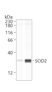
Western Blot: SOD2/Mn-SOD Antibody [NB100-1992]
Western Blot: SOD2/Mn-SOD Antibody [NB100-1992] - Western blot analysis of Rat Tissue lysates showing detection of SOD2/Mn-SOD protein using Rabbit Anti-SOD2/Mn-SOD Polyclonal Antibody (NB100-1992). Load: 15 ugprotein. Block: 1.5% BSA for 30 minutes at RT. Primary Antibody: Rabbit Anti-SOD2/Mn-SOD Polyclonal Antibody (NB100-1992) at 1:1000 for 2 hours at RT. Secondary Antibody: Donkey Anti-Rabbit IgG: HRP for 1 hour at RT.![Immunocytochemistry/ Immunofluorescence: SOD2/Mn-SOD Antibody [NB100-1992]](https://resources.rndsystems.com/images/products/SOD2-Mn-SOD-Antibody-Immunocytochemistry-Immunofluorescence-NB100-1992-img0012.jpg "Immunocytochemistry/ Immunofluorescence: SOD2/Mn-SOD Antibody [NB100-1992]")
Immunocytochemistry/ Immunofluorescence: SOD2/Mn-SOD Antibody [NB100-1992]
Immunocytochemistry/Immunofluorescence: SOD2/Mn-SOD Antibody [NB100-1992] - Analysis using Rabbit Anti-SOD (Mn) Polyclonal Antibody. Tissue: HeLa Cells. Species: Human. Fixation: 2% Formaldehyde for 20 min at RT. Primary Antibody: Rabbit Anti-SOD (Mn) Polyclonal Antibody at 1:120 for 12 hours at 4 degrees C. Secondary Antibody: APC Goat Anti-Rabbit (red) at 1:200 for 120 min at RT. Counterstain: DAPI (blue) nuclear stain at 1:40000 for 120 min at RT. Localization: Mitochondrion matrix. Magnification: 20x. (A) DAPI (blue) nuclear stain. (B) Anti-SOD (Mn) Antibody. (C) Composite.![Immunohistochemistry: SOD2/Mn-SOD Antibody [NB100-1992]](https://resources.rndsystems.com/images/products/SOD2-Mn-SOD-Antibody-Immunohistochemistry-NB100-1992-img0017.jpg "Immunohistochemistry: SOD2/Mn-SOD Antibody [NB100-1992]")
Immunohistochemistry: SOD2/Mn-SOD Antibody [NB100-1992]
Immunohistochemistry: SOD2/Mn-SOD Antibody [NB100-1992] - Immunohistochemistry analysis using Rabbit Anti-SOD2/Mn-SOD Polyclonal Antibody (NB100-1992). Tissue: backskin. Species: Mouse. Fixation: Bouin's Fixative Solution. Primary Antibody: Rabbit Anti-SOD2/Mn-SOD Polyclonal Antibody (NB100-1992) at 1:100 for 1 hour at RT. Secondary Antibody: FITC Goat Anti-Rabbit (green) at 1:50 for 1 hour at RT. Localization: Mitochondrion matrix.
Immunohistochemistry: SOD2/Mn-SOD Antibody [NB100-1992] -
Immunohistochemistry: SOD2/Mn-SOD Antibody [NB100-1992] - SOD2 mRNA & protein expression. RT-PCR: (A) SOD2 mRNA expression is higher in adults, & highest in the adult parenchyma. (B) No exposure effects on SOD2 mRNA were observed in neonates. (C) Adult SOD2 mRNA was decreased in PFP48. Data are presented as mean+SEM (n=5-7 rats/group, in each compartment), * significantly different compared to neonates in the same compartment, † significantly different compared to airways in the same age, ‡ significantly different compared to FA in the same compartment. Western blotting: (D) Scan of representative SOD2 & actin blots. (E) Neonatal whole lung SOD2 protein expression was unchanged with exposure, & (F) adult whole lung SOD2 protein trended upwards at PFP2, but was statistically insignificant. (G-J) Immunohistochemical localization of SOD2 in lung (n=6 rats/group). SOD2 protein was more abundant in adults compared to neonates, but no exposure specific differences were observed. Scale bar is 50 μm. Image collected & cropped by CiteAb from the following publication (https://particleandfibretoxicology.biomedcentral.com/articles/10.1186/1…), licensed under a CC-BY license. Not internally tested by Novus Biologicals.
Western Blot: SOD2/Mn-SOD Antibody [NB100-1992] -
Western Blot: SOD2/Mn-SOD Antibody [NB100-1992] - CAV-1 preserves mitochondrial respiratory function through regulation of OXPHOS expression.(A) OXPHOS complex subunits were detected through western blotting with appropriate antibodies. CAV-1 treatment significantly restored hypercholesterolemia-associated increased ETC complex I–V proteins levels. (B) Percentage of the OXPHOS complex (I–V) band intensities is presented in the graph. (C) Representative immunoblot displaying levels of CAV-1, CyPA, mitochondrial biogenesis markers, & antioxidant enzymes in the NC, HC, & HCAV-1 groups. (D) Columns represent average values over three independent experiments. The density for the NC group was set at 1; * & † are significantly different for the NC & HC groups, respectively, at P < 0.05. beta -actin was used as a loading control. Values are means ± SD. Image collected & cropped by CiteAb from the following publication (https://dx.plos.org/10.1371/journal.pone.0154210), licensed under a CC-BY license. Not internally tested by Novus Biologicals.
Western Blot: SOD2/Mn-SOD Antibody [NB100-1992] -
Western Blot: SOD2/Mn-SOD Antibody [NB100-1992] - SOD2 mRNA & protein expression. RT-PCR: (A) SOD2 mRNA expression is higher in adults, & highest in the adult parenchyma. (B) No exposure effects on SOD2 mRNA were observed in neonates. (C) Adult SOD2 mRNA was decreased in PFP48. Data are presented as mean+SEM (n=5-7 rats/group, in each compartment), * significantly different compared to neonates in the same compartment, † significantly different compared to airways in the same age, ‡ significantly different compared to FA in the same compartment. Western blotting: (D) Scan of representative SOD2 & actin blots. (E) Neonatal whole lung SOD2 protein expression was unchanged with exposure, & (F) adult whole lung SOD2 protein trended upwards at PFP2, but was statistically insignificant. (G-J) Immunohistochemical localization of SOD2 in lung (n=6 rats/group). SOD2 protein was more abundant in adults compared to neonates, but no exposure specific differences were observed. Scale bar is 50 μm. Image collected & cropped by CiteAb from the following publication (https://particleandfibretoxicology.biomedcentral.com/articles/10.1186/1…), licensed under a CC-BY license. Not internally tested by Novus Biologicals.
Immunohistochemistry: SOD2/Mn-SOD Antibody [NB100-1992] -
Immunohistochemistry: SOD2/Mn-SOD Antibody [NB100-1992] - SOD2 mRNA & protein expression. RT-PCR: (A) SOD2 mRNA expression is higher in adults, & highest in the adult parenchyma. (B) No exposure effects on SOD2 mRNA were observed in neonates. (C) Adult SOD2 mRNA was decreased in PFP48. Data are presented as mean+SEM (n=5-7 rats/group, in each compartment), * significantly different compared to neonates in the same compartment, † significantly different compared to airways in the same age, ‡ significantly different compared to FA in the same compartment. Western blotting: (D) Scan of representative SOD2 & actin blots. (E) Neonatal whole lung SOD2 protein expression was unchanged with exposure, & (F) adult whole lung SOD2 protein trended upwards at PFP2, but was statistically insignificant. (G-J) Immunohistochemical localization of SOD2 in lung (n=6 rats/group). SOD2 protein was more abundant in adults compared to neonates, but no exposure specific differences were observed. Scale bar is 50 μm. Image collected & cropped by CiteAb from the following publication (https://particleandfibretoxicology.biomedcentral.com/articles/10.1186/1…), licensed under a CC-BY license. Not internally tested by Novus Biologicals.
Western Blot: SOD2/Mn-SOD Antibody [NB100-1992] -
Western Blot: SOD2/Mn-SOD Antibody [NB100-1992] - SOD2 mRNA & protein expression. RT-PCR: (A) SOD2 mRNA expression is higher in adults, & highest in the adult parenchyma. (B) No exposure effects on SOD2 mRNA were observed in neonates. (C) Adult SOD2 mRNA was decreased in PFP48. Data are presented as mean+SEM (n=5-7 rats/group, in each compartment), * significantly different compared to neonates in the same compartment, † significantly different compared to airways in the same age, ‡ significantly different compared to FA in the same compartment. Western blotting: (D) Scan of representative SOD2 & actin blots. (E) Neonatal whole lung SOD2 protein expression was unchanged with exposure, & (F) adult whole lung SOD2 protein trended upwards at PFP2, but was statistically insignificant. (G-J) Immunohistochemical localization of SOD2 in lung (n=6 rats/group). SOD2 protein was more abundant in adults compared to neonates, but no exposure specific differences were observed. Scale bar is 50 μm. Image collected & cropped by CiteAb from the following publication (https://particleandfibretoxicology.biomedcentral.com/articles/10.1186/1…), licensed under a CC-BY license. Not internally tested by Novus Biologicals.
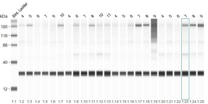
Simple Western: Rabbit Polyclonal SOD2/Mn-SOD Antibody [NB100-1992]
SOD2 increase after oxidative stress in mouse lung tissue. 1:20 in antibody diluent 2, 60min of incubation. Image from a verified customer review.Applications for SOD2/Mn-SOD Antibody - BSA Free
ELISA
Immunocytochemistry/ Immunofluorescence
Immunohistochemistry
Immunohistochemistry-Paraffin
Immunoprecipitation
Simple Western
Western Blot
Reviewed Applications
Read 2 reviews rated 5 using NB100-1992 in the following applications:
Formulation, Preparation, and Storage
Purification
Formulation
Format
Preservative
Concentration
Shipping
Stability & Storage
Background: SOD2/Mn-SOD
Long Name
Alternate Names
Gene Symbol
UniProt
Additional SOD2/Mn-SOD Products
Product Documents for SOD2/Mn-SOD Antibody - BSA Free
Certificate of Analysis
To download a Certificate of Analysis, please enter a lot or batch number in the search box below.
Product Specific Notices for SOD2/Mn-SOD Antibody - BSA Free
This product is for research use only and is not approved for use in humans or in clinical diagnosis. Primary Antibodies are guaranteed for 1 year from date of receipt.
Related Research Areas
Citations for SOD2/Mn-SOD Antibody - BSA Free
 Powered by Bioz
Customer Reviews for SOD2/Mn-SOD Antibody - BSA Free (2)
Have you used SOD2/Mn-SOD Antibody - BSA Free?
Submit a review and receive an Amazon gift card!
$25/€18/£15/$25CAN/¥2500 Yen for a review with an image
$10/€7/£6/$10CAN/¥1110 Yen for a review without an image
Submit a review
Customer Images
-
Application: Simple WesternSample Tested: lung homogenateSpecies: MouseVerified Customer | Posted 09/08/2025SOD2 protein expression in experimental mouse lung homogenates was determined with JESS simple westernprotein expression of sod2 in the mouse lung homogenate treated with smoke

-
Application: Simple WesternSample Tested: Lung tissueSpecies: MouseVerified Customer | Posted 06/03/2025SOD2 increase after oxidative stress1:20 in antibody diluent 2, 60min of incubation
Bio-Techne ResponseThis review reflects a new species or application tested on a primary antibody.
There are no reviews that match your criteria.
Protocols
Find general support by application which include: protocols, troubleshooting, illustrated assays, videos and webinars.
- Antigen Retrieval Protocol (PIER)
- Antigen Retrieval for Frozen Sections Protocol
- Appropriate Fixation of IHC/ICC Samples
- Cellular Response to Hypoxia Protocols
- Chromogenic IHC Staining of Formalin-Fixed Paraffin-Embedded (FFPE) Tissue Protocol
- Chromogenic Immunohistochemistry Staining of Frozen Tissue
- ClariTSA™ Fluorophore Kits
- Detection & Visualization of Antibody Binding
- ELISA Sample Preparation & Collection Guide
- ELISA Troubleshooting Guide
- Fluorescent IHC Staining of Frozen Tissue Protocol
- Graphic Protocol for Heat-induced Epitope Retrieval
- Graphic Protocol for the Preparation and Fluorescent IHC Staining of Frozen Tissue Sections
- Graphic Protocol for the Preparation and Fluorescent IHC Staining of Paraffin-embedded Tissue Sections
- Graphic Protocol for the Preparation of Gelatin-coated Slides for Histological Tissue Sections
- How to Run an R&D Systems DuoSet ELISA
- How to Run an R&D Systems Quantikine ELISA
- How to Run an R&D Systems Quantikine™ QuicKit™ ELISA
- ICC Cell Smear Protocol for Suspension Cells
- ICC Immunocytochemistry Protocol Videos
- ICC for Adherent Cells
- IHC Sample Preparation (Frozen sections vs Paraffin)
- Immunocytochemistry (ICC) Protocol
- Immunocytochemistry Troubleshooting
- Immunofluorescence of Organoids Embedded in Cultrex Basement Membrane Extract
- Immunofluorescent IHC Staining of Formalin-Fixed Paraffin-Embedded (FFPE) Tissue Protocol
- Immunohistochemistry (IHC) and Immunocytochemistry (ICC) Protocols
- Immunohistochemistry Frozen Troubleshooting
- Immunohistochemistry Paraffin Troubleshooting
- Immunoprecipitation Protocol
- Preparing Samples for IHC/ICC Experiments
- Preventing Non-Specific Staining (Non-Specific Binding)
- Primary Antibody Selection & Optimization
- Protocol for Heat-Induced Epitope Retrieval (HIER)
- Protocol for Making a 4% Formaldehyde Solution in PBS
- Protocol for VisUCyte™ HRP Polymer Detection Reagent
- Protocol for the Fluorescent ICC Staining of Cell Smears - Graphic
- Protocol for the Fluorescent ICC Staining of Cultured Cells on Coverslips - Graphic
- Protocol for the Preparation & Fixation of Cells on Coverslips
- Protocol for the Preparation and Chromogenic IHC Staining of Frozen Tissue Sections
- Protocol for the Preparation and Chromogenic IHC Staining of Frozen Tissue Sections - Graphic
- Protocol for the Preparation and Chromogenic IHC Staining of Paraffin-embedded Tissue Sections
- Protocol for the Preparation and Chromogenic IHC Staining of Paraffin-embedded Tissue Sections - Graphic
- Protocol for the Preparation and Fluorescent ICC Staining of Cells on Coverslips
- Protocol for the Preparation and Fluorescent ICC Staining of Non-adherent Cells
- Protocol for the Preparation and Fluorescent ICC Staining of Stem Cells on Coverslips
- Protocol for the Preparation and Fluorescent IHC Staining of Frozen Tissue Sections
- Protocol for the Preparation and Fluorescent IHC Staining of Paraffin-embedded Tissue Sections
- Protocol for the Preparation of Gelatin-coated Slides for Histological Tissue Sections
- Protocol for the Preparation of a Cell Smear for Non-adherent Cell ICC - Graphic
- Quantikine HS ELISA Kit Assay Principle, Alkaline Phosphatase
- Quantikine HS ELISA Kit Principle, Streptavidin-HRP Polymer
- R&D Systems Quality Control Western Blot Protocol
- Sandwich ELISA (Colorimetric) – Biotin/Streptavidin Detection Protocol
- Sandwich ELISA (Colorimetric) – Direct Detection Protocol
- TUNEL and Active Caspase-3 Detection by IHC/ICC Protocol
- The Importance of IHC/ICC Controls
- Troubleshooting Guide: ELISA
- Troubleshooting Guide: Immunohistochemistry
- Troubleshooting Guide: Western Blot Figures
- Western Blot Conditions
- Western Blot Protocol
- Western Blot Protocol for Cell Lysates
- Western Blot Troubleshooting
- Western Blot Troubleshooting Guide
- View all Protocols, Troubleshooting, Illustrated assays and Webinars
FAQs for SOD2/Mn-SOD Antibody - BSA Free
-
Q: I want to measure SOD and Catalase on different erythroid subpopulation by using flow cytometry.I want to know whether these SOD and Catalase molecules are directly conjugated with fluorescence molecules or I have to do indirect staining.I have to use Ter 119 and CD71 molecule along with catalase or SO antibody.Can you please provide me detail protocol for using these antibodis or send me some references, if anyone used these antibodies by utilizing multicolour flow cytometry.
A: For our testing of NB100-1992, we used conjugated secondary antibodies. We have not yet tried to directly conjugate this primary, although this is certainly possible and would be expected to give the same results. We do not routinely test our antibodies in FACS, and in most cases, this application is validated by a trusted collaborator. For NB100-1992, viability for FACS was reported by a customer, and we do not have a detailed protocol for this application and product.
-
Q: What is your recommended working dilution for IHC?
A: In regards to the IHC dilution, we would recommend starting at about 1:10-1:2000. Obviously this will depend on your samples and how highly expressed the protein is in them. You may need to optimize, but this is a good starting point.
-
Q: I want to measure SOD and Catalase on different erythroid subpopulation by using flow cytometry.I want to know whether these SOD and Catalase molecules are directly conjugated with fluorescence molecules or I have to do indirect staining.I have to use Ter 119 and CD71 molecule along with catalase or SO antibody.Can you please provide me detail protocol for using these antibodis or send me some references, if anyone used these antibodies by utilizing multicolour flow cytometry.
A: For our testing of NB100-1992, we used conjugated secondary antibodies. We have not yet tried to directly conjugate this primary, although this is certainly possible and would be expected to give the same results. We do not routinely test our antibodies in FACS, and in most cases, this application is validated by a trusted collaborator. For NB100-1992, viability for FACS was reported by a customer, and we do not have a detailed protocol for this application and product.
-
Q: What is your recommended working dilution for IHC?
A: In regards to the IHC dilution, we would recommend starting at about 1:10-1:2000. Obviously this will depend on your samples and how highly expressed the protein is in them. You may need to optimize, but this is a good starting point.