Key Product Details
Validated by
Orthogonal Validation
Species Reactivity
Human
Applications
Immunohistochemistry, Immunohistochemistry-Paraffin, Immunocytochemistry/ Immunofluorescence
Label
Unconjugated
Antibody Source
Monoclonal Mouse IgG2B Clone # CL5690
Format
BSA Free
Loading...
Product Specifications
Immunogen
This antibody was developed using a synthetic peptide derived from P35712, with the exact immunogen sequence remaining proprietary.
Reactivity Notes
Mouse (84%), Rat (89%).
Clonality
Monoclonal
Host
Mouse
Isotype
IgG2B
Scientific Data Images for SOX6 Antibody (CL5690) - BSA Free
 [NBP2-61423] -")
![Immunohistochemistry-Paraffin: SOX6 Antibody (CL5690) [NBP2-61423]](https://resources.rndsystems.com/images/products/SOX6-Antibody-CL5690-Immunohistochemistry-Paraffin-NBP2-61423-img0005.jpg "Immunohistochemistry-Paraffin: SOX6 Antibody (CL5690) [NBP2-61423]")
Immunohistochemistry-Paraffin: SOX6 Antibody (CL5690) [NBP2-61423]
Immunohistochemistry-Paraffin: SOX6 Antibody (CL5690) [NBP2-61423] - Staining of human tonsil shows no nuclear immunoreactivity in lymphoid cells as expected (negative control).![Immunohistochemistry-Paraffin: SOX6 Antibody (CL5690) [NBP2-61423]](https://resources.rndsystems.com/images/products/SOX6-Antibody-CL5690-Immunohistochemistry-Paraffin-NBP2-61423-img0002.jpg "Immunohistochemistry-Paraffin: SOX6 Antibody (CL5690) [NBP2-61423]")
Immunohistochemistry-Paraffin: SOX6 Antibody (CL5690) [NBP2-61423]
Immunohistochemistry-Paraffin: SOX6 Antibody (CL5690) [NBP2-61423] - Staining of human glioma shows moderate to strong nuclear positivity in tumor cells.![Immunohistochemistry-Paraffin: SOX6 Antibody (CL5690) [NBP2-61423]](https://resources.rndsystems.com/images/products/SOX6-Antibody-CL5690-Immunohistochemistry-Paraffin-NBP2-61423-img0003.jpg "Immunohistochemistry-Paraffin: SOX6 Antibody (CL5690) [NBP2-61423]")
Immunohistochemistry-Paraffin: SOX6 Antibody (CL5690) [NBP2-61423]
Immunohistochemistry-Paraffin: SOX6 Antibody (CL5690) [NBP2-61423] - Staining of human testis shows moderate nuclear immunoreactivity in a subset of cells in seminiferous tubules. [NBP2-61423] -")
Immunohistochemistry-Paraffin: SOX6 Antibody (CL5690) [NBP2-61423] -
Immunohistochemistry-Paraffin: SOX6 Antibody (CL5690) [NBP2-61423] - Staining of human small intestine shows moderate nuclear positivity in glandular cells.![SOX6 Antibody (CL5690) - BSA Free Immunocytochemistry/ Immunofluorescence: SOX6 Antibody (CL5690) [NBP2-61423] -](https://resources.rndsystems.com/images/products/nbp2-61423_-immunocytochemistry-immunofluorescence-639174077038986127.jpg "Immunocytochemistry/ Immunofluorescence: SOX6 Antibody (CL5690) [NBP2-61423] -")
Immunocytochemistry/ Immunofluorescence: SOX6 Antibody (CL5690) [NBP2-61423] -
Staining of CACO-2 cells, showing specific staining in the nucleoplasm in green. Microtubule- and nuclear probes are visualized in red and blue, respectively (where available).Applications for SOX6 Antibody (CL5690) - BSA Free
Application
Recommended Usage
Immunocytochemistry/ Immunofluorescence
2-10 ug/ml
Immunohistochemistry
1:200 - 1:500
Immunohistochemistry-Paraffin
1:200 - 1:500
Application Notes
For IHC-Paraffin, HIER pH 6 retrieval is recommended. ICC/IF Fixation Permeabilization: PFA/Triton X-100
Reviewed Applications
Read 1 review rated 3 using NBP2-61423 in the following applications:
Formulation, Preparation, and Storage
Purification
Protein A purified
Formulation
PBS (pH 7.2) and 40% Glycerol
Format
BSA Free
Preservative
0.02% Sodium Azide
Concentration
1 mg/ml
Shipping
The product is shipped with polar packs. Upon receipt, store it immediately at the temperature recommended below.
Stability & Storage
Store at 4C short term. Aliquot and store at -20C long term. Avoid freeze-thaw cycles.
Background: SOX6
Long Name
SRY-related HMG-box 6
Alternate Names
SOXD
Gene Symbol
SOX6
Additional SOX6 Products
Product Documents for SOX6 Antibody (CL5690) - BSA Free
Certificate of Analysis
To download a Certificate of Analysis, please enter a lot or batch number in the search box below.
Product Specific Notices for SOX6 Antibody (CL5690) - BSA Free
This product is for research use only and is not approved for use in humans or in clinical diagnosis. Primary Antibodies are guaranteed for 1 year from date of receipt.
Related Research Areas
Customer Reviews for SOX6 Antibody (CL5690) - BSA Free (1)
3 out of 5
1 Customer Rating
Have you used SOX6 Antibody (CL5690) - BSA Free?
Submit a review and receive an Amazon gift card!
$25/€18/£15/$25CAN/¥2500 Yen for a review with an image
$10/€7/£6/$10CAN/¥1110 Yen for a review without an image
Submit a review
Customer Images
Showing
1
-
1 of
1 review
Showing All
Filter By:
-
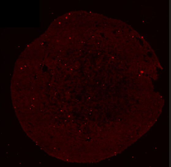
Application: Immunohistochemistry-FrozenSample Tested: Midbrain organoid tissueSpecies: HumanVerified Customer | Posted 11/21/2025Image of a slice of human midbrain organoid stained with SOX6 (in red)I used this antibody (dilution 1:100) on slices of frozen human midbrain organoids (derived from human iPSC). Unfortunately the staining didn't work in my case.
 Bio-Techne ResponseThank you for reviewing our product. We are sorry to hear that this product did not perform as expected. We have been in touch with the customer to resolve this issue according to our Product Guarantee and to the customer’s satisfaction.
There are no reviews that match your criteria.
Protocols
Find general support by application which include: protocols, troubleshooting, illustrated assays, videos and webinars.
- Antigen Retrieval Protocol (PIER)
- Antigen Retrieval for Frozen Sections Protocol
- Appropriate Fixation of IHC/ICC Samples
- Cellular Response to Hypoxia Protocols
- Chromogenic IHC Staining of Formalin-Fixed Paraffin-Embedded (FFPE) Tissue Protocol
- Chromogenic Immunohistochemistry Staining of Frozen Tissue
- ClariTSA™ Fluorophore Kits
- Detection & Visualization of Antibody Binding
- Fluorescent IHC Staining of Frozen Tissue Protocol
- Graphic Protocol for Heat-induced Epitope Retrieval
- Graphic Protocol for the Preparation and Fluorescent IHC Staining of Frozen Tissue Sections
- Graphic Protocol for the Preparation and Fluorescent IHC Staining of Paraffin-embedded Tissue Sections
- Graphic Protocol for the Preparation of Gelatin-coated Slides for Histological Tissue Sections
- ICC Cell Smear Protocol for Suspension Cells
- ICC Immunocytochemistry Protocol Videos
- ICC for Adherent Cells
- IHC Sample Preparation (Frozen sections vs Paraffin)
- Immunocytochemistry (ICC) Protocol
- Immunocytochemistry Troubleshooting
- Immunofluorescence of Organoids Embedded in Cultrex Basement Membrane Extract
- Immunofluorescent IHC Staining of Formalin-Fixed Paraffin-Embedded (FFPE) Tissue Protocol
- Immunohistochemistry (IHC) and Immunocytochemistry (ICC) Protocols
- Immunohistochemistry Frozen Troubleshooting
- Immunohistochemistry Paraffin Troubleshooting
- Preparing Samples for IHC/ICC Experiments
- Preventing Non-Specific Staining (Non-Specific Binding)
- Primary Antibody Selection & Optimization
- Protocol for Heat-Induced Epitope Retrieval (HIER)
- Protocol for Making a 4% Formaldehyde Solution in PBS
- Protocol for VisUCyte™ HRP Polymer Detection Reagent
- Protocol for the Fluorescent ICC Staining of Cell Smears - Graphic
- Protocol for the Fluorescent ICC Staining of Cultured Cells on Coverslips - Graphic
- Protocol for the Preparation & Fixation of Cells on Coverslips
- Protocol for the Preparation and Chromogenic IHC Staining of Frozen Tissue Sections
- Protocol for the Preparation and Chromogenic IHC Staining of Frozen Tissue Sections - Graphic
- Protocol for the Preparation and Chromogenic IHC Staining of Paraffin-embedded Tissue Sections
- Protocol for the Preparation and Chromogenic IHC Staining of Paraffin-embedded Tissue Sections - Graphic
- Protocol for the Preparation and Fluorescent ICC Staining of Cells on Coverslips
- Protocol for the Preparation and Fluorescent ICC Staining of Non-adherent Cells
- Protocol for the Preparation and Fluorescent ICC Staining of Stem Cells on Coverslips
- Protocol for the Preparation and Fluorescent IHC Staining of Frozen Tissue Sections
- Protocol for the Preparation and Fluorescent IHC Staining of Paraffin-embedded Tissue Sections
- Protocol for the Preparation of Gelatin-coated Slides for Histological Tissue Sections
- Protocol for the Preparation of a Cell Smear for Non-adherent Cell ICC - Graphic
- TUNEL and Active Caspase-3 Detection by IHC/ICC Protocol
- The Importance of IHC/ICC Controls
- Troubleshooting Guide: Immunohistochemistry
- View all Protocols, Troubleshooting, Illustrated assays and Webinars
Loading...
Associated Pathways
