SOX9 Antibody - BSA Free
Novus Biologicals | Catalog # NBP1-85551
![Immunohistochemistry-Paraffin: SOX9 Antibody [NBP1-85551]](https://resources.rndsystems.com/images/products/SOX9-Antibody-Immunohistochemistry-Paraffin-NBP1-85551-img0034.jpg "Immunohistochemistry-Paraffin: SOX9 Antibody [NBP1-85551]")
Loading...
Key Product Details
Validated by
Knockout/Knockdown, Orthogonal Validation
Species Reactivity
Validated:
Human, Mouse, Rat
Cited:
Human, Mouse, Rat, Porcine, Canine
Applications
Validated:
Immunohistochemistry, Immunohistochemistry-Paraffin, Western Blot, Immunocytochemistry/ Immunofluorescence
Cited:
Immunohistochemistry, Immunohistochemistry-Paraffin, Immunohistochemistry-Frozen, Western Blot, EMSA, IF/IHC, IHF-Fr
Label
Unconjugated
Antibody Source
Polyclonal Rabbit IgG
Format
BSA Free
Loading...
Product Specifications
Immunogen
This antibody was developed against Recombinant Protein corresponding to amino acids: SQRTHIKTEQLSPSHYSEQQQHSPQQIAYSPFNLPHYSPSYPPITRSQYDYTDHQNSSSYYSHAAGQGTGLYSTFTYMNPAQRPMYTPIADTSGVPSIPQTHSPQHWEQPVYTQLTR
Marker
Sertoli Cell Marker
Clonality
Polyclonal
Host
Rabbit
Isotype
IgG
Scientific Data Images for SOX9 Antibody - BSA Free
![Knockdown Validated: SOX9 Antibody [NBP1-85551]](https://resources.rndsystems.com/images/products/SOX9-Antibody-Western-Blot-NBP1-85551-img0035.jpg "Western Blot: SOX9 Antibody [NBP1-85551]")
![Immunocytochemistry/ Immunofluorescence: SOX9 Antibody [NBP1-85551]](https://resources.rndsystems.com/images/products/SOX9-Antibody-Immunocytochemistry-Immunofluorescence-NBP1-85551-img0037.jpg "Immunocytochemistry/ Immunofluorescence: SOX9 Antibody [NBP1-85551]")
Immunocytochemistry/ Immunofluorescence: SOX9 Antibody [NBP1-85551]
SOX9-Antibody-Immunocytochemistry-Immunofluorescence-NBP1-85551-img0037.jpg![Western Blot: SOX9 Antibody [NBP1-85551]](https://resources.rndsystems.com/images/products/SOX9-Antibody-Western-Blot-NBP1-85551-img0025.jpg "Western Blot: SOX9 Antibody [NBP1-85551]")
Western Blot: SOX9 Antibody [NBP1-85551]
Western Blot: SOX9 Antibody [NBP1-85551] - Analysis in mouse cell line NIH-3T3 and rat cell line NBT-II.![Immunocytochemistry/ Immunofluorescence: SOX9 Antibody [NBP1-85551]](https://resources.rndsystems.com/images/products/SOX9-Antibody-Immunocytochemistry-Immunofluorescence-NBP1-85551-img0036.jpg "Immunocytochemistry/ Immunofluorescence: SOX9 Antibody [NBP1-85551]")
Immunocytochemistry/ Immunofluorescence: SOX9 Antibody [NBP1-85551]
Immunocytochemistry/Immunofluorescence: SOX9 Antibody [NBP1-85551] - Staining of human cell line U-251 MG shows localization to nucleoplasm. Antibody staining is shown in green.![Immunohistochemistry-Paraffin: SOX9 Antibody [NBP1-85551]](https://resources.rndsystems.com/images/products/SOX9-Antibody-Immunohistochemistry-Paraffin-NBP1-85551-img0029.jpg "Immunohistochemistry-Paraffin: SOX9 Antibody [NBP1-85551]")
Immunohistochemistry-Paraffin: SOX9 Antibody [NBP1-85551]
Immunohistochemistry-Paraffin: SOX9 Antibody [NBP1-85551] - Staining of human colorectal cancer shows moderate to strong nuclear positivity in tumor cells.![Immunohistochemistry-Paraffin: SOX9 Antibody [NBP1-85551]](https://resources.rndsystems.com/images/products/SOX9-Antibody-Immunohistochemistry-Paraffin-NBP1-85551-img0030.jpg "Immunohistochemistry-Paraffin: SOX9 Antibody [NBP1-85551]")
Immunohistochemistry-Paraffin: SOX9 Antibody [NBP1-85551]
Immunohistochemistry-Paraffin: SOX9 Antibody [NBP1-85551] - Staining of human glioma shows moderate to strong nuclear positivity in tumor cells.![Immunohistochemistry-Paraffin: SOX9 Antibody [NBP1-85551]](https://resources.rndsystems.com/images/products/SOX9-Antibody-Immunohistochemistry-Paraffin-NBP1-85551-img0031.jpg "Immunohistochemistry-Paraffin: SOX9 Antibody [NBP1-85551]")
Immunohistochemistry-Paraffin: SOX9 Antibody [NBP1-85551]
Immunohistochemistry-Paraffin: SOX9 Antibody [NBP1-85551] - Staining of human skeletal muscle shows no nuclear positivity in striated muscle fibers as expected.![Immunohistochemistry-Paraffin: SOX9 Antibody [NBP1-85551]](https://resources.rndsystems.com/images/products/SOX9-Antibody-Immunohistochemistry-Paraffin-NBP1-85551-img0032.jpg "Immunohistochemistry-Paraffin: SOX9 Antibody [NBP1-85551]")
Immunohistochemistry-Paraffin: SOX9 Antibody [NBP1-85551]
Immunohistochemistry-Paraffin: SOX9 Antibody [NBP1-85551] - Staining of human small intestine shows moderate nuclear positivity in a subset of glandular cells.![Immunohistochemistry-Paraffin: SOX9 Antibody [NBP1-85551]](https://resources.rndsystems.com/images/products/SOX9-Antibody-Immunohistochemistry-Paraffin-NBP1-85551-img0033.jpg "Immunohistochemistry-Paraffin: SOX9 Antibody [NBP1-85551]")
Immunohistochemistry-Paraffin: SOX9 Antibody [NBP1-85551]
Immunohistochemistry-Paraffin: SOX9 Antibody [NBP1-85551] - Staining of human testis shows moderate nuclear positivity in a subset of cells in seminiferous ducts.![Simple Western: SOX9 Antibody [NBP1-85551]](https://resources.rndsystems.com/images/products/SOX9-Antibody-Simple-Western-NBP1-85551-img0006.jpg "Simple Western: SOX9 Antibody [NBP1-85551]")
Simple Western: SOX9 Antibody [NBP1-85551]
Simple Western: SOX9 Antibody [NBP1-85551] - Simple Western lane view shows a specific band for SOX9 in 0.1 mg/ml of U-251MG sp (left) and HepG2 (right) lysate. This experiment was performed under reducing conditions using the 12-230 kDa separation system.![Simple Western: SOX9 Antibody [NBP1-85551]](https://resources.rndsystems.com/images/products/SOX9-Antibody-Simple-Western-NBP1-85551-img0012.jpg "Simple Western: SOX9 Antibody [NBP1-85551]")
Simple Western: SOX9 Antibody [NBP1-85551]
Simple Western: SOX9 Antibody [NBP1-85551] - Electropherogram image(s) of corresponding Simple Western lane view. SOX9 antibody was used at 1:100 dilution on U-251MG sp and HepG2 lysates(s).
Immunohistochemistry: SOX9 Antibody [NBP1-85551] -
Immunohistochemistry of candidate biomarkers in prostate cancer. Representative immunohistochemical staining of ACPP, ADAM9, ALDH1A2, CASR, CCND1, CCPG1, CD34, CD44, CD44v6, CHGA, CHMP1A, EI24, ENO2, GADD45B, HA, HAS2, HES6, HMMR, HOXC6, HYAL1, IGF1, IQCK, MAP4K4, MKI67, PAGE4, PLIN2, PTEN, SIAH2, SMAD4, SOX9, SPP1, SYP, & TP53 from prostate cancer tissue microarrays. Scale bar represents 50 μm. Image collected & cropped by CiteAb from the following publication (https://bmccancer.biomedcentral.com/articles/10.1186/1471-2407-14-244), licensed under a CC-BY license. Not internally tested by Novus Biologicals.Applications for SOX9 Antibody - BSA Free
Application
Recommended Usage
Immunocytochemistry/ Immunofluorescence
0.25-2 ug/ml
Immunohistochemistry
1:500 - 1:1000
Immunohistochemistry-Paraffin
1:500 - 1:1000
Western Blot
0.04-0.4 ug/ml
Application Notes
IHC-Paraffin, HIER pH 6 retrieval is recommended. ICC/IF, Fixation/Permeabilization: PFA/Triton X-100br/>In Simple Western only 10 - 15 uL of the recommended dilution is used per data point.
See Simple Western Antibody Database for Simple Western validation: Tested in U-251MG sp and HepG2, separated by Size, antibody dilution of 1:100, apparent MW was 59 kDa. Separated by Size-Wes, Sally Sue/Peggy Sue.
See Simple Western Antibody Database for Simple Western validation: Tested in U-251MG sp and HepG2, separated by Size, antibody dilution of 1:100, apparent MW was 59 kDa. Separated by Size-Wes, Sally Sue/Peggy Sue.
Reviewed Applications
Read 1 review rated 5 using NBP1-85551 in the following applications:
Formulation, Preparation, and Storage
Purification
Affinity purified
Formulation
PBS (pH 7.2) and 40% Glycerol
Format
BSA Free
Preservative
0.02% Sodium Azide
Concentration
Concentrations vary lot to lot. See vial label for concentration. If unlisted please contact technical services.
Shipping
The product is shipped with polar packs. Upon receipt, store it immediately at the temperature recommended below.
Stability & Storage
Store at 4C short term. Aliquot and store at -20C long term. Avoid freeze-thaw cycles.
Background: SOX9
Long Name
Transcription Factor SOX9
Alternate Names
campomelic dysplasia, autosomal sex-reversal, CMD 1, CMD1, CMPD1, SRA1SRY (sex-determining region Y)-box 9 protein, SRY (sex determining region Y)-box 9, SRY-related HMG-box, gene 9, transcription factor SOX-9
Entrez Gene IDs
6662 (Human)
Gene Symbol
SOX9
UniProt
Additional SOX9 Products
Product Documents for SOX9 Antibody - BSA Free
Certificate of Analysis
To download a Certificate of Analysis, please enter a lot or batch number in the search box below.
Product Specific Notices for SOX9 Antibody - BSA Free
This product is for research use only and is not approved for use in humans or in clinical diagnosis. Primary Antibodies are guaranteed for 1 year from date of receipt.
Related Research Areas
Citations for SOX9 Antibody - BSA Free
 Powered by Bioz
Customer Reviews for SOX9 Antibody - BSA Free (1)
5 out of 5
1 Customer Rating
Have you used SOX9 Antibody - BSA Free?
Submit a review and receive an Amazon gift card!
$25/€18/£15/$25CAN/¥2500 Yen for a review with an image
$10/€7/£6/$10CAN/¥1110 Yen for a review without an image
Submit a review
Customer Images
Showing
1
-
1 of
1 review
Showing All
Filter By:
-
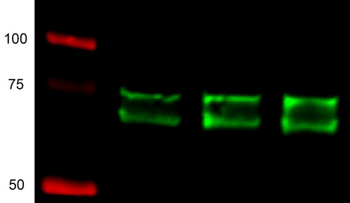
Application: Western BlotSample Tested: 293T lysatesSpecies: HumanVerified Customer | Posted 01/30/2019There are two bands around 75kd in 293t cells lysates. Green is Sox9 and red is marker.

There are no reviews that match your criteria.
Protocols
Find general support by application which include: protocols, troubleshooting, illustrated assays, videos and webinars.
- Antigen Retrieval Protocol (PIER)
- Antigen Retrieval for Frozen Sections Protocol
- Appropriate Fixation of IHC/ICC Samples
- Cellular Response to Hypoxia Protocols
- Chromogenic IHC Staining of Formalin-Fixed Paraffin-Embedded (FFPE) Tissue Protocol
- Chromogenic Immunohistochemistry Staining of Frozen Tissue
- ClariTSA™ Fluorophore Kits
- Detection & Visualization of Antibody Binding
- Fluorescent IHC Staining of Frozen Tissue Protocol
- Graphic Protocol for Heat-induced Epitope Retrieval
- Graphic Protocol for the Preparation and Fluorescent IHC Staining of Frozen Tissue Sections
- Graphic Protocol for the Preparation and Fluorescent IHC Staining of Paraffin-embedded Tissue Sections
- Graphic Protocol for the Preparation of Gelatin-coated Slides for Histological Tissue Sections
- ICC Cell Smear Protocol for Suspension Cells
- ICC Immunocytochemistry Protocol Videos
- ICC for Adherent Cells
- IHC Sample Preparation (Frozen sections vs Paraffin)
- Immunocytochemistry (ICC) Protocol
- Immunocytochemistry Troubleshooting
- Immunofluorescence of Organoids Embedded in Cultrex Basement Membrane Extract
- Immunofluorescent IHC Staining of Formalin-Fixed Paraffin-Embedded (FFPE) Tissue Protocol
- Immunohistochemistry (IHC) and Immunocytochemistry (ICC) Protocols
- Immunohistochemistry Frozen Troubleshooting
- Immunohistochemistry Paraffin Troubleshooting
- Preparing Samples for IHC/ICC Experiments
- Preventing Non-Specific Staining (Non-Specific Binding)
- Primary Antibody Selection & Optimization
- Protocol for Heat-Induced Epitope Retrieval (HIER)
- Protocol for Making a 4% Formaldehyde Solution in PBS
- Protocol for VisUCyte™ HRP Polymer Detection Reagent
- Protocol for the Fluorescent ICC Staining of Cell Smears - Graphic
- Protocol for the Fluorescent ICC Staining of Cultured Cells on Coverslips - Graphic
- Protocol for the Preparation & Fixation of Cells on Coverslips
- Protocol for the Preparation and Chromogenic IHC Staining of Frozen Tissue Sections
- Protocol for the Preparation and Chromogenic IHC Staining of Frozen Tissue Sections - Graphic
- Protocol for the Preparation and Chromogenic IHC Staining of Paraffin-embedded Tissue Sections
- Protocol for the Preparation and Chromogenic IHC Staining of Paraffin-embedded Tissue Sections - Graphic
- Protocol for the Preparation and Fluorescent ICC Staining of Cells on Coverslips
- Protocol for the Preparation and Fluorescent ICC Staining of Non-adherent Cells
- Protocol for the Preparation and Fluorescent ICC Staining of Stem Cells on Coverslips
- Protocol for the Preparation and Fluorescent IHC Staining of Frozen Tissue Sections
- Protocol for the Preparation and Fluorescent IHC Staining of Paraffin-embedded Tissue Sections
- Protocol for the Preparation of Gelatin-coated Slides for Histological Tissue Sections
- Protocol for the Preparation of a Cell Smear for Non-adherent Cell ICC - Graphic
- R&D Systems Quality Control Western Blot Protocol
- TUNEL and Active Caspase-3 Detection by IHC/ICC Protocol
- The Importance of IHC/ICC Controls
- Troubleshooting Guide: Immunohistochemistry
- Troubleshooting Guide: Western Blot Figures
- Western Blot Conditions
- Western Blot Protocol
- Western Blot Protocol for Cell Lysates
- Western Blot Troubleshooting
- Western Blot Troubleshooting Guide
- View all Protocols, Troubleshooting, Illustrated assays and Webinars


