SPINK1 Antibody (4D4) - Azide and BSA Free
Novus Biologicals | Catalog # H00006690-M01
![Western Blot: SPINK1 Antibody (4D4) [H00006690-M01]](https://resources.rndsystems.com/images/products/SPINK1-Antibody-4D4-Western-Blot-H00006690-M01-img0010.jpg "Western Blot: SPINK1 Antibody (4D4) [H00006690-M01]")
Loading...
Key Product Details
Validated by
Biological Validation
Species Reactivity
Validated:
Human, Mouse
Cited:
Human, Mouse
Applications
Validated:
Immunohistochemistry, Immunohistochemistry-Paraffin, Immunohistochemistry-Frozen, Western Blot, ELISA, Immunocytochemistry/ Immunofluorescence, Immunoprecipitation
Cited:
Immunohistochemistry, Immunohistochemistry-Paraffin, Immunohistochemistry-Frozen, Immunocytochemistry/ Immunofluorescence, IF/IHC
Label
Unconjugated
Antibody Source
Monoclonal Mouse IgG2a Kappa Clone # 4D4
Format
Azide and BSA Free
Loading...
Product Specifications
Immunogen
SPINK1 (AAH25790, 24 a.a. ~ 79 a.a) partial recombinant protein with GST tag. MW of the GST tag alone is 26 KDa. DSLGREAKCYNELNGCTKIYDPVCGTDGNTYPNECVLCFENRKRQTSILIQKSGPC
Reactivity Notes
Mouse reactivity reported in scientific literature (PMID: 32929152)
Localization
Secreted
Specificity
SPINK1 - serine protease inhibitor, Kazal type 1
Clonality
Monoclonal
Host
Mouse
Isotype
IgG2a Kappa
Description
Quality control test: Antibody Reactive Against Recombinant Protein.
Scientific Data Images for SPINK1 Antibody (4D4) - Azide and BSA Free
Western Blot: SPINK1 Antibody (4D4) [H00006690-M01]
Western Blot: SPINK1 Antibody (4D4) [H00006690-M01] - Detection against Immunogen (31.9 kDa). Antibody reactive against recombinant protein.![Immunohistochemistry-Paraffin: SPINK1 Antibody (4D4) [H00006690-M01]](https://resources.rndsystems.com/images/products/SPINK1-Antibody-4D4-Immunohistochemistry-Paraffin-H00006690-M01-img0016.jpg "Immunohistochemistry-Paraffin: SPINK1 Antibody (4D4) [H00006690-M01]")
Immunohistochemistry-Paraffin: SPINK1 Antibody (4D4) [H00006690-M01]
Immunohistochemistry-Paraffin: SPINK1 Antibody (4D4) [H00006690-M01] - Analysis of SPINK1 antibody (4D4) on mouse pancreas tissue. Antibody concentration 1 ug/ml. Image from verified customer review.![Western Blot: SPINK1 Antibody (4D4) [H00006690-M01]](https://resources.rndsystems.com/images/products/SPINK1-Antibody-4D4-Western-Blot-H00006690-M01-img0009.jpg "Western Blot: SPINK1 Antibody (4D4) [H00006690-M01]")
Western Blot: SPINK1 Antibody (4D4) [H00006690-M01]
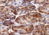
Western Blot: SPINK1 Antibody (4D4) [H00006690-M01] - SPINK1 monoclonal antibody (M01), clone 4D4. Analysis of SPINK1 expression in human pancreas.![Immunohistochemistry-Paraffin: SPINK1 Antibody (4D4) [H00006690-M01]](https://resources.rndsystems.com/images/products/SPINK1-Antibody-4D4-Immunohistochemistry-Paraffin-H00006690-M01-img0006.jpg "Immunohistochemistry-Paraffin: SPINK1 Antibody (4D4) [H00006690-M01]")
Immunohistochemistry-Paraffin: SPINK1 Antibody (4D4) [H00006690-M01]
Immunohistochemistry-Paraffin: SPINK1 Antibody (4D4) [H00006690-M01] - Analysis of monoclonal antibody to SPINK1 on formalin-fixed paraffin-embedded human pancreas. Antibody concentration 1 ug/ml.![Immunohistochemistry: SPINK1 Antibody (4D4) [H00006690-M01]](https://resources.rndsystems.com/images/products/SPINK1-Antibody-4D4-Immunohistochemistry-H00006690-M01-img0012.jpg "Immunohistochemistry: SPINK1 Antibody (4D4) [H00006690-M01]")
![Immunohistochemistry: SPINK1 Antibody (4D4) [H00006690-M01]](https://resources.rndsystems.com/images/products/SPINK1-Antibody-4D4-Immunohistochemistry-H00006690-M01-img0013.jpg "Immunohistochemistry: SPINK1 Antibody (4D4) [H00006690-M01]")
Immunohistochemistry: SPINK1 Antibody (4D4) [H00006690-M01]
SPINK1-Antibody-4D4-Immunohistochemistry-H00006690-M01-img0013.jpg![Immunohistochemistry: SPINK1 Antibody (4D4) [H00006690-M01]](https://resources.rndsystems.com/images/products/SPINK1-Antibody-4D4-Immunohistochemistry-H00006690-M01-img0014.jpg "Immunohistochemistry: SPINK1 Antibody (4D4) [H00006690-M01]")
Immunohistochemistry: SPINK1 Antibody (4D4) [H00006690-M01]
SPINK1-Antibody-4D4-Immunohistochemistry-H00006690-M01-img0014.jpg![Immunohistochemistry: SPINK1 Antibody (4D4) [H00006690-M01]](https://resources.rndsystems.com/images/products/SPINK1-Antibody-4D4-Immunohistochemistry-H00006690-M01-img0015.jpg "Immunohistochemistry: SPINK1 Antibody (4D4) [H00006690-M01]")
Immunohistochemistry: SPINK1 Antibody (4D4) [H00006690-M01]
SPINK1-Antibody-4D4-Immunohistochemistry-H00006690-M01-img0015.jpg![Immunoprecipitation: SPINK1 Antibody (4D4) [H00006690-M01]](https://resources.rndsystems.com/images/products/SPINK1-Antibody-4D4-Immunoprecipitation-H00006690-M01-img0007.jpg "Immunoprecipitation: SPINK1 Antibody (4D4) [H00006690-M01]")
Immunoprecipitation: SPINK1 Antibody (4D4) [H00006690-M01]
Immunoprecipitation: SPINK1 Antibody (4D4) [H00006690-M01] - Analysis of SPINK1 transfected lysate using anti-SPINK1 monoclonal antibody and Protein A Magnetic Bead, and immunoblotted with SPINK1 MaxPab rabbit polyclonal antibody.![ELISA: SPINK1 Antibody (4D4) [H00006690-M01]](https://resources.rndsystems.com/images/products/SPINK1-Antibody-4D4-ELISA-H00006690-M01-img0011.jpg "ELISA: SPINK1 Antibody (4D4) [H00006690-M01]")
ELISA: SPINK1 Antibody (4D4) [H00006690-M01]
ELISA: SPINK1 Antibody (4D4) [H00006690-M01] - Detection limit for recombinant GST tagged SPINK1 is 0.03 ng/ml as a capture antibody. [H00006690-M01] -")
Immunohistochemistry: SPINK1 Antibody (4D4) [H00006690-M01] -
Immunohistochemistry: SPINK1 Antibody (4D4) [H00006690-M01] - ADT induced SPINK1 upregulation associates with NE-phenotype in mice & NEPC patients.a Box plots depicting relative expression of SPINK1, SYP, CHGA, TUBB3, & VIM transcripts (read counts) in VCaP tumors implanted orthotopically in orchiectomized mice & subjected to vehicle (n = 4) or anti-androgens [enzalutamide (n = 4) or ARN-509 (n = 4)] treatment for 4 weeks (GSE95413). b Representative images of immunohistochemical staining for the same markers shown in a using VCaP xenograft tumors as described in a. Scale bar represents 100 μm. c Box plots depicting quantification of the immunohistochemical staining in VCaP xenografts for the markers shown in b. d Representative images for immunohistochemical staining of SPINK1, SYP, CHGA, & TUBB3 in 22RV1 xenograft tumors excised from orchiectomized mice treated with enzalutamide (20 mg/kg body weight) or vehicle control (n = 5 each). Intact group represents non-castrated control mice (n = 5). Scale bar represents 50 μm. e Box plots depicting quantification of the immunohistochemical staining in 22RV1 xenografts for the markers shown in d. f Representative images showing H&E staining (×200 magnification) & immunostaining (×200 magnification) for AR, synaptophysin, & SPINK1 in tumor specimens obtained from NEPC patients’, namely WCM12, WCM155 (an organoid), & WCM677. Scale bar represents 100 µm. Data are presented as box-and-whisker plots with median, where the box extends from 25th–75th percentile, & whiskers ranges from minimum & maximum values. For panels a, c, e one-way ANOVA, Dunnett’s multiple-comparisons test was applied. Image collected & cropped by CiteAb from the following publication (https://pubmed.ncbi.nlm.nih.gov/31959826), licensed under a CC-BY license. Not internally tested by Novus Biologicals. [H00006690-M01] -")
Immunohistochemistry: SPINK1 Antibody (4D4) [H00006690-M01] -
Immunohistochemistry: SPINK1 Antibody (4D4) [H00006690-M01] - SPINK1 is negatively correlated with AR in PCa patients.a Heatmap depicting AR & SPINK1 expression in TCGA-PRAD cohort (n = 180). Shades of yellow & blue represents expression values in log2 (RPM+1). b Representative micrographs depicting PCa tissue microarray (TMA) cores (n = 237), immunostained for SPINK1 & AR expression by immunohistochemistry (IHC). Top panel shows representative IHC for SPINK1 in SPINK1-negative (SPINK1−) & SPINK1-positive (SPINK1+) patients. Bottom panel represents IHC for AR expression in the tumor core from same patients. Scale bar represents 500 µm & 100 µm for the entire core & the inset images, respectively. c Bar plot showing percentage of IHC scoring for AR in the SPINK1+ & SPINK1− patients’ specimens. P-value for the Chi-Square test is indicated. d Contingency table for the AR & SPINK1 status. Patients showing high or medium expression of AR were grouped as AR-(Hi/Med), while patients with low or null AR expression were indicated as AR-(Low/Neg). P-value for Fisher’s exact test is indicated. Image collected & cropped by CiteAb from the following publication (https://pubmed.ncbi.nlm.nih.gov/31959826), licensed under a CC-BY license. Not internally tested by Novus Biologicals. [H00006690-M01] -")
Immunohistochemistry: SPINK1 Antibody (4D4) [H00006690-M01] -
Immunohistochemistry: SPINK1 Antibody (4D4) [H00006690-M01] - Spatial gene expression heterogeneity within the 1.2 cancer tissue sample. a Factor activity maps for selected factors corresponding to epithelial, stromal, cancerous, PIN, or inflamed regions. Remaining factors’ activity maps in Supplementary Figure 2 & Supplementary Data 1. b Annotated brightfield image of H&E-stained tissue section. c Heatmap of the 20 most variable genes between cancer, PIN & normal gland regions, using spot sets from Supplementary Fig. 4b. Centered rlog: difference of rlog (variance-stabilized transform of ST expression data) & gene-wise mean rlog. Arrows highlight genes of interest validated by immunohistochemistry (IHC). d First two principal components of spot sets from c separate cancer, PIN & normal regions. e Array dot plots for SPINK1 & NPY. Circle size in array dot plots indicates normalized ST counts. f IHC staining for SPINK1 & NPY of an adjacent section on the ST array. Nuclei are stained with DAPI (blue). Scale bar indicates 1 mm Image collected & cropped by CiteAb from the following publication (https://pubmed.ncbi.nlm.nih.gov/29925878), licensed under a CC-BY license. Not internally tested by Novus Biologicals. [H00006690-M01] -")
Immunohistochemistry: SPINK1 Antibody (4D4) [H00006690-M01] -
Immunohistochemistry: SPINK1 Antibody (4D4) [H00006690-M01] - ADT induced SPINK1 upregulation associates with NE-phenotype in mice & NEPC patients.a Box plots depicting relative expression of SPINK1, SYP, CHGA, TUBB3, & VIM transcripts (read counts) in VCaP tumors implanted orthotopically in orchiectomized mice & subjected to vehicle (n = 4) or anti-androgens [enzalutamide (n = 4) or ARN-509 (n = 4)] treatment for 4 weeks (GSE95413). b Representative images of immunohistochemical staining for the same markers shown in a using VCaP xenograft tumors as described in a. Scale bar represents 100 μm. c Box plots depicting quantification of the immunohistochemical staining in VCaP xenografts for the markers shown in b. d Representative images for immunohistochemical staining of SPINK1, SYP, CHGA, & TUBB3 in 22RV1 xenograft tumors excised from orchiectomized mice treated with enzalutamide (20 mg/kg body weight) or vehicle control (n = 5 each). Intact group represents non-castrated control mice (n = 5). Scale bar represents 50 μm. e Box plots depicting quantification of the immunohistochemical staining in 22RV1 xenografts for the markers shown in d. f Representative images showing H&E staining (×200 magnification) & immunostaining (×200 magnification) for AR, synaptophysin, & SPINK1 in tumor specimens obtained from NEPC patients’, namely WCM12, WCM155 (an organoid), & WCM677. Scale bar represents 100 µm. Data are presented as box-and-whisker plots with median, where the box extends from 25th–75th percentile, & whiskers ranges from minimum & maximum values. For panels a, c, e one-way ANOVA, Dunnett’s multiple-comparisons test was applied. Image collected & cropped by CiteAb from the following publication (https://pubmed.ncbi.nlm.nih.gov/31959826), licensed under a CC-BY license. Not internally tested by Novus Biologicals. [H00006690-M01] -")
Immunohistochemistry: SPINK1 Antibody (4D4) [H00006690-M01] -
Immunohistochemistry: SPINK1 Antibody (4D4) [H00006690-M01] - ADT induced SPINK1 upregulation associates with NE-phenotype in mice & NEPC patients.a Box plots depicting relative expression of SPINK1, SYP, CHGA, TUBB3, & VIM transcripts (read counts) in VCaP tumors implanted orthotopically in orchiectomized mice & subjected to vehicle (n = 4) or anti-androgens [enzalutamide (n = 4) or ARN-509 (n = 4)] treatment for 4 weeks (GSE95413). b Representative images of immunohistochemical staining for the same markers shown in a using VCaP xenograft tumors as described in a. Scale bar represents 100 μm. c Box plots depicting quantification of the immunohistochemical staining in VCaP xenografts for the markers shown in b. d Representative images for immunohistochemical staining of SPINK1, SYP, CHGA, & TUBB3 in 22RV1 xenograft tumors excised from orchiectomized mice treated with enzalutamide (20 mg/kg body weight) or vehicle control (n = 5 each). Intact group represents non-castrated control mice (n = 5). Scale bar represents 50 μm. e Box plots depicting quantification of the immunohistochemical staining in 22RV1 xenografts for the markers shown in d. f Representative images showing H&E staining (×200 magnification) & immunostaining (×200 magnification) for AR, synaptophysin, & SPINK1 in tumor specimens obtained from NEPC patients’, namely WCM12, WCM155 (an organoid), & WCM677. Scale bar represents 100 µm. Data are presented as box-and-whisker plots with median, where the box extends from 25th–75th percentile, & whiskers ranges from minimum & maximum values. For panels a, c, e one-way ANOVA, Dunnett’s multiple-comparisons test was applied. Image collected & cropped by CiteAb from the following publication (https://pubmed.ncbi.nlm.nih.gov/31959826), licensed under a CC-BY license. Not internally tested by Novus Biologicals.Applications for SPINK1 Antibody (4D4) - Azide and BSA Free
Application
Recommended Usage
ELISA
Optimal dilutions of this antibody should be experimentally determined.
Immunocytochemistry/ Immunofluorescence
Optimal dilutions of this antibody should be experimentally determined.
Immunohistochemistry
Optimal dilutions of this antibody should be experimentally determined.
Immunohistochemistry-Frozen
Optimal dilutions of this antibody should be experimentally determined.
Immunohistochemistry-Paraffin
Optimal dilutions of this antibody should be experimentally determined.
Immunoprecipitation
Optimal dilutions of this antibody should be experimentally determined.
Western Blot
Optimal dilutions of this antibody should be experimentally determined.
Reviewed Applications
Read 1 review rated 5 using H00006690-M01 in the following applications:
Formulation, Preparation, and Storage
Purification
IgG purified
Formulation
PBS, pH 7.4
Format
Azide and BSA Free
Preservative
No Preservative
Concentration
Concentrations vary lot to lot. See vial label for concentration. If unlisted please contact technical services.
Shipping
The product is shipped with polar packs. Upon receipt, store it immediately at the temperature recommended below.
Stability & Storage
Aliquot and store at -20C or -80C. Avoid freeze-thaw cycles.
Background: SPINK1
Long Name
Serine Peptidase Inhibitor, Kazal Type 1
Alternate Names
PCTT, PSTI, Spink3, TATI
Entrez Gene IDs
6690 (Human)
Gene Symbol
SPINK1
OMIM
167790 (Human)
UniProt
Additional SPINK1 Products
Product Documents for SPINK1 Antibody (4D4) - Azide and BSA Free
Certificate of Analysis
To download a Certificate of Analysis, please enter a lot or batch number in the search box below.
Product Specific Notices for SPINK1 Antibody (4D4) - Azide and BSA Free
This product is produced by and distributed for Abnova, a company based in Taiwan.
This product is for research use only and is not approved for use in humans or in clinical diagnosis. Primary Antibodies are guaranteed for 1 year from date of receipt.
Related Research Areas
Citations for SPINK1 Antibody (4D4) - Azide and BSA Free
 Powered by Bioz
Customer Reviews for SPINK1 Antibody (4D4) - Azide and BSA Free (1)
5 out of 5
1 Customer Rating
Have you used SPINK1 Antibody (4D4) - Azide and BSA Free?
Submit a review and receive an Amazon gift card!
$25/€18/£15/$25CAN/¥2500 Yen for a review with an image
$10/€7/£6/$10CAN/¥1110 Yen for a review without an image
Submit a review
Customer Images
Showing
1
-
1 of
1 review
Showing All
Filter By:
-
Application: Immunohistochemistry-ParaffinSample Tested: Pancreas tissueSpecies: MouseVerified Customer | Posted 02/10/2022Pancreas tissueAntibody concentration 1 ug/ml

There are no reviews that match your criteria.
Protocols
Find general support by application which include: protocols, troubleshooting, illustrated assays, videos and webinars.
- Antigen Retrieval Protocol (PIER)
- Antigen Retrieval for Frozen Sections Protocol
- Appropriate Fixation of IHC/ICC Samples
- Cellular Response to Hypoxia Protocols
- Chromogenic IHC Staining of Formalin-Fixed Paraffin-Embedded (FFPE) Tissue Protocol
- Chromogenic Immunohistochemistry Staining of Frozen Tissue
- ClariTSA™ Fluorophore Kits
- Detection & Visualization of Antibody Binding
- ELISA Sample Preparation & Collection Guide
- ELISA Troubleshooting Guide
- Fluorescent IHC Staining of Frozen Tissue Protocol
- Graphic Protocol for Heat-induced Epitope Retrieval
- Graphic Protocol for the Preparation and Fluorescent IHC Staining of Frozen Tissue Sections
- Graphic Protocol for the Preparation and Fluorescent IHC Staining of Paraffin-embedded Tissue Sections
- Graphic Protocol for the Preparation of Gelatin-coated Slides for Histological Tissue Sections
- How to Run an R&D Systems DuoSet ELISA
- How to Run an R&D Systems Quantikine ELISA
- How to Run an R&D Systems Quantikine™ QuicKit™ ELISA
- ICC Cell Smear Protocol for Suspension Cells
- ICC Immunocytochemistry Protocol Videos
- ICC for Adherent Cells
- IHC Sample Preparation (Frozen sections vs Paraffin)
- Immunocytochemistry (ICC) Protocol
- Immunocytochemistry Troubleshooting
- Immunofluorescence of Organoids Embedded in Cultrex Basement Membrane Extract
- Immunofluorescent IHC Staining of Formalin-Fixed Paraffin-Embedded (FFPE) Tissue Protocol
- Immunohistochemistry (IHC) and Immunocytochemistry (ICC) Protocols
- Immunohistochemistry Frozen Troubleshooting
- Immunohistochemistry Paraffin Troubleshooting
- Immunoprecipitation Protocol
- Preparing Samples for IHC/ICC Experiments
- Preventing Non-Specific Staining (Non-Specific Binding)
- Primary Antibody Selection & Optimization
- Protocol for Heat-Induced Epitope Retrieval (HIER)
- Protocol for Making a 4% Formaldehyde Solution in PBS
- Protocol for VisUCyte™ HRP Polymer Detection Reagent
- Protocol for the Fluorescent ICC Staining of Cell Smears - Graphic
- Protocol for the Fluorescent ICC Staining of Cultured Cells on Coverslips - Graphic
- Protocol for the Preparation & Fixation of Cells on Coverslips
- Protocol for the Preparation and Chromogenic IHC Staining of Frozen Tissue Sections
- Protocol for the Preparation and Chromogenic IHC Staining of Frozen Tissue Sections - Graphic
- Protocol for the Preparation and Chromogenic IHC Staining of Paraffin-embedded Tissue Sections
- Protocol for the Preparation and Chromogenic IHC Staining of Paraffin-embedded Tissue Sections - Graphic
- Protocol for the Preparation and Fluorescent ICC Staining of Cells on Coverslips
- Protocol for the Preparation and Fluorescent ICC Staining of Non-adherent Cells
- Protocol for the Preparation and Fluorescent ICC Staining of Stem Cells on Coverslips
- Protocol for the Preparation and Fluorescent IHC Staining of Frozen Tissue Sections
- Protocol for the Preparation and Fluorescent IHC Staining of Paraffin-embedded Tissue Sections
- Protocol for the Preparation of Gelatin-coated Slides for Histological Tissue Sections
- Protocol for the Preparation of a Cell Smear for Non-adherent Cell ICC - Graphic
- Quantikine HS ELISA Kit Assay Principle, Alkaline Phosphatase
- Quantikine HS ELISA Kit Principle, Streptavidin-HRP Polymer
- R&D Systems Quality Control Western Blot Protocol
- Sandwich ELISA (Colorimetric) – Biotin/Streptavidin Detection Protocol
- Sandwich ELISA (Colorimetric) – Direct Detection Protocol
- TUNEL and Active Caspase-3 Detection by IHC/ICC Protocol
- The Importance of IHC/ICC Controls
- Troubleshooting Guide: ELISA
- Troubleshooting Guide: Immunohistochemistry
- Troubleshooting Guide: Western Blot Figures
- Western Blot Conditions
- Western Blot Protocol
- Western Blot Protocol for Cell Lysates
- Western Blot Troubleshooting
- Western Blot Troubleshooting Guide
- View all Protocols, Troubleshooting, Illustrated assays and Webinars
Loading...