SR-BI Antibody - BSA Free
Novus Biologicals | Catalog # NB400-104
![Simple Western: SR-BI AntibodyBSA Free [NB400-104]](https://resources.rndsystems.com/images/products/SR-BI-Antibody-Simple-Western-NB400-104-img0014.jpg "Simple Western: SR-BI AntibodyBSA Free [NB400-104]")
Key Product Details
Validated by
Knockout/Knockdown
Species Reactivity
Validated:
Human, Mouse, Rat, Porcine, Chinese Hamster, Golden Syrian Hamster, Hamster, Mustelid, Primate, Rabbit
Cited:
Human, Mouse, Rat, Porcine, Golden Syrian Hamster, Hamster, Hamster - Cricetulus (Chinese Hamster), Primate, Rabbit
Applications
Validated:
Knockout Validated, Immunohistochemistry, Immunohistochemistry-Paraffin, Immunohistochemistry-Frozen, Western Blot, Block/Neutralize, Flow Cytometry, Flow (Intracellular), Immunocytochemistry/ Immunofluorescence, Simple Western, Immunoprecipitation, Proximity Ligation Assay, Knockdown Validated
Cited:
Knockout Validated, Immunohistochemistry-Paraffin, Immunohistochemistry-Frozen, Western Blot, Flow Cytometry, Immunocytochemistry/ Immunofluorescence, Immunoprecipitation, BN, Proximity Ligation Assay, IF/IHC, Knockdown Validated
Label
Unconjugated
Antibody Source
Polyclonal Rabbit IgG
Format
BSA Free
Loading...
Product Specifications
Immunogen
A C-terminal peptide containing residues from mouse SR-BI (within residues 450-509). [UniProt Q61009]
Reactivity Notes
Use in Mouse reported in scientific literature (PMID:33719499).
Localization
Cell Membrane
Clonality
Polyclonal
Host
Rabbit
Isotype
IgG
Theoretical MW
82 kDa.
Disclaimer note: The observed molecular weight of the protein may vary from the listed predicted molecular weight due to post translational modifications, post translation cleavages, relative charges, and other experimental factors.
Disclaimer note: The observed molecular weight of the protein may vary from the listed predicted molecular weight due to post translational modifications, post translation cleavages, relative charges, and other experimental factors.
Scientific Data Images for SR-BI Antibody - BSA Free
Simple Western: SR-BI AntibodyBSA Free [NB400-104]
Simple Western: SR-BI Antibody [NB400-104] - Image shows a specific band for SR-BI in 0.5 mg/mL of HeLa lysate. This experiment was performed under reducing conditions using the 12-230 kDa separation system.![Western Blot: SR-BI AntibodyBSA Free [NB400-104]](https://resources.rndsystems.com/images/products/SR-BI-Antibody-Western-Blot-NB400-104-img0003.jpg "Western Blot: SR-BI AntibodyBSA Free [NB400-104]")
Western Blot: SR-BI AntibodyBSA Free [NB400-104]
Western Blot: SR-BI Antibody [NB400-104] - SR-BI antibody was tested in human adrenal cell lysate.![Immunocytochemistry/ Immunofluorescence: SR-BI Antibody - BSA Free [NB400-104]](https://resources.rndsystems.com/images/products/SR-BI-Antibody-Immunocytochemistry-Immunofluorescence-NB400-104-img0030.jpg "Immunocytochemistry/ Immunofluorescence: SR-BI Antibody - BSA Free [NB400-104]")
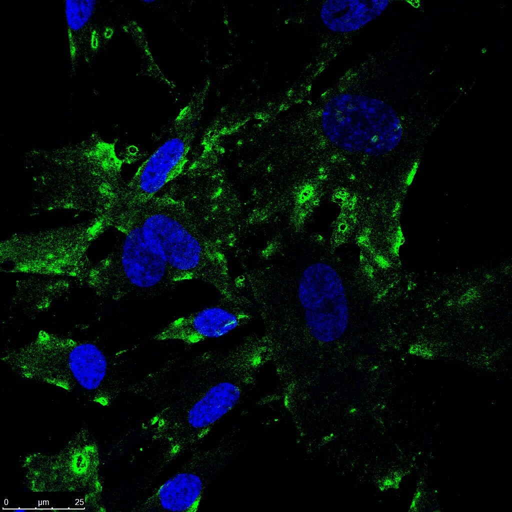
Immunocytochemistry/ Immunofluorescence: SR-BI Antibody - BSA Free [NB400-104]
Immunocytochemistry/Immunofluorescence: SR-BI Antibody [NB400-104] - SR-BI antibody was tested in human fibroblast samples fixed in 4% PFA and permeabilized in PBS (0.2% Tween). Primary incubation overnight at 4C using a 1:100 dilution in PBS (0.1% Tween) with 1% BSA. Secondary antibody is anti-rabbit conjugate to Alexa Fluor 488. SR-BI is shown in green and nucleus in blue (Hoescht 33342 stain).![Flow Cytometry: SR-BI Antibody - BSA Free [NB400-104]](https://resources.rndsystems.com/images/products/SR-BI-Antibody-Flow-Cytometry-NB400-104-img0026.jpg "Flow Cytometry: SR-BI Antibody - BSA Free [NB400-104]")
Flow Cytometry: SR-BI Antibody - BSA Free [NB400-104]
Flow Cytometry: SR-BI Antibody [NB400-104] - An intracellular stain was performed on HeLa cells with SR-BI antibody NB400-104AF488 (blue) and a matched isotype control NBP2-24893AF488 (orange). Cells were fixed with 4% PFA and then permeablized with 0.1% saponin. Cells were incubated in an antibody dilution of 5 ug/mL for 30 minutes at room temperature. Both antibodies were conjugated to Alexa Fluor 488.![Western Blot: SR-BI AntibodyBSA Free [NB400-104]](https://resources.rndsystems.com/images/products/SR-BI-Antibody-Western-Blot-NB400-104-img0013.jpg "Western Blot: SR-BI AntibodyBSA Free [NB400-104]")
Western Blot: SR-BI AntibodyBSA Free [NB400-104]
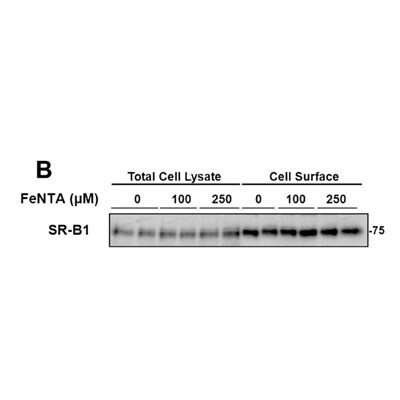
Western Blot: SR-BI Antibody [NB400-104] - Detection of SR-BI in rat H4IIE total cell lysates and plasma membrane proteins. Photo courtesy of product review by verified customer.![Immunohistochemistry: SR-BI Antibody - BSA Free [NB400-104]](https://resources.rndsystems.com/images/products/SR-BI-Antibody-Immunohistochemistry-NB400-104-img0011.jpg "Immunohistochemistry: SR-BI Antibody - BSA Free [NB400-104]")
Immunohistochemistry: SR-BI Antibody - BSA Free [NB400-104]
Immunohistochemistry: SR-BI Antibody [NB400-104] - Immunolocalization of SR-BI in adult mink testis using NB400-104. SR-BI labeling is visible at the surface and along the outline of the large vacuole. Photo courtesy of R.M. Pelletier, University of Montreal.![Immunohistochemistry-Paraffin: SR-BI Antibody - BSA Free [NB400-104]](https://resources.rndsystems.com/images/products/SR-BI-Antibody-Immunohistochemistry-Paraffin-NB400-104-img0022.jpg "Immunohistochemistry-Paraffin: SR-BI Antibody - BSA Free [NB400-104]")
Immunohistochemistry-Paraffin: SR-BI Antibody - BSA Free [NB400-104]
Immunohistochemistry-Paraffin: SR-BI Antibody [NB400-104] - FFPE tissue section of mouse liver using SR-BI antibody (Lot 8310) at 1:300 dilution with HRP-DAB detection and hematoxylin counterstaining. The antibody generated a specific membrane signal of SR-BI protein in the murine hepatocytes.![Knockdown Validated: SR-BI Antibody - BSA Free [NB400-104]](https://resources.rndsystems.com/images/products/SR-BI-Antibody-Western-Blot-NB400-104-img0034.jpg "Western Blot: SR-BI Antibody - BSA Free [NB400-104]")
![Immunohistochemistry-Paraffin: SR-BI Antibody - BSA Free [NB400-104]](https://resources.rndsystems.com/images/products/SR-BI-Antibody-Immunohistochemistry-Paraffin-NB400-104-img0012.jpg "Immunohistochemistry-Paraffin: SR-BI Antibody - BSA Free [NB400-104]")
Immunohistochemistry-Paraffin: SR-BI Antibody - BSA Free [NB400-104]
Immunohistochemistry-Paraffin: SR-BI Antibody [NB400-104] - Staining of human adrenal cortex.![Immunocytochemistry/ Immunofluorescence: SR-BI Antibody - BSA Free [NB400-104]](https://resources.rndsystems.com/images/products/SR-BI-Antibody-Immunocytochemistry-Immunofluorescence-NB400-104-img0029.jpg "Immunocytochemistry/ Immunofluorescence: SR-BI Antibody - BSA Free [NB400-104]")
Immunocytochemistry/ Immunofluorescence: SR-BI Antibody - BSA Free [NB400-104]
Immunocytochemistry/Immunofluorescence: SR-BI Antibody [NB400-104] - HeLa cells were fixed and permeabilized for 10 minutes using -20C MeOH. The cells were incubated with anti-SR-BI at 2 ug/ml overnight at 4C and detected with an anti-rabbit Dylight 488 (Green) at a 1:500 dilution. Alpha tubulin (DM1A) NB100-690 was used as a co-stain at a 1:1000 dilution and detected with an anti-mouse Dylight 550 (Red) at a 1:500 dilution. Nuclei were counterstained with DAPI (Blue). Cells were imaged using a 40X objective.![Immunohistochemistry-Paraffin: SR-BI Antibody - BSA Free [NB400-104]](https://resources.rndsystems.com/images/products/SR-BI-Antibody-Immunohistochemistry-Paraffin-NB400-104-img0023.jpg "Immunohistochemistry-Paraffin: SR-BI Antibody - BSA Free [NB400-104]")
Immunohistochemistry-Paraffin: SR-BI Antibody - BSA Free [NB400-104]
Immunohistochemistry-Paraffin: SR-BI Antibody [NB400-104] - FFPE tissue section of mouse liver using SR-BI antibody (Lot R-4) at 1:300 dilution with HRP-DAB detection and hematoxylin counterstaining. The antibody generated mainly a membranous signal of SR-BI protein in the murine hepatocytes.![Flow Cytometry: SR-BI Antibody - BSA Free [NB400-104]](https://resources.rndsystems.com/images/products/SR-BI-Antibody-Flow-Cytometry-NB400-104-img0004.jpg "Flow Cytometry: SR-BI Antibody - BSA Free [NB400-104]")
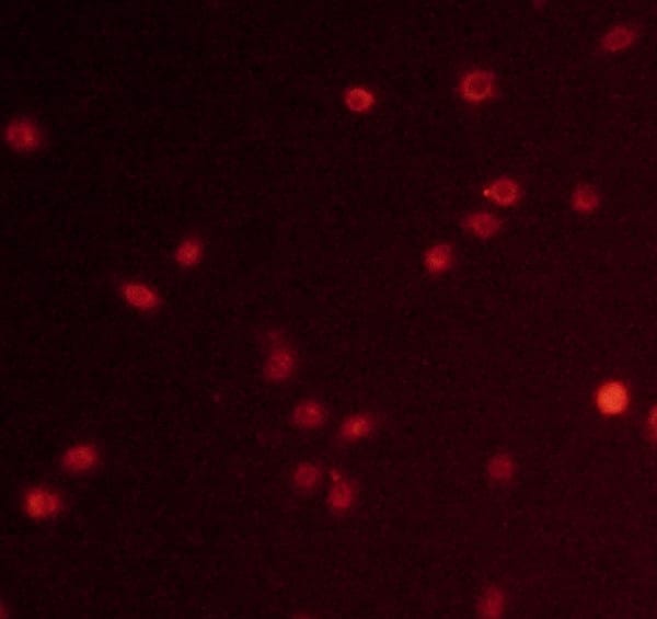
Flow Cytometry: SR-BI Antibody - BSA Free [NB400-104]
Flow Cytometry: SR-BI Antibody [NB400-104] - Analysis of Huh7 and HepG2 cells using SR-B1 antibody NB400-104. Courtesy of Bruno Sainz, Jr., PhD, University of Illinois at Chicago.
Western Blot: SR-BI Antibody - BSA Free [NB400-104] -
ABCA1, ABCG1 & SR-BI protein expressions. a & b Hepatic protein expressions of ABCA1, ABCG1 & SR-BI were significantly decreased in COMT−/− mice at GD 18.5, compared to C57BL/6 J mice. Decreased hepatic ABCA1 expression was also observed at 10 days postpartum. ATI-5261 increased ABCA1 & ABCG1 expression in the liver at 10 days postpartum. c Placental protein expressions of ABCA1 & ABCG1 was reduced in COMT−/− mice, compared to C57BL/6 J mice. ATI-5261 treatment significantly increased ABCA1 levels in the placenta of COMT−/− mice. d Representative immunoblots of the corresponding proteins in the placenta with mouse RAW264.7 cell lysate included as positive control. Similar results were obtained when the experiment was repeated with lysates prepared from three batches of tissues. Data are presented as mean ± SEM. Groups (n = 8 in all groups) were compared using one-way ANOVA with post-hoc analysis (Tukey’s procedure). *, p < 0.05 Image collected & cropped by CiteAb from the following publication (https://pubmed.ncbi.nlm.nih.gov/30237900), licensed under a CC-BY license. Not internally tested by Novus Biologicals.
Immunocytochemistry/ Immunofluorescence: SR-BI Antibody - BSA Free [NB400-104] -
Expression of scavenger receptors & immune lectins in rat LSECs & KCs. a. Unscaled heatmaps of normalized log2 expression values (log2 (RPKM+ 1), & log2 (iBAQ+ 1)) for scavenger receptors (SR) & C-type lectins in the KC & LSEC transcriptomes & proteomes. Underlined: Genes expressed in the transcriptome that were also present in the proteome. b. Absolute abundance of selected SR gene products in the KC & LSEC transcriptomes & proteomes. The bar height reflects good correlation between the transcriptome & proteome data for gene products of Clec4g, Clec4m, Stab1, & Stab2 in both cell types. The abundance of gene products of Marco & Cd5l were well correlated between the KC transcriptome & proteome, while LSECs showed high abundance of these gene products only at mRNA level. c. Immune labeling of non-parenchymal liver cell (NPC) cultures for selected SRs & C-type lectins. NPCs from the 25–45% interface on the Percoll gradient were incubated for 1 h, then fixed 15 min in 4% paraformaldehyde, & double immune-labeled with antibodies to Fc gamma RIIb2 (SE-1; red fluorescence; left column), or CD68 (red fluorescence; right column), & to either stabilin-2 (STAB2; green), mannose receptor (MRC1; green), SR-A1 (green), or SR-B1 (green). Overlap of green & red fluorescence is seen as yellow staining in the overlay images. Antibodies are listed in Table 1. Cell nuclei were stained with DAPI (blue). Arrow heads point to CD68 positive KCs. Antibodies to stabilin-2 & Fc gamma RIIb2 (SE-1) specifically labeled LSECs & the CD68-antibody specifically labeled KCs, whereas positive labeling for the mannose receptor, SR-A1, & SR-B1 was observed in both LSECs & KCs Image collected & cropped by CiteAb from the following publication (https://pubmed.ncbi.nlm.nih.gov/33246411), licensed under a CC-BY license. Not internally tested by Novus Biologicals.
Western Blot: SR-BI Antibody - BSA Free [NB400-104] -
Western Blot: SR-BI Antibody - BSA Free [NB400-104] - Protein expressions of ABCA1, SR-BI & ABCG1 in liver & macrophage by Western blot. Simvastatin increased the expressions of ABCA1 & ABCG1 in liver & ABCA1 in macrophage, Both L-4F & the combination group improved the expressions of ABCA1, SR-BI & ABCG1 in liver & ABCA1 & ABCG1 in macrophage. 1P < 0.05, 2P < 0.001, vs. AS group; aP < 0.05, bP < 0.001, vs. Simva group;*P < 0.05, **P < 0.001, vs. L-4F group. Image collected & cropped by CiteAb from the following publication (https://lipidworld.biomedcentral.com/articles/10.1186/1476-511X-12-180), licensed under a CC-BY license. Not internally tested by Novus Biologicals.
Western Blot: SR-BI Antibody - BSA Free [NB400-104] -
Western Blot: SR-BI Antibody - BSA Free [NB400-104] - Expression of cellular HCV receptors at the mRNA & protein levels.(a) mRNA copy numbers of CD81, occluding (OCLN), claudin-1 (CLDN1) & SR-BI were quantified in OASF, RASF & Huh-7.5 cells, normalized to the reference gene GAPDH. (b) Detection of CD81 on Huh-7.5 cells, OASF, & RASF by FACS analysis. (c–d) Detection of OCLN, CLDN1 & SR-BI in OASF & RASF by immunoblot analysis. Huh-7.5 cells were used as positive control & Hep-56.1D as negative control. In both the real-time PCR & the FACS analysis experiments the number of biological replicates was 3 & the number of experimental replicates was 2 (total n = 6). In immunoblotting detection, the number of biological replicates was 2 & the number of experimental replicates was 2 (n = 4). Image collected & cropped by CiteAb from the following publication (https://pubmed.ncbi.nlm.nih.gov/26643193), licensed under a CC-BY license. Not internally tested by Novus Biologicals.
Western Blot: SR-BI Antibody - BSA Free [NB400-104] -
Western Blot: SR-BI Antibody - BSA Free [NB400-104] - Protein expressions of ABCA1, SR-BI & ABCG1 in liver & macrophage by Western blot. Simvastatin increased the expressions of ABCA1 & ABCG1 in liver & ABCA1 in macrophage, Both L-4F & the combination group improved the expressions of ABCA1, SR-BI & ABCG1 in liver & ABCA1 & ABCG1 in macrophage. 1P < 0.05, 2P < 0.001, vs. AS group; aP < 0.05, bP < 0.001, vs. Simva group;*P < 0.05, **P < 0.001, vs. L-4F group. Image collected & cropped by CiteAb from the following publication (https://lipidworld.biomedcentral.com/articles/10.1186/1476-511X-12-180), licensed under a CC-BY license. Not internally tested by Novus Biologicals.
Western Blot: SR-BI Antibody - BSA Free [NB400-104] -
Western Blot: SR-BI Antibody - BSA Free [NB400-104] - 6‐Dihydroparadol increases A) ABCA1 & B) ABCG1, but not C) SR‐BI protein levels in cholesterol‐loaded THP‐1‐derived macrophages. THP‐1 cells were differentiated as described in Figure 2, & then loaded with unlabeled cholesterol for 24 h. Cells were treated with increasing concentrations of 6‐dihydroparadol (1–30 μm) for another 24 h. The protein levels of ABCA1, ABCG1, & SR‐B1 were detected by Western blot analysis. The control was treated with solvent vehicle (0.1% DMSO). As a positive control, TO901317 (5 μm, 24 h) was used. The bar graphs represent mean ± SD from three independent experiments. *p < 0.05, **p < 0.01, & ***p < 0.001 versus control (determined by Student's t‐test or ANOVA with Bonferroni's post hoc test). Image collected & cropped by CiteAb from the following publication (https://pubmed.ncbi.nlm.nih.gov/29802792), licensed under a CC-BY license. Not internally tested by Novus Biologicals.
Immunocytochemistry/ Immunofluorescence: SR-BI Antibody - BSA Free [NB400-104] -
Immunocytochemistry/ Immunofluorescence: SR-BI Antibody - BSA Free [NB400-104] - Identification of Plasmodium sporozoite-associated receptors on immortalized hepatocyte-like cells (imHCs). The expression of Plasmodium sporozoite-associated receptors (CD81, EphA2 & SR-BI) on imHCs & HC-04 cells was determined using immunofluorescent staining. Prior to analysis, the hepatocytes were grown in MEM/F12 medium containing 10% fetal bovine serum until confluence was reached. The expression of CD81 (a), EphA2 (b) & SR-BI (c) on imHCs & HC-04 cells was compared (d, e, f, respectively). Hepatocyte nuclei were stained with Hoechst 33342 dye. Fluorescence images were captured & analysed using an Operetta High-Content Imaging System (PerkinElmer) with a ×40 objective lens. Scale bar = 50 μm. Cells expressing CD81, EphA2 or SR-BI were quantified from 15 randomly selected image fields (total number of analysed cells > 2000) (g, h, i, respectively). Bar graph shows the mean percentage of positively stained cells. Error bars depict standard deviations. ****p < 0.0001, Student’s t test Image collected & cropped by CiteAb from the following publication (https://pubmed.ncbi.nlm.nih.gov/29370800), licensed under a CC-BY license. Not internally tested by Novus Biologicals.
Immunocytochemistry/ Immunofluorescence: SR-BI Antibody - BSA Free [NB400-104] -
Immunocytochemistry/ Immunofluorescence: SR-BI Antibody - BSA Free [NB400-104] - Immunofluorescence assay on GIM biopsies of HCV-transplanted patients.Immunofluorescence assay on GIM polyp biopsies of HCV-transplanted patients, using antibodies against HCV receptors: CD81, SR-B1, Claudin-1, Occludin (Original magnification X400). (A, E) CD81 in colon (patient 26); (B, F) Claudin-1 in colon (patient 26); (C, G) Occludin in colon (C patient 28, G patient 26); (D) SR-B1 in antrum (patient 28); (H) SR-B1 in colon (patient 26). All the sections were clearly positive for the analyzed HCV receptors before (A-D) & after transplantation (E-H). Negative controls (I-N) were performed on GIM biopsies of HCV positive patients by omitting the primary antibodies, & by using polyclonal FITC-conjugated Donkey anti-mouse (I) & anti-rabbit (L, M, N) as secondary antibodies. (I CD81 in antrum; L Claudin-1 in polyp colon; M Occludin in antrum; N SR-B1 in polyp colon. Nuclei were counterstained with DAPI (blue). Scale bar 50 μm. Image collected & cropped by CiteAb from the following publication (https://dx.plos.org/10.1371/journal.pone.0181683), licensed under a CC-BY license. Not internally tested by Novus Biologicals.
Immunocytochemistry/ Immunofluorescence: SR-BI Antibody - BSA Free [NB400-104] -
Immunocytochemistry/ Immunofluorescence: SR-BI Antibody - BSA Free [NB400-104] - ALDH2 rs671 modulates HDL-C levels in mice & human liver through increasing poly(ADP-ribosyl)ation of LXR alpha due to attenuated ALDH2/PARP1 interaction.(A) Representative H&E & Oil Red O staining for mouse liver fed with a Western diet (WD) for 8 weeks (WT & ALDH2 rs671-KI mice, referred to as rs671). Scale bar: 100 μm. (B) HDL-C in plasma at 16th week (WD for 8 weeks). WT, n = 9; rs671, n = 10. (C) Western blotting analysis of ABCA1, ALDH2, LXR alpha, & SR-B1 expression in mouse liver tissue. WT, n = 3; rs671, n = 3. (D) IP results of WT ALDH2 or ALDH2 rs671, PARP1. (E) IP results of ALDH2 & PARP1 in human liver tissues. WT, n = 3; rs671, n = 3. Experiments were repeated 3 times. (F) ALDH2 rs671 significantly increased nuclear translocation of PARP1 in human liver tissue. WT, n = 3; rs671, n = 3. Statistical comparisons were made using a 2-tailed Student’s t test. All data are mean ± SD. *P < 0.05, **P < 0.01, ***P < 0.001. Image collected & cropped by CiteAb from the following publication (https://pubmed.ncbi.nlm.nih.gov/35393951), licensed under a CC-BY license. Not internally tested by Novus Biologicals.
Western Blot: SR-BI Antibody - BSA Free [NB400-104] -
Western Blot: SR-BI Antibody - BSA Free [NB400-104] - siRNA-mediated knock down of SR-B1 impairs HDL-mediated protection of HIVCM against OGD-induced necrosis.(A) Representative immunoblot & (B) quantification (n = 3) of SR-B1 knockdown by siRNA in HIVCM. UT: untransfected cells; siCntrl: transfected with control siRNA; siSR-B1: transfected with siRNA targeting SR-B1. (C) Quantification of the percentage of OGD-induced cell death in siCntrl- or siSR-B1-transfected HIVCMs treated with or without HDL (100 μg/ml) prior to 4 h of OGD. Cell viability was measured using the Cell Titer Blue assay & the reduction in viability in siCntrl-transfected cells treated without HDL was set as 100% OGD-induced cell death (n = 9). (D) Proportion of PI-stained cells when HIVCMs were transfected with siCntrl or siSR-B1, treated for 30 min with or without HDL, & exposed to OGD for 4 h as above (n = 3). (E) Representative immunoblots of SR-B1, AKT, & beta -actin & quantification of (F) SR-B1/ beta -actin (n = 5, 6, 6) & (G) AKT/ beta -actin (n = 3), expressed as fold change in arbitrary relative units. ***P = 0.0003; **P < 0.005; *P < 0.03; ns, not statistically significant (P > 0.3) by one-way ANOVA. Image collected & cropped by CiteAb from the following publication (https://pubmed.ncbi.nlm.nih.gov/29523748), licensed under a CC-BY license. Not internally tested by Novus Biologicals.
Western Blot: SR-BI Antibody - BSA Free [NB400-104] -
Western Blot: SR-BI Antibody - BSA Free [NB400-104] - Effects of 13-HODE & LA in the presence & absence of PPAR alpha & PPAR gamma selective antagonists on molecular markers of cholesterol homeostasis in RAW264.7 macrophages. RAW264.7 cells were pre-treated without or with the PPAR alpha selective antagonist GW6471 or the PPAR gamma selective antagonist GW9662 & subsequently treated without (vehicle control) or with 2.5 μmol/L 13-HODE or 100 μmol/L LA for 24 h. Afterwards, cells were lysed & subsequently processed for western blotting as described in the materials & methods section. A, Representative immunoblots specific for ABCA1, ABCG1, SR-BI, LXR alpha, & beta -actin which was used for normalization are shown. B, Bars represent data from densitometric analysis & are means ± SD from three independent experiments (n = 3). Data are expressed as percentage of protein concentration of vehicle control cells. Results from statistical analysis are indicated: Significant effects are denoted with superscript letters. Bars marked without a common superscript letter significantly differ (P < 0.05). Image collected & cropped by CiteAb from the following publication (https://lipidworld.biomedcentral.com/articles/10.1186/1476-511X-10-222), licensed under a CC-BY license. Not internally tested by Novus Biologicals.
Immunocytochemistry/ Immunofluorescence: SR-BI Antibody - BSA Free [NB400-104] -
Immunocytochemistry/ Immunofluorescence: SR-BI Antibody - BSA Free [NB400-104] - Identification of Plasmodium sporozoite-associated receptors on immortalized hepatocyte-like cells (imHCs). The expression of Plasmodium sporozoite-associated receptors (CD81, EphA2 & SR-BI) on imHCs & HC-04 cells was determined using immunofluorescent staining. Prior to analysis, the hepatocytes were grown in MEM/F12 medium containing 10% fetal bovine serum until confluence was reached. The expression of CD81 (a), EphA2 (b) & SR-BI (c) on imHCs & HC-04 cells was compared (d, e, f, respectively). Hepatocyte nuclei were stained with Hoechst 33342 dye. Fluorescence images were captured & analysed using an Operetta High-Content Imaging System (PerkinElmer) with a ×40 objective lens. Scale bar = 50 μm. Cells expressing CD81, EphA2 or SR-BI were quantified from 15 randomly selected image fields (total number of analysed cells > 2000) (g, h, i, respectively). Bar graph shows the mean percentage of positively stained cells. Error bars depict standard deviations. ****p < 0.0001, Student’s t test Image collected & cropped by CiteAb from the following publication (https://pubmed.ncbi.nlm.nih.gov/29370800), licensed under a CC-BY license. Not internally tested by Novus Biologicals.
Western Blot: SR-BI Antibody - BSA Free [NB400-104] -
Western Blot: SR-BI Antibody - BSA Free [NB400-104] - siRNA-mediated knock down of SR-B1 impairs HDL-mediated protection of HIVCM against OGD-induced necrosis.(A) Representative immunoblot & (B) quantification (n = 3) of SR-B1 knockdown by siRNA in HIVCM. UT: untransfected cells; siCntrl: transfected with control siRNA; siSR-B1: transfected with siRNA targeting SR-B1. (C) Quantification of the percentage of OGD-induced cell death in siCntrl- or siSR-B1-transfected HIVCMs treated with or without HDL (100 μg/ml) prior to 4 h of OGD. Cell viability was measured using the Cell Titer Blue assay & the reduction in viability in siCntrl-transfected cells treated without HDL was set as 100% OGD-induced cell death (n = 9). (D) Proportion of PI-stained cells when HIVCMs were transfected with siCntrl or siSR-B1, treated for 30 min with or without HDL, & exposed to OGD for 4 h as above (n = 3). (E) Representative immunoblots of SR-B1, AKT, & beta -actin & quantification of (F) SR-B1/ beta -actin (n = 5, 6, 6) & (G) AKT/ beta -actin (n = 3), expressed as fold change in arbitrary relative units. ***P = 0.0003; **P < 0.005; *P < 0.03; ns, not statistically significant (P > 0.3) by one-way ANOVA. Image collected & cropped by CiteAb from the following publication (https://pubmed.ncbi.nlm.nih.gov/29523748), licensed under a CC-BY license. Not internally tested by Novus Biologicals.
Western Blot: SR-BI Antibody - BSA Free [NB400-104] -
Western Blot: SR-BI Antibody - BSA Free [NB400-104] - Differentiation & characteristics of iHLCs. (a) Cell morphology & culture conditions at different stages of differentiation from iPSCs into iHLCs. Bright-field images were taken at the indicated time points (scale bar 100 µm). (b) Sequential expression & repression of transcription factors marking successful differentiation. Cells were immunostained with antibodies against Oct 3/4 (pluripotency marker), the endodermal transcription factor GATA-4, & the hepatic marker HNF4 alpha at day 0, 5, & 10 of differentiation (scale bars 20 µm). Shown are representative fluorescence & DIC images. (c) mRNA expression levels of different hepatocyte-specific factors in iHLCs, Huh7.5 cells, & primary human hepatocytes (PHHs) were analysed via qRT-PCR. Expression levels are displayed as fold over iPSCs normalised to GAPDH & 18S rRNA (mean ± SEM, n = 3–4, *p < 0.05). (d) Western blot analysis of protein expression in HCV-infected iHLCs compared to Huh7.5 cells of different host factors crucial for HCV infection. Tubulin served as loading control. Full-length blots are presented in Supplementary Figure 1. (e) iHLCs & iPSCs were immunostained for the hepatic marker HNF4 alpha & the tight junction proteins occludin & claudin-1 (nuclei were visualised with Draq5) (scale bar 20 µm). (f) Metabolic functionality of mature iHLCs was evaluated by analysing glycogen storage by Periodic acid-Schiff (PAS) staining & indocyanine green (ICG) uptake visualised by bright-field microscopy (scale bar 100 µm). Shown are representative bright-field images. Image collected & cropped by CiteAb from the following publication (https://pubmed.ncbi.nlm.nih.gov/29497123), licensed under a CC-BY license. Not internally tested by Novus Biologicals.
Western Blot: SR-BI Antibody - BSA Free [NB400-104] -
Western Blot: SR-BI Antibody - BSA Free [NB400-104] - Evodiamine enhances apo A1-mediated ChE from THP-1 macrophages & increases ABCA1 protein level. (a) Differentiated THP-1 cells were loaded with [3H]-cholesterol together with the indicated treatments for 24 h. On the next day, the cells were washed twice with PBS & incubated with the same compounds [solvent vehicle control (Veh; ≤0.1% DMSO), evodiamine (1–20 μM), & the PPAR gamma agonist pioglitazone (10 μM) as positive control] with or without 10 µg/mL apo A1. Extracellular as well as intracellular radioactivities were quantified with scintillation counter. Differentiated THP-1-derived macrophages were treated with solvent vehicle control (Veh; ≤0.1% DMSO), evodiamine (10 μM), & the PPAR gamma agonist pioglitazone (10 μM) as positive control. After 24 h incubation, the cells were lysed & 20 μg protein was resolved via SDS-PAGE. Immunodetection was performed with antibodies against the indicated proteins, ABCA1 (b), ABCG1 (c), & SR-B1 (d), & visualized by chemiluminescence detection. All experiments were performed at least three times & data are presented as means ± S.D. vs. solvent vehicle control, *p < 0.05, **p < 0.01, ***p < 0.001, n.s. no significance (ANOVA/Bonferroni). Image collected & cropped by CiteAb from the following publication (https://pubmed.ncbi.nlm.nih.gov/30038271), licensed under a CC-BY license. Not internally tested by Novus Biologicals.
Western Blot: SR-BI Antibody - BSA Free [NB400-104] -
Western Blot: SR-BI Antibody - BSA Free [NB400-104] - siRNA-mediated knock down of SR-B1 impairs HDL-stimulated AKT phosphorylation in HIVCMs.(A) Representative immunoblots of phospho-AKT (pAKT), total AKT (tAKT), & beta -actin (loading control) & (B) quantification of the ratio of pAKT/tAKT (n = 3) in HIVCMs incubated with HDL (100 μg protein/ml) for different times. (C) Representative immunoblots of pAKT & tAKT & (D) quantification of the ratio of pAKT/tAKT (n = 3) in HIVCMs incubated with HDL (100 μg protein/ml) for 30 min prior to & during exposure to OGD or maintenance under normal conditions (‘Normoxia’). (E) Representative immunoblots of pAKT, tAKT, SR-B1, & beta -actin (loading control) in siCntrl & siSR-B1-transfected HIVCMs incubated with or without HDL for 30 min. (F) Quantification of the ratio of pAKT/tAKT (expressed as fold change relative to untreated; n = 4). Data are means ± SEM. **P < 0.004; *P < 0.05; ns, not statistically significant (P > 0.9) by one-way ANOVA. §P < 0.02 by unpaired Student's t-test, relative to 0 h time point. Image collected & cropped by CiteAb from the following publication (https://pubmed.ncbi.nlm.nih.gov/29523748), licensed under a CC-BY license. Not internally tested by Novus Biologicals.Applications for SR-BI Antibody - BSA Free
Application
Recommended Usage
Block/Neutralize
reported in scientific literature (PMID 24859737)
Flow Cytometry
1:10 - 1:1000
Immunocytochemistry/ Immunofluorescence
1:50 - 1:1000
Immunohistochemistry
2.5 - 5 ug/mL
Immunohistochemistry-Frozen
reported in scientific literature (PMID 26865459)
Immunohistochemistry-Paraffin
2.5 - 5 ug/mL
Immunoprecipitation
1:10 - 1:500
Knockout Validated
reported in scientific literature (PMID 31462534)
Proximity Ligation Assay
reported in scientific literature (10.1016/j.jbc.2021.100828)
Simple Western
1:100
Western Blot
1:1000 - 1:5000
Application Notes
In Western blot a band is observed at approx. 82 kDa in tissues that express SR-BI such as liver, ovary and adrenals and to a lesser extent testis, heart and mammary gland. In Simple Western only 10-15 uL of the recommended dilution is used per data point.
See Simple Western Antibody Database for Simple Western validation: Tested in HeLa lysate 0.5 mg/mL, separated by Size, antibody dilution of 1:100, apparent MW was 78 kDa. Separated by Size-Wes, Sally Sue/Peggy Sue.
See Simple Western Antibody Database for Simple Western validation: Tested in HeLa lysate 0.5 mg/mL, separated by Size, antibody dilution of 1:100, apparent MW was 78 kDa. Separated by Size-Wes, Sally Sue/Peggy Sue.
Reviewed Applications
Read 5 reviews rated 4.2 using NB400-104 in the following applications:
Flow Cytometry Panel Builder
Bio-Techne Knows Flow Cytometry
Save time and reduce costly mistakes by quickly finding compatible reagents using the Panel Builder Tool.
Advanced Features
- Spectra Viewer - Custom analysis of spectra from multiple fluorochromes
- Spillover Popups - Visualize the spectra of individual fluorochromes
- Antigen Density Selector - Match fluorochrome brightness with antigen density

Formulation, Preparation, and Storage
Purification
Immunogen affinity purified
Formulation
PBS
Format
BSA Free
Preservative
0.02% Sodium Azide
Concentration
1.0 mg/ml
Shipping
The product is shipped with polar packs. Upon receipt, store it immediately at the temperature recommended below.
Stability & Storage
Store at 4C short term. Store at -20C long term. Avoid freeze-thaw cycles.
Background: SR-BI
At least 5 different human splice variants have been identified with theoretical molecular weights ranging from 60.9 kDa for the canonical sequence to 45 kDa for an N-terminally truncated variant (SR-BII) (3). The extracellular domain of human SR-BI (CLA-1) shares 80%, 80%, 89%, 86% and 84% aa sequence identity with mouse, rat, porcine, rabbit, and bovine SR-BI, respectively. SR-BI is highly glycosylated and removal of N-linked glycosylation reduces lipid transport.
References
1. Linton MF, Tao H, Linton EF, Yancey PG. (2017) SR-BI: A Multifunctional Receptor in Cholesterol Homeostasis and Atherosclerosis. Trends Endocrinol Metab. 28(6):461-472. PMID: 28259375
2. Hoekstra M, Sorci-Thomas M. (2017) Rediscovering scavenger receptor type BI: surprising new roles for the HDL receptor. Curr Opin Lipidol. 28(3):255-260. PMID: 28301373
3. Shen WJ, Asthana S, Kraemer FB, Azhar S. (2018) Scavenger receptor B type 1: expression, molecular regulation, and cholesterol transport function.
Long Name
Scavenger Receptor Class B, Member I
Alternate Names
CD36L1, CLA1, HDLQTL6, SCARB1, SR-B1, SRBI
Gene Symbol
SCARB1
UniProt
Additional SR-BI Products
Product Documents for SR-BI Antibody - BSA Free
Certificate of Analysis
To download a Certificate of Analysis, please enter a lot or batch number in the search box below.
Product Specific Notices for SR-BI Antibody - BSA Free
This product is for research use only and is not approved for use in humans or in clinical diagnosis. Primary Antibodies are guaranteed for 1 year from date of receipt.
Related Research Areas
Citations for SR-BI Antibody - BSA Free
 Powered by Bioz
Customer Reviews for SR-BI Antibody - BSA Free (5)
4.2 out of 5
5 Customer Ratings
Have you used SR-BI Antibody - BSA Free?
Submit a review and receive an Amazon gift card!
$25/€18/£15/$25CAN/¥2500 Yen for a review with an image
$10/€7/£6/$10CAN/¥1110 Yen for a review without an image
Submit a review
Customer Images
Showing
1
-
5 of
5 reviews
Showing All
Filter By:
-
Application: ImmunocytochemistrySample Tested: mouse macrophageSpecies: MouseVerified Customer | Posted 10/02/2020IF stain in mouse macrophages

-
Application: ImmunocytochemistrySample Tested: Human fibroblastSpecies: HumanVerified Customer | Posted 04/05/2017SR-BI antibody was tested in human fibroblast samples fixed in 4% PFA and permeabilized in PBS (0.2% Tween). Primary incubation was performed overnight at 4C using a 1:100 dilution in PBS (0.1% Tween) with 1% bovine serum albumin. Secondary antibody is anti-rabbit conjugate to Alexa Fluor 488. SR-BI is shown in green and nucleus in blue (Hoescht 33342 stain).
-
Application: Flow CytometrySample Tested: HepG2 liver cellsSpecies: HumanVerified Customer | Posted 11/06/2012
-
Application: Western BlotSample Tested: H4IIE total cell lysates and plasma membrane proteinsSpecies: RatVerified Customer | Posted 10/23/2012
-
Application: Flow CytometrySample Tested: MouseSpecies: MouseVerified Customer | Posted 11/23/2011
There are no reviews that match your criteria.
Protocols
View specific protocols for SR-BI Antibody - BSA Free (NB400-104):
Protocol for Flow Cytometry Intracellular Staining
Sample Preparation.
1. Grow cells to 60-85% confluency. Flow cytometry requires between 2 x 10⁵ and 1 x 10⁶ cells for optimal performance.
2. If cells are adherent, harvest gently by washing once with staining buffer and then scraping. Avoid using trypsin as this can disrupt certain epitopes of interest. If enzymatic harvest is required, use Accutase, Collagenase, or TrypLE Express for a less damaging option.
3. Reserve 100 uL for counting, then transfer cell volume into a 50 mL conical tube and centrifuge for 8 minutes at 400 RCF.
a. Count cells using a hemocytometer and a 1:1 trypan blue exclusion stain to determine cell viability before starting the flow protocol. If cells appear blue, do not proceed.
4. Re-suspend cells to a concentration of 1 x 10⁶ cells/mL in staining buffer (NBP2-26247).
5. Aliquot out 100 uL samples in accordance with your experimental samples.
Tip: When cell surface and intracellular staining are required in the same sample, it is advisable that the cell surface staining be performed first since the fixation and permeabilization steps might reduce the availability of surface antigens.
Intracellular Staining.
Tip: When performing intracellular staining, it is important to use appropriate fixation and permeabilization reagents based upon the target and its subcellular location. Generally, our Intracellular Flow Assay Kit (NBP2-29450) is a good place to start as it contains an optimized combination of reagents for intracellular staining as well as an inhibitor of intracellular protein transport (necessary if staining secreted proteins). Certain targets may require more gentle or transient permeabilization protocols such as the commonly employed methanol or saponin-based methods.
Protocol for Cytoplasmic Targets:
1. Fix the cells by adding 100 uL fixation solution (such as 4% PFA) to each sample for 10-15 minutes.
2. Permeabilize cells by adding 100 uL of a permeabilization buffer to every 1 x 10⁶ cells present in the sample. Mix well and incubate at room temperature for 15 minutes.
a. For cytoplasmic targets, use a gentle permeabilization solution such as 1X PBS + 0.5% Saponin or 1X PBS + 0.5% Tween-20.
b. To maintain the permeabilized state throughout your experiment, use staining buffer + 0.1% of the permeabilization reagent (i.e. 0.1% Tween-20 or 0.1% Saponin).
3. Following the 15 minute incubation, add 2 mL of the staining buffer + 0.1% permeabilizer to each sample.
4. Centrifuge for 1 minute at 400 RCF.
5. Discard supernatant and re-suspend in 100 uL of staining buffer + 0.1% permeabilizer.
6. Add appropriate amount of each antibody (eg. 1 test or 1 ug per sample, as experimentally determined).
7. Mix well and incubate at room temperature for 30 minutes- 1 hour. Gently mix samples every 10-15 minutes.
8. Following the primary/conjugate incubation, add 1-2 mL/sample of staining buffer +0.1% permeabilizer and centrifuge for 1 minute at 400 RCF.
9. Wash twice by re-suspending cells in staining buffer (2 mL for tubes or 200 uL for wells) and centrifuging at 400 RCF for 5 minutes. Discard supernatant.
10. Add appropriate amount of secondary antibody (as experimentally determined) to each sample.
11. Incubate at room temperature in dark for 20 minutes.
12. Add 1-2 mL of staining buffer and centrifuge at 400 RCF for 1 minute and discard supernatant.
13. Wash twice by re-suspending cells in staining buffer (2 mL for tubes or 200 uL for wells) and centrifuging at 400 RCF for 5 minutes. Discard supernatant.
14. Resuspend in an appropriate volume of staining buffer (usually 500 uL per sample) and proceed with analysis on your flow cytometer.
Immunocytochemistry Protocol
Culture cells to appropriate density in 35 mm culture dishes or 6-well plates.
1. Remove culture medium and wash the cells briefly in PBS. Add 10% formalin to the dish and fix at room temperature for 10 minutes.
2. Remove the formalin and wash the cells in PBS.
3. Permeablize the cells with 0.1% Triton X100 or other suitable detergent for 10 min.
4. Remove the permeablization buffer and wash three times for 10 minutes each in PBS. Be sure to not let the specimen dry out.
5. To block nonspecific antibody binding, incubate in 10% normal goat serum from 1 hour to overnight at room temperature.
6. Add primary antibody at appropriate dilution and incubate overnight at 4C.
7. Remove primary antibody and replace with PBS. Wash three times for 10 minutes each.
8. Add secondary antibody at appropriate dilution. Incubate for 1 hour at room temperature.
9. Remove secondary antibody and replace with PBS. Wash three times for 10 minutes each.
10. Counter stain DNA with DAPi if required.
Immunohistochemistry-Paraffin Embedded Sections
Antigen Unmasking:
Bring slides to a boil in 10 mM sodium citrate buffer (pH 6.0) then maintain at a sub-boiling temperature for 10 minutes. Cool slides on bench-top for 30 minutes (keep slides in the sodium citrate buffer at all times).
Staining:
1. Wash sections in deionized water three times for 5 minutes each.
2. Wash sections in PBS for 5 minutes.
3. Block each section with 100-400 ul blocking solution (1% BSA in PBS) for 1 hour at room temperature.
4. Remove blocking solution and add 100-400 ul diluted primary antibody. Incubate overnight at 4 C.
5. Remove antibody solution and wash sections in wash buffer three times for 5 minutes each.
6. Add 100-400 ul HRP polymer conjugated secondary antibody. Incubate 30 minutes at room temperature.
7. Wash sections three times in wash buffer for 5 minutes each.
8. Add 100-400 ul DAB substrate to each section and monitor staining closely.
9. As soon as the sections develop, immerse slides in deionized water.
10. Counterstain sections in hematoxylin.
11. Wash sections in deionized water two times for 5 minutes each.
12. Dehydrate sections.
13. Mount coverslips.
Western Blot Protocol
1. Perform SDS-PAGE on samples to be analyzed, loading 10-25 ug of total protein per lane.
2. Transfer proteins to PVDF membrane according to the instructions provided by the manufacturer of the membrane and transfer apparatus.
3. Stain the membrane with Ponceau S (or similar product) to assess transfer success, and mark molecular weight standards where appropriate.
4. Rinse the blot TBS -0.05% Tween 20 (TBST).
5. Block the membrane in 5% Non-fat milk in TBST (blocking buffer) for at least 1 hour.
6. Wash the membrane in TBST three times for 10 minutes each.
7. Dilute primary antibody in blocking buffer and incubate overnight at 4C with gentle rocking.
8. Wash the membrane in TBST three times for 10 minutes each.
9. Incubate the membrane in diluted HRP conjugated secondary antibody in blocking buffer (as per manufacturer's instructions) for 1 hour at room temperature.
10. Wash the blot in TBST three times for 10 minutes each (this step can be repeated as required to reduce background).
11. Apply the detection reagent of choice in accordance with the manufacturer's instructions.
Find general support by application which include: protocols, troubleshooting, illustrated assays, videos and webinars.
- 7-Amino Actinomycin D (7-AAD) Cell Viability Flow Cytometry Protocol
- Antigen Retrieval Protocol (PIER)
- Antigen Retrieval for Frozen Sections Protocol
- Appropriate Fixation of IHC/ICC Samples
- Cellular Response to Hypoxia Protocols
- Chromogenic IHC Staining of Formalin-Fixed Paraffin-Embedded (FFPE) Tissue Protocol
- Chromogenic Immunohistochemistry Staining of Frozen Tissue
- ClariTSA™ Fluorophore Kits
- Detection & Visualization of Antibody Binding
- Extracellular Membrane Flow Cytometry Protocol
- Flow Cytometry Protocol for Cell Surface Markers
- Flow Cytometry Protocol for Staining Membrane Associated Proteins
- Flow Cytometry Staining Protocols
- Flow Cytometry Troubleshooting Guide
- Fluorescent IHC Staining of Frozen Tissue Protocol
- Graphic Protocol for Heat-induced Epitope Retrieval
- Graphic Protocol for the Preparation and Fluorescent IHC Staining of Frozen Tissue Sections
- Graphic Protocol for the Preparation and Fluorescent IHC Staining of Paraffin-embedded Tissue Sections
- Graphic Protocol for the Preparation of Gelatin-coated Slides for Histological Tissue Sections
- ICC Cell Smear Protocol for Suspension Cells
- ICC Immunocytochemistry Protocol Videos
- ICC for Adherent Cells
- IHC Sample Preparation (Frozen sections vs Paraffin)
- Immunocytochemistry (ICC) Protocol
- Immunocytochemistry Troubleshooting
- Immunofluorescence of Organoids Embedded in Cultrex Basement Membrane Extract
- Immunofluorescent IHC Staining of Formalin-Fixed Paraffin-Embedded (FFPE) Tissue Protocol
- Immunohistochemistry (IHC) and Immunocytochemistry (ICC) Protocols
- Immunohistochemistry Frozen Troubleshooting
- Immunohistochemistry Paraffin Troubleshooting
- Immunoprecipitation Protocol
- Intracellular Flow Cytometry Protocol Using Alcohol (Methanol)
- Intracellular Flow Cytometry Protocol Using Detergents
- Intracellular Nuclear Staining Flow Cytometry Protocol Using Detergents
- Intracellular Staining Flow Cytometry Protocol Using Alcohol Permeabilization
- Intracellular Staining Flow Cytometry Protocol Using Detergents to Permeabilize Cells
- Preparing Samples for IHC/ICC Experiments
- Preventing Non-Specific Staining (Non-Specific Binding)
- Primary Antibody Selection & Optimization
- Propidium Iodide Cell Viability Flow Cytometry Protocol
- Protocol for Heat-Induced Epitope Retrieval (HIER)
- Protocol for Liperfluo
- Protocol for Making a 4% Formaldehyde Solution in PBS
- Protocol for VisUCyte™ HRP Polymer Detection Reagent
- Protocol for the Characterization of Human Th22 Cells
- Protocol for the Characterization of Human Th9 Cells
- Protocol for the Fluorescent ICC Staining of Cell Smears - Graphic
- Protocol for the Fluorescent ICC Staining of Cultured Cells on Coverslips - Graphic
- Protocol for the Preparation & Fixation of Cells on Coverslips
- Protocol for the Preparation and Chromogenic IHC Staining of Frozen Tissue Sections
- Protocol for the Preparation and Chromogenic IHC Staining of Frozen Tissue Sections - Graphic
- Protocol for the Preparation and Chromogenic IHC Staining of Paraffin-embedded Tissue Sections
- Protocol for the Preparation and Chromogenic IHC Staining of Paraffin-embedded Tissue Sections - Graphic
- Protocol for the Preparation and Fluorescent ICC Staining of Cells on Coverslips
- Protocol for the Preparation and Fluorescent ICC Staining of Non-adherent Cells
- Protocol for the Preparation and Fluorescent ICC Staining of Stem Cells on Coverslips
- Protocol for the Preparation and Fluorescent IHC Staining of Frozen Tissue Sections
- Protocol for the Preparation and Fluorescent IHC Staining of Paraffin-embedded Tissue Sections
- Protocol for the Preparation of Gelatin-coated Slides for Histological Tissue Sections
- Protocol for the Preparation of a Cell Smear for Non-adherent Cell ICC - Graphic
- Protocol: Annexin V and PI Staining by Flow Cytometry
- Protocol: Annexin V and PI Staining for Apoptosis by Flow Cytometry
- R&D Systems Quality Control Western Blot Protocol
- TUNEL and Active Caspase-3 Detection by IHC/ICC Protocol
- The Importance of IHC/ICC Controls
- Troubleshooting Guide: Fluorokine Flow Cytometry Kits
- Troubleshooting Guide: Immunohistochemistry
- Troubleshooting Guide: Western Blot Figures
- Western Blot Conditions
- Western Blot Protocol
- Western Blot Protocol for Cell Lysates
- Western Blot Troubleshooting
- Western Blot Troubleshooting Guide
- View all Protocols, Troubleshooting, Illustrated assays and Webinars
FAQs for SR-BI Antibody - BSA Free
Showing
1
-
4 of
4 FAQs
Showing All
-
Q: Do the cells have to be permeabilized before using this antibody for flow cytometry?
A:
For flow cytometry, we do recommend fixation and permeabilization of the cell prior to incubation with this antibody. The product NB400-104 recognizes an epitope in the cytoplasmic domain of this membrane protein. Usually our lab will recommend using 0.1-0.5% Tween-20 for membrane permeabilization. Sometimes, you can also achieve ample permeabilization of the membrane using methanol fixation. Our lab's flow protocol can be found here: Flow Cytometry protocol.
-
Q: Hi. I'm interested in scarb1 antibody. Can you tell me what is the difference between NB400-101 and 104?
A: SR-BI antibody NB400-101 has been validated for Block/Neutralize, while NB400-104 has not been.
-
Q: What is the difference between NB400-101 and NB400-104? The same IF image is shown on both product pages, so which antibody was really used?
A: Regarding your inquiry, NB400-101 and NB400-104 are the same antibodies and the reason why they have different catalog numbers is, in the first stage of production one of these antibodies was purified and the other was not but now both are purified and the same.
-
Q: Would you please help us to confirm the application of NB400-101? Can this antibody be used for IHC-P or IHC-Fr? If yes, what is the dilution for the two application? The customer finds that NB400-104 and NB400-101 have the same immunogen, but the molecular band is not the same, they are approx. 82kDa and approx. 75kDa separately. We are confused with this and would you please tell us the dilution for NB400-104 in IHC-P detection?
A: Both NB400-100 and NB400104 can be used with IHC paraffin embedded sections at 2.5-5 ug/ml. With regards to the WB image, since the markers are different on those gels, a 5 kDa difference between markers would not be out of the range of possibility but as far as the immunogen is concerned, both antibodies share the same region, meanwhile please keep in mind that NB400-104 can be used in human, mouse and rat tissues but NB400-101 can only be used in human and mouse.
-
Q: Do the cells have to be permeabilized before using this antibody for flow cytometry?
A:
For flow cytometry, we do recommend fixation and permeabilization of the cell prior to incubation with this antibody. The product NB400-104 recognizes an epitope in the cytoplasmic domain of this membrane protein. Usually our lab will recommend using 0.1-0.5% Tween-20 for membrane permeabilization. Sometimes, you can also achieve ample permeabilization of the membrane using methanol fixation. Our lab's flow protocol can be found here: Flow Cytometry protocol.
-
Q: Hi. I'm interested in scarb1 antibody. Can you tell me what is the difference between NB400-101 and 104?
A: SR-BI antibody NB400-101 has been validated for Block/Neutralize, while NB400-104 has not been.
-
Q: What is the difference between NB400-101 and NB400-104? The same IF image is shown on both product pages, so which antibody was really used?
A: Regarding your inquiry, NB400-101 and NB400-104 are the same antibodies and the reason why they have different catalog numbers is, in the first stage of production one of these antibodies was purified and the other was not but now both are purified and the same.
-
Q: Would you please help us to confirm the application of NB400-101? Can this antibody be used for IHC-P or IHC-Fr? If yes, what is the dilution for the two application? The customer finds that NB400-104 and NB400-101 have the same immunogen, but the molecular band is not the same, they are approx. 82kDa and approx. 75kDa separately. We are confused with this and would you please tell us the dilution for NB400-104 in IHC-P detection?
A: Both NB400-100 and NB400104 can be used with IHC paraffin embedded sections at 2.5-5 ug/ml. With regards to the WB image, since the markers are different on those gels, a 5 kDa difference between markers would not be out of the range of possibility but as far as the immunogen is concerned, both antibodies share the same region, meanwhile please keep in mind that NB400-104 can be used in human, mouse and rat tissues but NB400-101 can only be used in human and mouse.
Loading...