STING/TMEM173 Antibody - BSA Free
Novus Biologicals | Catalog # NBP2-24683
![Western Blot: STING/TMEM173 AntibodyBSA Free [NBP2-24683]](https://resources.rndsystems.com/images/products/STING-TMEM173-Antibody-Western-Blot-NBP2-24683-img0014.jpg "Western Blot: STING/TMEM173 AntibodyBSA Free [NBP2-24683]")
Key Product Details
Validated by
Knockout/Knockdown
Species Reactivity
Validated:
Human, Mouse, Primate, Rhesus Macaque
Cited:
Human, Mouse, Rat
Predicted:
Bovine (94%). Backed by our 100% Guarantee.
Applications
Validated:
Knockout Validated, Immunohistochemistry, Immunohistochemistry-Paraffin, Immunohistochemistry-Frozen, Western Blot, ELISA, Flow Cytometry, Immunocytochemistry/ Immunofluorescence, Immunoprecipitation
Cited:
Immunohistochemistry-Paraffin, Immunohistochemistry-Frozen, Western Blot, ELISA, Flow Cytometry, Immunocytochemistry/ Immunofluorescence, Immunoprecipitation, IF/IHC
Label
Unconjugated
Antibody Source
Polyclonal Rabbit IgG
Format
BSA Free
Loading...
Product Specifications
Immunogen
Partial synthetic peptide made to an internal portion of human STING/TMEM173 (between amino acids 310-360) [UniProt Q86WV6]
Reactivity Notes
Opossum, Zebrafish (83%), Xenopus (72%), Rat (88%).
Clonality
Polyclonal
Host
Rabbit
Isotype
IgG
Theoretical MW
42 kDa.
Disclaimer note: The observed molecular weight of the protein may vary from the listed predicted molecular weight due to post translational modifications, post translation cleavages, relative charges, and other experimental factors.
Disclaimer note: The observed molecular weight of the protein may vary from the listed predicted molecular weight due to post translational modifications, post translation cleavages, relative charges, and other experimental factors.
Scientific Data Images for STING/TMEM173 Antibody - BSA Free
Western Blot: STING/TMEM173 AntibodyBSA Free [NBP2-24683]
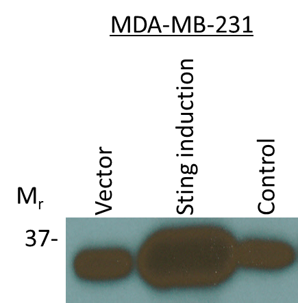
Western Blot: STING/TMEM173 Antibody [NBP2-24683] - STING/TMEM173 expression was induced in human breast MDA-MB-231 cells followed by Western blotting using STING/TMEM173 Antibody antibody (1:1000). Only one specific band at an apparent molecular mass of 37 kDa was observed. Image from verified customer review.![Immunocytochemistry/ Immunofluorescence: STING/TMEM173 Antibody - BSA Free [NBP2-24683]](https://resources.rndsystems.com/images/products/STING-TMEM173-Antibody-Immunocytochemistry-Immunofluorescence-NBP2-24683-img0017.jpg "Immunocytochemistry/ Immunofluorescence: STING/TMEM173 Antibody - BSA Free [NBP2-24683]")
Immunocytochemistry/ Immunofluorescence: STING/TMEM173 Antibody - BSA Free [NBP2-24683]
Immunocytochemistry/Immunofluorescence: STING/TMEM173 Antibody [NBP2-24683] - RH-30 cells were fixed for 10 minutes using 10% formalin and then permeabilized for 5 minutes using 1X PBS + 0.05% Triton X-100. The cells were incubated with STING/TMEM173 Antibody at 2 ug/mL overnight at 4C and detected with an anti-rabbit DyLight 488 (Green) at a 1:500 dilution. Alpha Tubulin Antibody (DM1A) (NB100-690) was used as a co-stain at a 1:1000 dilution and detected with an anti-mouse Dylight 550 (Red) at a 1:500 dilution. Nuclei were counterstained with DAPI (Blue). Cells were imaged using a 40X objective.![Western Blot: STING/TMEM173 AntibodyBSA Free [NBP2-24683]](https://resources.rndsystems.com/images/products/STING-TMEM173-Antibody-Western-Blot-NBP2-24683-img0011.jpg "Western Blot: STING/TMEM173 AntibodyBSA Free [NBP2-24683]")
Western Blot: STING/TMEM173 AntibodyBSA Free [NBP2-24683]
Western Blot: STING/TMEM173 Antibody [NBP2-24683] - Total protein from THP-1, HT-29, U2OS cells and human spleen was separated on a 12% gel by SDS-PAGE, transferred to PVDF membrane and blocked in 5% non-fat milk in TBST. The membrane was probed with 2.0 ug/ml STING/TMEM173 Antibody in 1% non-fat milk in TBST and detected with an anti-rabbit HRP secondary antibody using chemiluminescence.![Immunohistochemistry-Paraffin: STING/TMEM173 Antibody - BSA Free [NBP2-24683]](https://resources.rndsystems.com/images/products/STING-TMEM173-Antibody-Immunohistochemistry-Paraffin-NBP2-24683-img0006.jpg "Immunohistochemistry-Paraffin: STING/TMEM173 Antibody - BSA Free [NBP2-24683]")
Immunohistochemistry-Paraffin: STING/TMEM173 Antibody - BSA Free [NBP2-24683]
Immunohistochemistry-Paraffin: STING/TMEM173 Antibody [NBP2-24683] - Human colon cancer tissue section using STING/TMEM173 Antibody at 1:100 dilution with detection employing HRP-conjugated secondary antibody. The signal was developed using DAB reagent and the nuclei were counterstained with hematoxylin. The antibody generated very weak cytoplasmic staining in columnar epithelial cells with a very strong signal in the secretory/goblet cells.![Immunohistochemistry-Paraffin: STING/TMEM173 Antibody - BSA Free [NBP2-24683]](https://resources.rndsystems.com/images/products/STING-TMEM173-Antibody-Immunohistochemistry-Paraffin-NBP2-24683-img0007.jpg "Immunohistochemistry-Paraffin: STING/TMEM173 Antibody - BSA Free [NBP2-24683]")
Immunohistochemistry-Paraffin: STING/TMEM173 Antibody - BSA Free [NBP2-24683]
Immunohistochemistry-Paraffin: STING/TMEM173 Antibody [NBP2-24683] - Mouse lung tissue section using STING/TMEM173 Antibody at 1:150 dilution with detection employing HRP-conjugated secondary antibody. The signal was developed using DAB reagent and the nuclei were counterstained with hematoxylin. The antibody generated mainly a cytoplasmic staining in the bronchiolar and alveolar epithelial cells.![Flow Cytometry: STING/TMEM173 Antibody - BSA Free [NBP2-24683]](https://resources.rndsystems.com/images/products/STING-TMEM173-Antibody-Flow-Cytometry-NBP2-24683-img0020.jpg "Flow Cytometry: STING/TMEM173 Antibody - BSA Free [NBP2-24683]")
Flow Cytometry: STING/TMEM173 Antibody - BSA Free [NBP2-24683]
Flow Cytometry: STING/TMEM173 Antibody [NBP2-24683] - An intracellular stain was performed on U937 cells with NBP2-24683AF594 (blue) and a matched isotype control (orange). Cells were fixed with 4% PFA and then permeabilized with 0.1% saponin. Cells were incubated in an antibody dilution of 2.5 ug/mL for 30 minutes at room temperature. Both antibodies were conjugated to Alexa Fluor 594.![Flow Cytometry: STING/TMEM173 Antibody - BSA Free [NBP2-24683]](https://resources.rndsystems.com/images/products/STING-TMEM173-Antibody---BSA-Free-Flow-Cytometry-NBP2-24683-img0021.jpg "Flow Cytometry: STING/TMEM173 Antibody - BSA Free [NBP2-24683]")
Flow Cytometry: STING/TMEM173 Antibody - BSA Free [NBP2-24683]
Flow Cytometry: STING/TMEM173 Antibody - BSA Free [NBP2-24683] - An intracellular stain was performed on THP-1 cells with STING/TMEM173 Antibody NBP2-24683AF647 (blue) and a matched isotype control NBP2-24891 (orange). Cells were fixed with 4% PFA and then permeabilized with 0.1% saponin. Cells were incubated in an antibody dilution of 2.5 ug/mL for 30 minutes at room temperature. Both antibodies were conjugated to Alexa Fluor 647.![Immunocytochemistry/ Immunofluorescence: STING/TMEM173 Antibody - BSA Free [NBP2-24683]](https://resources.rndsystems.com/images/products/STING-TMEM173-Antibody-Immunocytochemistry-Immunofluorescence-NBP2-24683-img0015.jpg "Immunocytochemistry/ Immunofluorescence: STING/TMEM173 Antibody - BSA Free [NBP2-24683]")
Immunocytochemistry/ Immunofluorescence: STING/TMEM173 Antibody - BSA Free [NBP2-24683]
Immunocytochemistry/Immunofluorescence: STING/TMEM173 Antibody [NBP2-24683] - HT-29 cells were fixed for 10 minutes using 10% formalin and then permeabilized for 5 minutes using 1X PBS + 0.05% Triton-X100. The cells were incubated with STING/TMEM173 Antibody at 10 ug/ml overnight at 4C and detected with an anti-rabbit Dylight 488 (Green) at a 1:500 dilution. Nuclei were counterstained with DAPI (Blue). Cells were imaged using a 40X objective.![Flow Cytometry: STING/TMEM173 Antibody - BSA Free [NBP2-24683]](https://resources.rndsystems.com/images/products/STING-TMEM173-Antibody-Flow-Cytometry-NBP2-24683-img0012.jpg "Flow Cytometry: STING/TMEM173 Antibody - BSA Free [NBP2-24683]")
Flow Cytometry: STING/TMEM173 Antibody - BSA Free [NBP2-24683]
Flow Cytometry: STING/TMEM173 Antibody [NBP2-24683] - An intracellular stain was performed on THP-1 cells with STING/TMEM173 Antibody and a matched isotype control. Cells were fixed with 4% PFA and then permeablized with 0.1% saponin. Cells were incubated in an antibody dilution of 2.5 ug/mL for 30 minutes at room temperature, followed by Rabbit IgG (H+L) Cross-Adsorbed Secondary Antibody.![Flow Cytometry: STING/TMEM173 Antibody - BSA Free [NBP2-24683]](https://resources.rndsystems.com/images/products/STING-TMEM173-Antibody-Flow-Cytometry-NBP2-24683-img0016.jpg "Flow Cytometry: STING/TMEM173 Antibody - BSA Free [NBP2-24683]")
Flow Cytometry: STING/TMEM173 Antibody - BSA Free [NBP2-24683]
Flow Cytometry: STING/TMEM173 Antibody [NBP2-24683] - An intracellular stain was performed on RH30 cells with STING/TMEM173 Antibody (blue) and a matched isotype control (orange). Cells were fixed with 4% PFA and then permeabilized with 0.1% saponin. Cells were incubated in an antibody dilution of 2.5 ug/mL for 30 minutes at room temperature, followed by Rabbit IgG (H+L) Cross-Adsorbed Secondary Antibody.
Western Blot Shows Human STING/TMEM173 Specificity Using Knockout Cell Line.
Western blot shows lysates of THP-1 cell line and STING/TMEM173 knockout THP-1 cell line (KO). Nitrocellulose membrane was probed with STING/TMEM173 Antibody (Catalog # NBP2-24683) followed by an HRP-conjugated secondary antibody. A specific band was detected for STING/TMEM173 at approximately 41 kDa (as indicated) in the parental THP-1 cell line, but is not detectable in knockout THP-1 cell line. Primary antibody dilution used: 1/1000. The Ponceau stained transfer of the blot is shown. This experiment was conducted under reducing conditions. Image, protocol, and testing courtesy of YCharOS Inc. See ycharos.com for additional details.
Detection of STING/TMEM173 by Immunoprecipitation.
PMA-treated THP-1 lysates were prepared and immunoprecipitation was performed using 2.0 µg of STING/TMEM173 Antibody (Catalog # NBP2-24683) pre-coupled to Dynabeads protein A. Immunoprecipitated STING/TMEM173 was detected with STING/TMEM173 Antibody (Catalog # NBP3-18816). For western blot, NBP3-18816 was used at 1/1000. The Ponceau stained transfer of the blot is shown. SM=4% starting material; UB=4% unbound fraction; IP=immunoprecipitate; HC=antibody heavy chain. Image, protocol and testing courtesy of YCharOS Inc. (ycharos.com).
STING/TMEM173 Specificity is Shown by Immunocytochemistry in Knockout Cell Line.
PMA-treated THP-1 WT and STING/TMEM173 KO cells were labelled with a green or a far-red fluorescent dye, respectively. Cells were stained with STING/TMEM173 Antibody (Catalog # NBP2-24683) and with an Alexa-fluor 555 coupled secondary antibody including DAPI. Acquisition of the blue (nucleus-DAPI), green (identification of WT cells), red (antibody staining) and far-red (identification of KO cells) channels was performed. Representative images of the blue and red (grayscale) channels are shown. WT and KO cells are outlined with green and magenta dashed line, respectively. Primary antibody dilution used: 1/1000. Image, protocol and testing courtesy of YCharOS Inc. (ycharos.com).
Western Blot: STING/TMEM173 Antibody - BSA Free [NBP2-24683] -
STING pathway of tumour cells is not required for cGAMP-induced tumour EC apoptosis.a, b mRNA and protein levels of STING in cultured LLC cells transduced with nothing (NOT), shControl (shCon) or shSTING. Each dot indicates a value from one sample and n = 3 from two independent experiments. Vertical bars indicate mean +/- SD. c–e Diagram depicting generation of implanted LLC tumour by injection of the LLC cells transduced with NOT, shCon or shSTING, and treatment schedule of i.t. PBS or cGAMP in B6 mice. Comparison of LLC tumour growths. n = 6 mice/group from three independent experiments. Dots and bars indicate mean +/- SD. Plot indicates each individual tumour growth. f–i Diagram depicting generation of implanted LLC tumour by injection of the LLC cells transduced with shCon or shSTING, i.t. PBS or cGAMP treatment, and sampling of tumours at 24 h later. Representative images and comparisons of apoptosis in tumour ECs and whole tumour cells (whole cells). White arrowheads indicate apoptotic ECs. Scale bars, 1.0 mm (yellow) and 100 μm (white). Each dot indicates a value from one mouse and n = 6 mice from four independent experiments. Vertical bars indicate mean +/- SD. P values by Welch’s one-way ANOVA test followed by Dunnett’s T3 test (a, d, i). ****P < 0.0001; ns, not significant. Source data are provided as a Source Data file. Image collected and cropped by CiteAb from the following open publication (https://pubmed.ncbi.nlm.nih.gov/34285232), licensed under a CC-BY license. Not internally tested by Novus Biologicals.
Western Blot: STING/TMEM173 Antibody - BSA Free [NBP2-24683] -
cGAS, STING and HMGB‐1 expressions in HT22 cells after OGD/R and melatonin treatment. (A) Quantitative evaluation and representative Western blots of cGAS, STING (B) and nuclear (C) and cytosolic (D) HMGB‐1 expressions in untreated HT22 cells (Ctrl), 8‐h OGD‐exposed cells (OGD) or followed by 15 min, 30 min or 1 h of reoxygenation (OGD/R) and 8‐h OGD‐exposed cells followed by 15 min, 30 min or 1 h of reoxygenation in the presence of 50 μM melatonin (OGD/R + Mel). Data normalised to the loading control beta ‐actin or lamin A are expressed as % of control and are the mean +/- SD (N = 3 independent experiments performed in triplicate); *p < 0.05, **p < 0.01, §p < 0.001 vs Ctrl; **p < 0.01, §p < 0.001, bars. Image collected and cropped by CiteAb from the following open publication (https://pubmed.ncbi.nlm.nih.gov/39707673), licensed under a CC-BY license. Not internally tested by Novus Biologicals.
Western Blot: STING/TMEM173 Antibody - BSA Free [NBP2-24683] -
Combination of diABZI and BRAF inhibitors prevent NRF2 activation and activate STING. (A) Immunoblot analysis of phospho STING upon treatment with diABZI and BRAFis, Dabrafenib (DF) and Vemurafenib (VF). (B) Densitometric quantification of pSTING (n = 3). (C) Immunoblot analysis of phospho IRF3 upon treatment with diABZI and BRAFis, Dabrafenib (DF) and Vemurafenib (VF). (D) Densitometric quantification of pIRF3 (n = 3). (E) Immunoblot analysis of NRF2 upon diABZI and BRAFi treatment in C32 cells. (F) Densitometric quantification of NRF2 (n = 3). (G) Immunoblot of cytosolic and nuclear cell fractions upon treatment with diABZI and BRAFis, Dabrafenib (DF) and Vemurafenib (VF). **P < 0.01, ***P < 0.001, ****P < 0.0001. Image collected and cropped by CiteAb from the following open publication (https://pubmed.ncbi.nlm.nih.gov/32477956), licensed under a CC-BY license. Not internally tested by Novus Biologicals.
Western Blot: STING/TMEM173 Antibody - BSA Free [NBP2-24683] -
Low-dose GCV inhibited cGAS-STING pathways in RAW264.7 cells. (A–C) Q-PCR analysis showed the effect of pretreatment of GCV on up-regulation of mRNA expression of Sting1, Il10, Ifnb1 induced by CMA- (A), DMXAA- (B), and cGAMP (C) in RAW264.7 cells. (D) Western blotting analysis showed the effect of GCV on CMA-induced expression changes of cGAS, STING, IFN-beta, and p-TBK1 in RAW264.7 cells. (E) Statistical analysis results for (D). (F) Western blotting analysis showed the effect of GCV on DMXAA-induced expression changes of cGAS, STING, IFN-beta, and p-TBK1 in RAW264.7 cells. (G) Statistical analysis results for (F). (H) Western blotting analysis showed the effect of GCV on cGAMP-induced expression changes of cGAS, STING, IFN-beta, and p-TBK1 in RAW264.7 cells. (I) Statistical analysis results for (H). (n = 4 each group, *p < 0.05, **p < 0.01, ***p < 0.001 vs. saline. &p < 0.05, &&p < 0.01, &&&p < 0.001 vs. STING agonists group, unpaired Student’s t-test). (n = 4 each group, *p < 0.05, **p < 0.01, ***p < 0.001, &&p < 0.01, &&&p < 0.001; unpaired Student’s t-test). All data was expressed as Mean +/- SEM. VEH, vehicle. Image collected and cropped by CiteAb from the following open publication (https://pubmed.ncbi.nlm.nih.gov/36467059), licensed under a CC-BY license. Not internally tested by Novus Biologicals.
Western Blot: STING/TMEM173 Antibody - BSA Free [NBP2-24683] -
STING was upregulated following DSS-induced chronic colitis in mice and in UC patients. (A) Body weight loss was induced by DSS-colitis in mice. (B) The quantification of area under the curve (AUC) for (A). (C) DSS-colitis induced changes of the Disease activity index (DAI) score. (D) The quantification of AUC for (C). (E) DSS-colitis induced mechanical pain hypersensitivity in the abdomen. (F) The quantification of AUC for (E). (n = 7-8 per group; *p < 0.05, **p < 0.05, ***p < 0.001, DSS vs. vehicle group; two-way ANOVA with post-hoc Bonferroni test). (G) Representative pictures showed colon shortening induced by DSS. (H) The quantification of colon shortening for (G). (n = 7-8 per group; ***p < 0.001, DSS vs. vehicle group; unpaired Student’s t-test). (I) Representative H&E staining of colon sections from DSS group and vehicle group. (J) Statistical analysis for (I). (n = 4 each group, ***p < 0.001, DSS vs. vehicle group; unpaired Student’s t-test). (K) Double immunostaining of STING and F4/80 in the colon tissue from DSS group and vehicle group. (L) Statistical analysis for (K), scale bar: 50 μm. (n = 4 each group, *p < 0.05, DSS vs. vehicle group; unpaired Student’s t-test). (M) Western blotting analysis of STING expression in colon of mice. (N) Statistical analysis of Western blotting. (n = 3 each group, *p < 0.05, DSS vs. vehicle group; unpaired Student’s t-test). (O) The expression of STING in the colon tissue was increased in patients with ulcerative colitis compared with adjacent tissue of colon cancer. (P) Statistical analysis results for (O). (n = 4; **p < 0.01 vs Normal; unpaired Student’s t-test). The scale bar: 20 μm. All data was expressed as Mean +/- SEM. DAI, disease activity index; DSS, dextran sulfate sodium. Image collected and cropped by CiteAb from the following open publication (https://pubmed.ncbi.nlm.nih.gov/36467059), licensed under a CC-BY license. Not internally tested by Novus Biologicals.
Western Blot: STING/TMEM173 Antibody - BSA Free [NBP2-24683] -
STING deficiency attenuated DSS-colitis in mice. (A) Representative Western blotting detected cGAS and STING expression in STINGgt/gt and WT mice. (B) Statistical analysis for (A). (C) Representative immunofluorescence detected STING and F4/80 expression in STINGgt/gt and WT mice. (D) Statistical analysis for (C). (n = 3 each group, **p < 0.01, STINGgt/gt vs. vehicle group; unpaired Student’s t-test). Wild-type (WT) and STINGgt/gt mice were given 3% dextran sulfate sodium (DSS) in drinking water for 8 days. (E) Body weight changed following DSS administration in mice. (F) The quantification of AUC for (E). (G) DSS-induced changes of DAI score in WT and STINGgt/gt mice. (H) The quantification of AUC for (G). (I) DSS-induced changes of mechanical pain sensitivity in the abdomen in STINGgt/gt mice and WT mice. (J) The quantification of AUC for (I). (n = 5-7 per group; **p < 0.01, ***p < 0.001, DSS vs. vehicle group; #p < 0.05, ##p < 0.01, ###p < 0.001, STINGgt/gt + DSS vs DSS group; two-way ANOVA with post-hoc Bonferroni test). (K) Representative pictures of colons from WT and STINGgt/gt mice on day 8. (L) Quantification of the colon length in (K). (n = 5-7 per group; ***p < 0.001, ###p < 0.001; unpaired Student’s t-test). (M) Representative photographies of H&E staining of colon sections from 4 different groups. (N) Statistical analysis for (M) (n = 4 each group; ***p < 0.001, #p < 0.05; unpaired Student’s t-test). (O) The Q-PCR analysis of mRNA expression of colonic Cgas, Il10, Ifnb1, Cxcl10, Il1b, and Tnf in mice from different group (n = 6 each group, **p < 0.01, ***p < 0.001, #p < 0.05, ##p < 0.01, ###p < 0.001; unpaired Student’s t-test). All data was expressed as Mean +/- SEM. DSS, dextran sulfate sodium; n.s., no significance. WT, wild type. Image collected and cropped by CiteAb from the following open publication (https://pubmed.ncbi.nlm.nih.gov/36467059), licensed under a CC-BY license. Not internally tested by Novus Biologicals.
Western Blot: STING/TMEM173 Antibody - BSA Free [NBP2-24683] -
Low-dose GCV attenuated cGAS-STING pathways in the colon of DSS-colitis in mice. (A) Western blotting analysis showed that protein expression of colonic STING, cGAS, p-TBK1, IFN-beta, IL-1 beta, and TNF-alpha in mice of different treatment group. (B) Statistical analysis for (A) (n = 4 per group, *p < 0.05, **p < 0.01, DSS vs. vehicle group; #p < 0.05, ##p < 0.01, ###p < 0.001, GCV + DSS vs. DSS group; unpaired Student’s t-test). (C) Double immunostaining of STING and F4/80 in the colon tissue. (D) Statistical results for (C). Scale bar = 50 μm. (n = 4 each group, ***p < 0.001, ##p < 0.01; unpaired Student’s t-test). All data was expressed as Mean +/- SEM. Image collected and cropped by CiteAb from the following open publication (https://pubmed.ncbi.nlm.nih.gov/36467059), licensed under a CC-BY license. Not internally tested by Novus Biologicals.
Western Blot: STING/TMEM173 Antibody - BSA Free [NBP2-24683] -
Low-dose GCV inhibited cGAS-STING pathways in RAW264.7 cells. (A–C) Q-PCR analysis showed the effect of pretreatment of GCV on up-regulation of mRNA expression of Sting1, Il10, Ifnb1 induced by CMA- (A), DMXAA- (B), and cGAMP (C) in RAW264.7 cells. (D) Western blotting analysis showed the effect of GCV on CMA-induced expression changes of cGAS, STING, IFN-beta, and p-TBK1 in RAW264.7 cells. (E) Statistical analysis results for (D). (F) Western blotting analysis showed the effect of GCV on DMXAA-induced expression changes of cGAS, STING, IFN-beta, and p-TBK1 in RAW264.7 cells. (G) Statistical analysis results for (F). (H) Western blotting analysis showed the effect of GCV on cGAMP-induced expression changes of cGAS, STING, IFN-beta, and p-TBK1 in RAW264.7 cells. (I) Statistical analysis results for (H). (n = 4 each group, *p < 0.05, **p < 0.01, ***p < 0.001 vs. saline. &p < 0.05, &&p < 0.01, &&&p < 0.001 vs. STING agonists group, unpaired Student’s t-test). (n = 4 each group, *p < 0.05, **p < 0.01, ***p < 0.001, &&p < 0.01, &&&p < 0.001; unpaired Student’s t-test). All data was expressed as Mean +/- SEM. VEH, vehicle. Image collected and cropped by CiteAb from the following open publication (https://pubmed.ncbi.nlm.nih.gov/36467059), licensed under a CC-BY license. Not internally tested by Novus Biologicals.
Western Blot: STING/TMEM173 Antibody - BSA Free [NBP2-24683] -
diABZI activates STING in melanoma cells. (A) Immunoblot analysis of STING pathway in untreated vs. diABZI treated samples (n = 3). (B) Densitometric analysis of phosphorylated STING (pSTING) and (C) Densitometric analysis of phosphorylated TBK1 (pTBK1) (n = 3). (D) Immunofluorescence staining and confocal microscopy analysis of pSTING in untreated vs. diABZI treatment. Arrows indicate perinuclear localization of pSTING. (E) Immunoblot of phosphorylated IRF3 in diABZI treated samples. Scale bar = 10 μm; **P < 0.01. Image collected and cropped by CiteAb from the following open publication (https://pubmed.ncbi.nlm.nih.gov/32477956), licensed under a CC-BY license. Not internally tested by Novus Biologicals.
Western Blot: STING/TMEM173 Antibody - BSA Free [NBP2-24683] -
Low-dose GCV inhibited cGAS-STING pathways in RAW264.7 cells. (A–C) Q-PCR analysis showed the effect of pretreatment of GCV on up-regulation of mRNA expression of Sting1, Il10, Ifnb1 induced by CMA- (A), DMXAA- (B), and cGAMP (C) in RAW264.7 cells. (D) Western blotting analysis showed the effect of GCV on CMA-induced expression changes of cGAS, STING, IFN-beta, and p-TBK1 in RAW264.7 cells. (E) Statistical analysis results for (D). (F) Western blotting analysis showed the effect of GCV on DMXAA-induced expression changes of cGAS, STING, IFN-beta, and p-TBK1 in RAW264.7 cells. (G) Statistical analysis results for (F). (H) Western blotting analysis showed the effect of GCV on cGAMP-induced expression changes of cGAS, STING, IFN-beta, and p-TBK1 in RAW264.7 cells. (I) Statistical analysis results for (H). (n = 4 each group, *p < 0.05, **p < 0.01, ***p < 0.001 vs. saline. &p < 0.05, &&p < 0.01, &&&p < 0.001 vs. STING agonists group, unpaired Student’s t-test). (n = 4 each group, *p < 0.05, **p < 0.01, ***p < 0.001, &&p < 0.01, &&&p < 0.001; unpaired Student’s t-test). All data was expressed as Mean +/- SEM. VEH, vehicle. Image collected and cropped by CiteAb from the following open publication (https://pubmed.ncbi.nlm.nih.gov/36467059), licensed under a CC-BY license. Not internally tested by Novus Biologicals.
Immunocytochemistry/ Immunofluorescence: STING/TMEM173 Antibody - BSA Free [NBP2-24683] -
Low-dose GCV attenuated cGAS-STING pathways in the colon of DSS-colitis in mice. (A) Western blotting analysis showed that protein expression of colonic STING, cGAS, p-TBK1, IFN-beta, IL-1 beta, and TNF-alpha in mice of different treatment group. (B) Statistical analysis for (A) (n = 4 per group, *p < 0.05, **p < 0.01, DSS vs. vehicle group; #p < 0.05, ##p < 0.01, ###p < 0.001, GCV + DSS vs. DSS group; unpaired Student’s t-test). (C) Double immunostaining of STING and F4/80 in the colon tissue. (D) Statistical results for (C). Scale bar = 50 μm. (n = 4 each group, ***p < 0.001, ##p < 0.01; unpaired Student’s t-test). All data was expressed as Mean +/- SEM. Image collected and cropped by CiteAb from the following open publication (https://pubmed.ncbi.nlm.nih.gov/36467059), licensed under a CC-BY license. Not internally tested by Novus Biologicals.
Immunocytochemistry/ Immunofluorescence: STING/TMEM173 Antibody - BSA Free [NBP2-24683] -
STING deficiency attenuated DSS-colitis in mice. (A) Representative Western blotting detected cGAS and STING expression in STINGgt/gt and WT mice. (B) Statistical analysis for (A). (C) Representative immunofluorescence detected STING and F4/80 expression in STINGgt/gt and WT mice. (D) Statistical analysis for (C). (n = 3 each group, **p < 0.01, STINGgt/gt vs. vehicle group; unpaired Student’s t-test). Wild-type (WT) and STINGgt/gt mice were given 3% dextran sulfate sodium (DSS) in drinking water for 8 days. (E) Body weight changed following DSS administration in mice. (F) The quantification of AUC for (E). (G) DSS-induced changes of DAI score in WT and STINGgt/gt mice. (H) The quantification of AUC for (G). (I) DSS-induced changes of mechanical pain sensitivity in the abdomen in STINGgt/gt mice and WT mice. (J) The quantification of AUC for (I). (n = 5-7 per group; **p < 0.01, ***p < 0.001, DSS vs. vehicle group; #p < 0.05, ##p < 0.01, ###p < 0.001, STINGgt/gt + DSS vs DSS group; two-way ANOVA with post-hoc Bonferroni test). (K) Representative pictures of colons from WT and STINGgt/gt mice on day 8. (L) Quantification of the colon length in (K). (n = 5-7 per group; ***p < 0.001, ###p < 0.001; unpaired Student’s t-test). (M) Representative photographies of H&E staining of colon sections from 4 different groups. (N) Statistical analysis for (M) (n = 4 each group; ***p < 0.001, #p < 0.05; unpaired Student’s t-test). (O) The Q-PCR analysis of mRNA expression of colonic Cgas, Il10, Ifnb1, Cxcl10, Il1b, and Tnf in mice from different group (n = 6 each group, **p < 0.01, ***p < 0.001, #p < 0.05, ##p < 0.01, ###p < 0.001; unpaired Student’s t-test). All data was expressed as Mean +/- SEM. DSS, dextran sulfate sodium; n.s., no significance. WT, wild type. Image collected and cropped by CiteAb from the following open publication (https://pubmed.ncbi.nlm.nih.gov/36467059), licensed under a CC-BY license. Not internally tested by Novus Biologicals.
Immunocytochemistry/ Immunofluorescence: STING/TMEM173 Antibody - BSA Free [NBP2-24683] -
Expression of STING and cGAS mRNA and protein in adipocytes. (A) Venn diagram summarizing the number of equally and differently expressed mRNA transcripts of young and adult mouse iAT. A gene network associated with Sting1 was equally expressed by young and adult iAT. A protein–protein interaction map, generated by STRING [30] is shown below the Venn diagram. Extended analysis presented in [24]. (B) Immunofluorescence of in vitro cultured adipocytes from young and adult mouse iAT; nc: nucleus, scale bar 20 μm. (C) Immunostaining of STING and cGAS proteins in the iAT of young mice, showing a region containing both multilocular and unilocular adipocytes. Arrowheads label nuclei; lp: lipid droplet; cyt: cytoplasm; scale bar: 50 μm. (D) Top: Expression of STING1 and CGAS mRNA in human inguinal and abdominal adipose tissue specimens. Linear regression analysis indicates a significant positive correlation between STING1 and CGAS mRNA levels. Each data point represents one tissue donor patient. Bottom: Correlation of donor age and the adipose tissue expression levels of STING1 and CGAS. (E) Immunohistochemistry of STING and cGAS proteins in human adipose tissue, collected from the inguinal-low abdominal region. Nineteen-month-old male infant; arrowheads label nuclei; lp: lipid droplet; cyt: cytoplasm; scale bar: 25 μm. Inlet shows nuclear STING labeling of an in vitro cultured human adipocyte. Scale bar: 20 μm. (F) Body mass index z-score (BMI z-score) and BMI standard deviation score (BMI-SDS) of adipose tissue donors involved in this study. Correlation of BMI z-score with adipose tissue STING1 and CGAS mRNA levels. Image collected and cropped by CiteAb from the following open publication (https://pubmed.ncbi.nlm.nih.gov/37830559), licensed under a CC-BY license. Not internally tested by Novus Biologicals.
Immunohistochemistry: STING/TMEM173 Antibody - BSA Free [NBP2-24683] -
STING deficiency attenuated DSS-colitis in mice. (A) Representative Western blotting detected cGAS and STING expression in STINGgt/gt and WT mice. (B) Statistical analysis for (A). (C) Representative immunofluorescence detected STING and F4/80 expression in STINGgt/gt and WT mice. (D) Statistical analysis for (C). (n = 3 each group, **p < 0.01, STINGgt/gt vs. vehicle group; unpaired Student’s t-test). Wild-type (WT) and STINGgt/gt mice were given 3% dextran sulfate sodium (DSS) in drinking water for 8 days. (E) Body weight changed following DSS administration in mice. (F) The quantification of AUC for (E). (G) DSS-induced changes of DAI score in WT and STINGgt/gt mice. (H) The quantification of AUC for (G). (I) DSS-induced changes of mechanical pain sensitivity in the abdomen in STINGgt/gt mice and WT mice. (J) The quantification of AUC for (I). (n = 5-7 per group; **p < 0.01, ***p < 0.001, DSS vs. vehicle group; #p < 0.05, ##p < 0.01, ###p < 0.001, STINGgt/gt + DSS vs DSS group; two-way ANOVA with post-hoc Bonferroni test). (K) Representative pictures of colons from WT and STINGgt/gt mice on day 8. (L) Quantification of the colon length in (K). (n = 5-7 per group; ***p < 0.001, ###p < 0.001; unpaired Student’s t-test). (M) Representative photographies of H&E staining of colon sections from 4 different groups. (N) Statistical analysis for (M) (n = 4 each group; ***p < 0.001, #p < 0.05; unpaired Student’s t-test). (O) The Q-PCR analysis of mRNA expression of colonic Cgas, Il10, Ifnb1, Cxcl10, Il1b, and Tnf in mice from different group (n = 6 each group, **p < 0.01, ***p < 0.001, #p < 0.05, ##p < 0.01, ###p < 0.001; unpaired Student’s t-test). All data was expressed as Mean +/- SEM. DSS, dextran sulfate sodium; n.s., no significance. WT, wild type. Image collected and cropped by CiteAb from the following open publication (https://pubmed.ncbi.nlm.nih.gov/36467059), licensed under a CC-BY license. Not internally tested by Novus Biologicals.
Immunohistochemistry: STING/TMEM173 Antibody - BSA Free [NBP2-24683] -
Expression of STING and cGAS mRNA and protein in adipocytes. (A) Venn diagram summarizing the number of equally and differently expressed mRNA transcripts of young and adult mouse iAT. A gene network associated with Sting1 was equally expressed by young and adult iAT. A protein–protein interaction map, generated by STRING [30] is shown below the Venn diagram. Extended analysis presented in [24]. (B) Immunofluorescence of in vitro cultured adipocytes from young and adult mouse iAT; nc: nucleus, scale bar 20 μm. (C) Immunostaining of STING and cGAS proteins in the iAT of young mice, showing a region containing both multilocular and unilocular adipocytes. Arrowheads label nuclei; lp: lipid droplet; cyt: cytoplasm; scale bar: 50 μm. (D) Top: Expression of STING1 and CGAS mRNA in human inguinal and abdominal adipose tissue specimens. Linear regression analysis indicates a significant positive correlation between STING1 and CGAS mRNA levels. Each data point represents one tissue donor patient. Bottom: Correlation of donor age and the adipose tissue expression levels of STING1 and CGAS. (E) Immunohistochemistry of STING and cGAS proteins in human adipose tissue, collected from the inguinal-low abdominal region. Nineteen-month-old male infant; arrowheads label nuclei; lp: lipid droplet; cyt: cytoplasm; scale bar: 25 μm. Inlet shows nuclear STING labeling of an in vitro cultured human adipocyte. Scale bar: 20 μm. (F) Body mass index z-score (BMI z-score) and BMI standard deviation score (BMI-SDS) of adipose tissue donors involved in this study. Correlation of BMI z-score with adipose tissue STING1 and CGAS mRNA levels. Image collected and cropped by CiteAb from the following open publication (https://pubmed.ncbi.nlm.nih.gov/37830559), licensed under a CC-BY license. Not internally tested by Novus Biologicals.
Immunohistochemistry: STING/TMEM173 Antibody - BSA Free [NBP2-24683] -
Expression of STING and cGAS mRNA and protein in adipocytes. (A) Venn diagram summarizing the number of equally and differently expressed mRNA transcripts of young and adult mouse iAT. A gene network associated with Sting1 was equally expressed by young and adult iAT. A protein–protein interaction map, generated by STRING [30] is shown below the Venn diagram. Extended analysis presented in [24]. (B) Immunofluorescence of in vitro cultured adipocytes from young and adult mouse iAT; nc: nucleus, scale bar 20 μm. (C) Immunostaining of STING and cGAS proteins in the iAT of young mice, showing a region containing both multilocular and unilocular adipocytes. Arrowheads label nuclei; lp: lipid droplet; cyt: cytoplasm; scale bar: 50 μm. (D) Top: Expression of STING1 and CGAS mRNA in human inguinal and abdominal adipose tissue specimens. Linear regression analysis indicates a significant positive correlation between STING1 and CGAS mRNA levels. Each data point represents one tissue donor patient. Bottom: Correlation of donor age and the adipose tissue expression levels of STING1 and CGAS. (E) Immunohistochemistry of STING and cGAS proteins in human adipose tissue, collected from the inguinal-low abdominal region. Nineteen-month-old male infant; arrowheads label nuclei; lp: lipid droplet; cyt: cytoplasm; scale bar: 25 μm. Inlet shows nuclear STING labeling of an in vitro cultured human adipocyte. Scale bar: 20 μm. (F) Body mass index z-score (BMI z-score) and BMI standard deviation score (BMI-SDS) of adipose tissue donors involved in this study. Correlation of BMI z-score with adipose tissue STING1 and CGAS mRNA levels. Image collected and cropped by CiteAb from the following open publication (https://pubmed.ncbi.nlm.nih.gov/37830559), licensed under a CC-BY license. Not internally tested by Novus Biologicals.Applications for STING/TMEM173 Antibody - BSA Free
Application
Recommended Usage
ELISA
reported in scientific literature (PMID 34905508)
Flow Cytometry
1-5 ug/million cells
Immunocytochemistry/ Immunofluorescence
5 - 10 ug/ml
Immunohistochemistry
1:100 - 1:300
Immunohistochemistry-Frozen
reported in scientific literature (PMID 33745949)
Immunohistochemistry-Paraffin
1:100 - 1:300
Immunoprecipitation
Validated for Immunoprecipitation from YCharOS Inc. (ycharos.com)
Knockout Validated
Validated for Knockout from YCharOS Inc. (ycharos.com)
Western Blot
1 - 2 ug/ml
Reviewed Applications
Read 1 review rated 5 using NBP2-24683 in the following applications:
Flow Cytometry Panel Builder
Bio-Techne Knows Flow Cytometry
Save time and reduce costly mistakes by quickly finding compatible reagents using the Panel Builder Tool.
Advanced Features
- Spectra Viewer - Custom analysis of spectra from multiple fluorochromes
- Spillover Popups - Visualize the spectra of individual fluorochromes
- Antigen Density Selector - Match fluorochrome brightness with antigen density

Formulation, Preparation, and Storage
Purification
Peptide affinity purified
Formulation
PBS
Format
BSA Free
Preservative
0.02% Sodium Azide
Concentration
1.0 mg/ml
Shipping
The product is shipped with polar packs. Upon receipt, store it immediately at the temperature recommended below.
Stability & Storage
Store at 4C short term. Aliquot and store at -20C long term. Avoid freeze-thaw cycles.
Background: STING/TMEM173
References
1. Patel, S., & Jin, L. (2019). TMEM173 variants and potential importance to human biology and disease. Genes and Immunity. https://doi.org/10.1038/s41435-018-0029-9
2. Jounai, N., Kobiyama, K., Takeshita, F., & Ishii, K. J. (2013). Recognition of damage-associated molecular patterns related to nucleic acids during inflammation and vaccination. Frontiers in Cellular and Infection Microbiology. https://doi.org/10.3389/fcimb.2012.00168
3. Xiao, T. S., & Fitzgerald, K. A. (2013). The cGAS-STING Pathway for DNA Sensing. Molecular Cell. https://doi.org/10.1016/j.molcel.2013.07.004
4. Kato, K., Omura, H., Ishitani, R., & Nureki, O. (2017). Cyclic GMP-AMP as an Endogenous Second Messenger in Innate Immune Signaling by Cytosolic DNA. Annual Review of Biochemistry. https://doi.org/10.1146/annurev-biochem-061516-044813
5. Crowl, J. T., Gray, E. E., Pestal, K., Volkman, H. E., & Stetson, D. B. (2017). Intracellular Nucleic Acid Detection in Autoimmunity. Annual Review of Immunology. https://doi.org/10.1146/annurev-immunol-051116-052331
Long Name
Stimulator of Interferon Genes Protein/Transmembrane protein 173
Alternate Names
ERIS, MITA, MPYS, NET23, TMEM173, anti-STING, human STING, mouse STING
Gene Symbol
STING1
UniProt
Additional STING/TMEM173 Products
Product Documents for STING/TMEM173 Antibody - BSA Free
Certificate of Analysis
To download a Certificate of Analysis, please enter a lot or batch number in the search box below.
Product Specific Notices for STING/TMEM173 Antibody - BSA Free
This product is for research use only and is not approved for use in humans or in clinical diagnosis. Primary Antibodies are guaranteed for 1 year from date of receipt.
Related Research Areas
Citations for STING/TMEM173 Antibody - BSA Free
 Powered by Bioz
Customer Reviews for STING/TMEM173 Antibody - BSA Free (1)
5 out of 5
1 Customer Rating
Have you used STING/TMEM173 Antibody - BSA Free?
Submit a review and receive an Amazon gift card!
$25/€18/£15/$25CAN/¥2500 Yen for a review with an image
$10/€7/£6/$10CAN/¥1110 Yen for a review without an image
Submit a review
Customer Images
Showing
1
-
1 of
1 review
Showing All
Filter By:
-
Application: Western BlotSample Tested: Human MDA-MB-231 cellsSpecies: HumanVerified Customer | Posted 10/29/2018Sting expression was induced in human breast MDA-MB-231 cells followed by Western blotting using Sting/TMEM173 antibody (1:1000, Novus). Only one specific band at an apparent molecular mass of 37 kDa was observed.Performed on multiple human cancer cells with and without Sting induction. Specific band detected ~37 kDa without other non-specific bands. Primary antibody was used at 1:1000 in 5% milk in IBS-T with traditional HRP detection.

There are no reviews that match your criteria.
Protocols
View specific protocols for STING/TMEM173 Antibody - BSA Free (NBP2-24683):
Protocol for Flow Cytometry Intracellular Staining
Sample Preparation.
1. Grow cells to 60-85% confluency. Flow cytometry requires between 2 x 10⁵ and 1 x 10⁶ cells for optimal performance.
2. If cells are adherent, harvest gently by washing once with staining buffer and then scraping. Avoid using trypsin as this can disrupt certain epitopes of interest. If enzymatic harvest is required, use Accutase, Collagenase, or TrypLE Express for a less damaging option.
3. Reserve 100 uL for counting, then transfer cell volume into a 50 mL conical tube and centrifuge for 8 minutes at 400 RCF.
a. Count cells using a hemocytometer and a 1:1 trypan blue exclusion stain to determine cell viability before starting the flow protocol. If cells appear blue, do not proceed.
4. Re-suspend cells to a concentration of 1 x 10⁶ cells/mL in staining buffer (NBP2-26247).
5. Aliquot out 100 uL samples in accordance with your experimental samples.
Tip: When cell surface and intracellular staining are required in the same sample, it is advisable that the cell surface staining be performed first since the fixation and permeablization steps might reduce the availability of surface antigens.
Intracellular Staining.
Tip: When performing intracellular staining, it is important to use appropriate fixation and permeabilization reagents based upon the target and its subcellular location. Generally, our Intracellular Flow Assay Kit (NBP2-29450) is a good place to start as it contains an optimized combination of reagents for intracellular staining as well as an inhibitor of intracellular protein transport (necessary if staining secreted proteins). Certain targets may require more gentle or transient permeabilization protocols such as the commonly employed methanol or saponin-based methods.
Protocol for Cytoplasmic Targets:
1. Fix the cells by adding 100 uL fixation solution (such as 4% PFA) to each sample for 10-15 minutes.
2. Permeabilize cells by adding 100 uL of a permeabization buffer to every 1 x 10⁶ cells present in the sample. Mix well and incubate at room temperature for 15 minutes.
a. For cytoplasmic targets, use a gentle permeabilization solution such as 1X PBS + 0.5% Saponin or 1X PBS + 0.5% Tween-20.
b. To maintain the permeabilized state throughout your experiment, use staining buffer + 0.1% of the permeabilization reagent (i.e. 0.1% Tween-20 or 0.1% Saponin).
3. Following the 15 minute incubation, add 2 mL of the staining buffer + 0.1% permeabilizer to each sample.
4. Centrifuge for 1 minute at 400 RCF.
5. Discard supernatant and re-suspend in 100 uL of staining buffer + 0.1% permeabilizer.
6. Add appropriate amount of each antibody (eg. 1 test or 1 ug per sample, as experimentally determined).
7. Mix well and incubate at room temperature for 30 minutes- 1 hour. Gently mix samples every 10-15 minutes.
8. Following the primary/conjugate incubation, add 1-2 mL/sample of staining buffer +0.1% permeabilizer and centrifuge for 1 minute at 400 RCF.
9. Wash twice by re-suspending cells in staining buffer (2 mL for tubes or 200 uL for wells) and centrifuging at 400 RCF for 5 minutes. Discard supernatant.
10. Add appropriate amount of secondary antibody (as experimentally determined) to each sample.
11. Incubate at room temperature in dark for 20 minutes.
12. Add 1-2 mL of staining buffer and centrifuge at 400 RCF for 1 minute and discard supernatant.
13. Wash twice by re-suspending cells in staining buffer (2 mL for tubes or 200 uL for wells) and centrifuging at 400 RCF for 5 minutes. Discard supernatant.
14. Resuspend in an appropriate volume of staining buffer (usually 500 uL per sample) and proceed with analysis on your flow cytometer.
Immunocytochemistry Protocol
Culture cells to appropriate density in 35 mm culture dishes or 6-well plates.
1. Remove culture medium and wash the cells briefly in PBS. Add 10% formalin to the dish and fix at room temperature for 10 minutes.
2. Remove the formalin and wash the cells in PBS.
3. Permeablize the cells with 0.1% Triton X100 or other suitable detergent for 10 min.
4. Remove the permeablization buffer and wash three times for 10 minutes each in PBS. Be sure to not let the specimen dry out.
5. To block nonspecific antibody binding, incubate in 10% normal goat serum from 1 hour to overnight at room temperature.
6. Add primary antibody at appropriate dilution and incubate overnight at 4C.
7. Remove primary antibody and replace with PBS. Wash three times for 10 minutes each.
8. Add secondary antibody at appropriate dilution. Incubate for 1 hour at room temperature.
9. Remove secondary antibody and replace with PBS. Wash three times for 10 minutes each.
10. Counter stain DNA with DAPi if required.
Immunohistochemistry-Paraffin Embedded Sections
Antigen Unmasking:
Bring slides to a boil in 10 mM sodium citrate buffer (pH 6.0) then maintain at a sub-boiling temperature for 10 minutes. Cool slides on bench-top for 30 minutes (keep slides in the sodium citrate buffer all the time).
Staining:
1. Wash sections in deionized water three times for 5 minutes each.
2. Wash sections in PBS for 5 minutes.
3. Block each section with 100-400 ul blocking solution (1% BSA in PBS) for 1 hour at room temperature.
4. Remove blocking solution and add 100-400 ul diluted primary antibody. Incubate overnight at 4 C.
5. Remove antibody solution and wash sections in wash buffer three times for 5 minutes each.
6. Add 100-400 ul HRP polymer conjugated secondary antibody. Incubate 30 minutes at room temperature.
7. Wash sections three times in wash buffer for 5 minutes each.
8. Add 100-400 ul DAB substrate to each section and monitor staining closely.
9. As soon as the sections develop, immerse slides in deionized water.
10. Counterstain sections in hematoxylin.
11. Wash sections in deionized water two times for 5 minutes each.
12. Dehydrate sections.
13. Mount coverslips.
Western Blot Protocol
1. Perform SDS-PAGE on samples to be analyzed, loading 10-25 ug of total protein per lane.
2. Transfer proteins to PVDF membrane according to the instructions provided by the manufacturer of the membrane and transfer apparatus.
3. Stain the membrane with Ponceau S (or similar product) to assess transfer success, and mark molecular weight standards where appropriate.
4. Rinse the blot TBS -0.05% Tween 20 (TBST).
5. Block the membrane in 5% Non-fat milk in TBST (blocking buffer) for at least 1 hour.
6. Wash the membrane in TBST three times for 10 minutes each.
7. Dilute primary antibody in blocking buffer and incubate overnight at 4C with gentle rocking.
8. Wash the membrane in TBST three times for 10 minutes each.
9. Incubate the membrane in diluted HRP conjugated secondary antibody in blocking buffer (as per manufacturer's instructions) for 1 hour at room temperature.
10. Wash the blot in TBST three times for 10 minutes each (this step can be repeated as required to reduce background).
11. Apply the detection reagent of choice in accordance with the manufacturers instructions.
Find general support by application which include: protocols, troubleshooting, illustrated assays, videos and webinars.
- 7-Amino Actinomycin D (7-AAD) Cell Viability Flow Cytometry Protocol
- Antigen Retrieval Protocol (PIER)
- Antigen Retrieval for Frozen Sections Protocol
- Appropriate Fixation of IHC/ICC Samples
- Cellular Response to Hypoxia Protocols
- Chromogenic IHC Staining of Formalin-Fixed Paraffin-Embedded (FFPE) Tissue Protocol
- Chromogenic Immunohistochemistry Staining of Frozen Tissue
- ClariTSA™ Fluorophore Kits
- Detection & Visualization of Antibody Binding
- ELISA Sample Preparation & Collection Guide
- ELISA Troubleshooting Guide
- Extracellular Membrane Flow Cytometry Protocol
- Flow Cytometry Protocol for Cell Surface Markers
- Flow Cytometry Protocol for Staining Membrane Associated Proteins
- Flow Cytometry Staining Protocols
- Flow Cytometry Troubleshooting Guide
- Fluorescent IHC Staining of Frozen Tissue Protocol
- Graphic Protocol for Heat-induced Epitope Retrieval
- Graphic Protocol for the Preparation and Fluorescent IHC Staining of Frozen Tissue Sections
- Graphic Protocol for the Preparation and Fluorescent IHC Staining of Paraffin-embedded Tissue Sections
- Graphic Protocol for the Preparation of Gelatin-coated Slides for Histological Tissue Sections
- How to Run an R&D Systems DuoSet ELISA
- How to Run an R&D Systems Quantikine ELISA
- How to Run an R&D Systems Quantikine™ QuicKit™ ELISA
- ICC Cell Smear Protocol for Suspension Cells
- ICC Immunocytochemistry Protocol Videos
- ICC for Adherent Cells
- IHC Sample Preparation (Frozen sections vs Paraffin)
- Immunocytochemistry (ICC) Protocol
- Immunocytochemistry Troubleshooting
- Immunofluorescence of Organoids Embedded in Cultrex Basement Membrane Extract
- Immunofluorescent IHC Staining of Formalin-Fixed Paraffin-Embedded (FFPE) Tissue Protocol
- Immunohistochemistry (IHC) and Immunocytochemistry (ICC) Protocols
- Immunohistochemistry Frozen Troubleshooting
- Immunohistochemistry Paraffin Troubleshooting
- Immunoprecipitation Protocol
- Intracellular Flow Cytometry Protocol Using Alcohol (Methanol)
- Intracellular Flow Cytometry Protocol Using Detergents
- Intracellular Nuclear Staining Flow Cytometry Protocol Using Detergents
- Intracellular Staining Flow Cytometry Protocol Using Alcohol Permeabilization
- Intracellular Staining Flow Cytometry Protocol Using Detergents to Permeabilize Cells
- Preparing Samples for IHC/ICC Experiments
- Preventing Non-Specific Staining (Non-Specific Binding)
- Primary Antibody Selection & Optimization
- Propidium Iodide Cell Viability Flow Cytometry Protocol
- Protocol for Heat-Induced Epitope Retrieval (HIER)
- Protocol for Liperfluo
- Protocol for Making a 4% Formaldehyde Solution in PBS
- Protocol for VisUCyte™ HRP Polymer Detection Reagent
- Protocol for the Characterization of Human Th22 Cells
- Protocol for the Characterization of Human Th9 Cells
- Protocol for the Fluorescent ICC Staining of Cell Smears - Graphic
- Protocol for the Fluorescent ICC Staining of Cultured Cells on Coverslips - Graphic
- Protocol for the Preparation & Fixation of Cells on Coverslips
- Protocol for the Preparation and Chromogenic IHC Staining of Frozen Tissue Sections
- Protocol for the Preparation and Chromogenic IHC Staining of Frozen Tissue Sections - Graphic
- Protocol for the Preparation and Chromogenic IHC Staining of Paraffin-embedded Tissue Sections
- Protocol for the Preparation and Chromogenic IHC Staining of Paraffin-embedded Tissue Sections - Graphic
- Protocol for the Preparation and Fluorescent ICC Staining of Cells on Coverslips
- Protocol for the Preparation and Fluorescent ICC Staining of Non-adherent Cells
- Protocol for the Preparation and Fluorescent ICC Staining of Stem Cells on Coverslips
- Protocol for the Preparation and Fluorescent IHC Staining of Frozen Tissue Sections
- Protocol for the Preparation and Fluorescent IHC Staining of Paraffin-embedded Tissue Sections
- Protocol for the Preparation of Gelatin-coated Slides for Histological Tissue Sections
- Protocol for the Preparation of a Cell Smear for Non-adherent Cell ICC - Graphic
- Protocol: Annexin V and PI Staining by Flow Cytometry
- Protocol: Annexin V and PI Staining for Apoptosis by Flow Cytometry
- Quantikine HS ELISA Kit Assay Principle, Alkaline Phosphatase
- Quantikine HS ELISA Kit Principle, Streptavidin-HRP Polymer
- R&D Systems Quality Control Western Blot Protocol
- Sandwich ELISA (Colorimetric) – Biotin/Streptavidin Detection Protocol
- Sandwich ELISA (Colorimetric) – Direct Detection Protocol
- TUNEL and Active Caspase-3 Detection by IHC/ICC Protocol
- The Importance of IHC/ICC Controls
- Troubleshooting Guide: ELISA
- Troubleshooting Guide: Fluorokine Flow Cytometry Kits
- Troubleshooting Guide: Immunohistochemistry
- Troubleshooting Guide: Western Blot Figures
- Western Blot Conditions
- Western Blot Protocol
- Western Blot Protocol for Cell Lysates
- Western Blot Troubleshooting
- Western Blot Troubleshooting Guide
- View all Protocols, Troubleshooting, Illustrated assays and Webinars
FAQs for STING/TMEM173 Antibody - BSA Free
Showing
1
-
1 of
1 FAQ
Showing All
-
Q: I’m curious about the mouse reactivity of your STING/TMEM173/MPYS antibody, Catalog Number NBP2-24683.
A: For our product NBP2-24683, the mouse reactivity for this product was determined by homology. The immunogen sequence for this antibody shares 94% homology to the mouse protein.
Loading...