TDP-43/TARDBP Antibody (2E2-D3) - Azide and BSA Free
Novus Biologicals | Catalog # H00023435-M01
![Western Blot: TDP-43/TARDBP Antibody (2E2-D3) [H00023435-M01]](https://resources.rndsystems.com/images/products/TDP-43-TARDBP-Antibody-2E2-D3-Western-Blot-H00023435-M01-img0025.jpg "Western Blot: TDP-43/TARDBP Antibody (2E2-D3) [H00023435-M01]")
Loading...
Key Product Details
Validated by
Knockout/Knockdown
Species Reactivity
Validated:
Human, Mouse, Rat, Bacteria, Firefly, Insect, Monkey
Cited:
Human, Mouse, Rat
Applications
Validated:
Immunohistochemistry, Immunohistochemistry-Paraffin, Western Blot, ELISA, Immunocytochemistry/ Immunofluorescence, Immunoprecipitation, Functional, Knockdown Validated
Cited:
Immunohistochemistry, Immunohistochemistry-Paraffin, Western Blot, Immunocytochemistry/ Immunofluorescence, Immunoprecipitation, IF/IHC
Label
Unconjugated
Antibody Source
Monoclonal Mouse IgG1 kappa Clone # 2E2-D3
Format
Azide and BSA Free
Loading...
Product Specifications
Immunogen
TARDBP (NP_031401.1, 1 a.a. ~ 260 a.a) partial recombinant protein with GST tag. MW of the GST tag alone is 26 KDa. MSEYIRVTEDENDEPIEIPSEDDGTVLLSTVTAQFPGACGLRYRNPVSQCMRGVRLVEGILHAPDAGWGNLVYVVNYPKDNKRKMDETDASSAVKVKRAVQKTSDLIVLGLPWKTTEQDLKEYFSTFGEVLMVQVKKDLKTGHSKGFGFVRFTEYETQVKVMSQRHMIDGRWCDCKLPNSKQSQDEPLRSRKVFVGRCTEDMTEDELREFFSQYGDVMDVFIPKPFRAFAFVTFADDQIAQSLCGEDLIIKGISVHISNA
Reactivity Notes
Cross reactivity in Mouse and Rat reported by customer review. Please note that this antibody is reactive to Mouse and derived from the same host, Mouse. Mouse-On-Mouse blocking reagent may be needed for IHC and ICC experiments to reduce high background signal. You can find these reagents under catalog numbers PK-2200-NB and MP-2400-NB. Please contact Technical Support if you have any questions.
Localization
H00023435-M01 Nucleus.
Specificity
TARDBP - TAR DNA binding protein
Clonality
Monoclonal
Host
Mouse
Isotype
IgG1 kappa
Theoretical MW
45 kDa.
Disclaimer note: The observed molecular weight of the protein may vary from the listed predicted molecular weight due to post translational modifications, post translation cleavages, relative charges, and other experimental factors.
Disclaimer note: The observed molecular weight of the protein may vary from the listed predicted molecular weight due to post translational modifications, post translation cleavages, relative charges, and other experimental factors.
Scientific Data Images for TDP-43/TARDBP Antibody (2E2-D3) - Azide and BSA Free
Western Blot: TDP-43/TARDBP Antibody (2E2-D3) [H00023435-M01]
Western Blot: TDP-43/TARDBP Antibody (2E2-D3) [H00023435-M01] - TARDBP monoclonal antibody (M01), clone 2E2-D3 Analysis of TARDBP expression in A-431.![Immunohistochemistry-Paraffin: TDP-43/TARDBP Antibody (2E2-D3) [H00023435-M01]](https://resources.rndsystems.com/images/products/TDP-43-TARDBP-Antibody-2E2-D3-Immunohistochemistry-Paraffin-H00023435-M01-img0011.jpg "Immunohistochemistry-Paraffin: TDP-43/TARDBP Antibody (2E2-D3) [H00023435-M01]")
Immunohistochemistry-Paraffin: TDP-43/TARDBP Antibody (2E2-D3) [H00023435-M01]
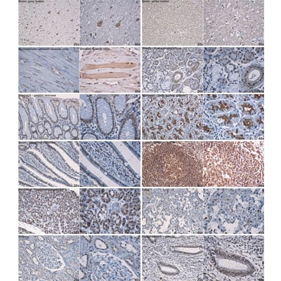
Immunohistochemistry-Paraffin: TDP-43/TARDBP Antibody (2E2-D3) [H00023435-M01] - TARDBP in rat tissue samples. Image courtsey of product review by Daniel Pirici.![Western Blot: TDP-43/TARDBP Antibody (2E2-D3) [H00023435-M01]](https://resources.rndsystems.com/images/products/TDP-43-TARDBP-Antibody-2E2-D3-Western-Blot-H00023435-M01-img0028.jpg "Western Blot: TDP-43/TARDBP Antibody (2E2-D3) [H00023435-M01]")
Western Blot: TDP-43/TARDBP Antibody (2E2-D3) [H00023435-M01]
TDP-43-TARDBP-Antibody-2E2-D3-Western-Blot-H00023435-M01-img0028.jpg![Immunocytochemistry/ Immunofluorescence: TDP-43/TARDBP Antibody (2E2-D3) [H00023435-M01]](https://resources.rndsystems.com/images/products/TDP-43-TARDBP-Antibody-2E2-D3-Immunocytochemistry-Immunofluorescence-H00023435-M01-img0019.jpg "Immunocytochemistry/ Immunofluorescence: TDP-43/TARDBP Antibody (2E2-D3) [H00023435-M01]")
Immunocytochemistry/ Immunofluorescence: TDP-43/TARDBP Antibody (2E2-D3) [H00023435-M01]
Immunocytochemistry/Immunofluorescence: TDP-43/TARDBP Antibody (2E2-D3) [H00023435-M01] - Analysis of monoclonal antibody to TARDBP on HeLa cell. Antibody concentration 10 ug/ml.![Immunohistochemistry-Paraffin: TDP-43/TARDBP Antibody (2E2-D3) [H00023435-M01]](https://resources.rndsystems.com/images/products/TDP-43-TARDBP-Antibody-2E2-D3-Immunohistochemistry-Paraffin-H00023435-M01-img0013.jpg "Immunohistochemistry-Paraffin: TDP-43/TARDBP Antibody (2E2-D3) [H00023435-M01]")
Immunohistochemistry-Paraffin: TDP-43/TARDBP Antibody (2E2-D3) [H00023435-M01]
Immunohistochemistry-Paraffin: TDP-43/TARDBP Antibody (2E2-D3) [H00023435-M01] - TARDBP on formalin-fixed paraffin-embedded human leiomyosarcoma. [antibody concentration 3 ug/ml]![Western Blot: TDP-43/TARDBP Antibody (2E2-D3) [H00023435-M01]](https://resources.rndsystems.com/images/products/TDP-43-TARDBP-Antibody-2E2-D3-Western-Blot-H00023435-M01-img0026.jpg "Western Blot: TDP-43/TARDBP Antibody (2E2-D3) [H00023435-M01]")
Western Blot: TDP-43/TARDBP Antibody (2E2-D3) [H00023435-M01]
Western Blot: TDP-43/TARDBP Antibody (2E2-D3) [H00023435-M01] - Analysis of TARDBP expression in transfected 293T cell line by TARDBP monoclonal antibody (M01), clone 2E2-D3.Lane 1: TARDBP transfected lysate(44.7 KDa).Lane 2: Non-transfected lysate.![Immunohistochemistry-Paraffin: TDP-43/TARDBP Antibody (2E2-D3) [H00023435-M01]](https://resources.rndsystems.com/images/products/TDP-43-TARDBP-Antibody-2E2-D3-Immunohistochemistry-Paraffin-H00023435-M01-img0021.jpg "Immunohistochemistry-Paraffin: TDP-43/TARDBP Antibody (2E2-D3) [H00023435-M01]")
Immunohistochemistry-Paraffin: TDP-43/TARDBP Antibody (2E2-D3) [H00023435-M01]
Immunohistochemistry-Paraffin: TDP-43/TARDBP Antibody (2E2-D3) [H00023435-M01] - Analysis of monoclonal antibody to TARDBP on formalin-fixed paraffin-embedded human leiomyosarcoma tissue Antibody concentration 3 ug/ml.![Knockdown Validated: TDP-43/TARDBP Antibody (2E2-D3) [H00023435-M01]](https://resources.rndsystems.com/images/products/TDP-43-TARDBP-Antibody-2E2-D3-Western-Blot-H00023435-M01-img0023.jpg "Western Blot: TDP-43/TARDBP Antibody (2E2-D3) [H00023435-M01]")
Western Blot: TDP-43/TARDBP Antibody (2E2-D3) [H00023435-M01]
Western Blot: TDP-43/TARDBP Antibody (2E2-D3) [H00023435-M01] - Analysis of TARDBP over-expressed 293 cell line, cotransfected with TARDBP Validated Chimera RNAi ( Cat # H00023435-R01V ) (Lane 2) or non-transfected control (Lane 1). Blot probed with TARDBP monoclonal antibody (M01), clone 2E2-D3 (Cat # H00023435-M01 ). GAPDH ( 36.1 kDa ) used as specificity and loading control.![ELISA: TDP-43/TARDBP Antibody (2E2-D3) [H00023435-M01]](https://resources.rndsystems.com/images/products/TDP-43-TARDBP-Antibody-2E2-D3-ELISA-H00023435-M01-img0027.jpg "ELISA: TDP-43/TARDBP Antibody (2E2-D3) [H00023435-M01]")
ELISA: TDP-43/TARDBP Antibody (2E2-D3) [H00023435-M01]
ELISA: TDP-43/TARDBP Antibody (2E2-D3) [H00023435-M01] - Detection limit for recombinant GST tagged TARDBP is 0.3 ng/ml as a capture antibody. [H00023435-M01] -")
Western Blot: TDP-43/TARDBP Antibody (2E2-D3) [H00023435-M01] -
Western Blot: TDP-43/TARDBP Antibody (2E2-D3) [H00023435-M01] - PDI co-precipitates with TDP-43 & is increased in mutant TDP-43 transgenic mouse spinal cords.(A) Co-immunoprecipitation of wildtype & mutant Q331K TDP-43-mCherry from Neuro2a cell lysates using an anti-PDI antibody followed by TDP-43 immunoblot. Control immunoprecipitations performed using untransfected (Untrans) or mCherry expressing cell lysates, buffer only with coprecipitating antibodies or TDP-43 Q331K cell lysates using an irrelevant, isotype-matched control antibody (IgG). Input control (2%) shows expression of mCherry-TDP43 in wildtype & Q331K cell lysates. Image collected & cropped by CiteAb from the following publication (https://dx.plos.org/10.1371/journal.pone.0081170), licensed under a CC-BY license. Not internally tested by Novus Biologicals. [H00023435-M01] -")
Western Blot: TDP-43/TARDBP Antibody (2E2-D3) [H00023435-M01] -
Western Blot: TDP-43/TARDBP Antibody (2E2-D3) [H00023435-M01] - CLIP shows that TDP-43 directly interacts with translational target mRNAs. (A) Schematic diagram of the CLIP experiment. (B) Immunoblot showing specific IP of TDP-43 from MN1 cells under CLIP conditions. A TDP-43 band is specifically observed in the input & TDP-43 IP lanes from both untreated & UV-treated cells, but not in the IgG or anti-RBP X control lanes. RBP X is an IP of a different RNA-binding protein, which serves as an additional specificity control here. (C and D) Plots showing percentage of input mRNA in the TDP-43 IP for the mRNAs indicated in three separate CLIP assays from MN1 cells. Performing CLIP from UV-treated cells leads to significant recovery & enrichment relative to Gapdh for all mRNAs examined (C). In the absence of UV treatment, much less mRNA is recovered & no significant enrichment relative to Gapdh mRNA is observed. Note that the Y-axis scale in D is 1000× smaller than in C, indicating strong crosslinking dependence of mRNA co-immunoprecipitation (n = 3, s.e.m. error bars, P-values indicated in the plot, unpaired two-tailed t-test). Image collected & cropped by CiteAb from the following publication (https://pubmed.ncbi.nlm.nih.gov/30357366), licensed under a CC-BY license. Not internally tested by Novus Biologicals. [H00023435-M01] -")
Western Blot: TDP-43/TARDBP Antibody (2E2-D3) [H00023435-M01] -
Western Blot: TDP-43/TARDBP Antibody (2E2-D3) [H00023435-M01] - SINE compounds partially rescue motor deficits in an animal model of ALS/FTD. (a) Schematic timeline for in vivo experiments. (b) Western blot demonstrating the expression of human TDP43 within the brains of AAV9-injected animals, which we have previously shown is largely restricted to neuronal cell types (Jackson et al.59). Full-length blots appear in Fig. S1. (c) The hang test measures the time it takes for animals to fall from grid-like scaffold when it is lifted & tilted. A single representative animal administered AAV9-TDP43 is shown; note the inability of the hind limbs to clasp the wire mesh. (d) TDP43 expression impaired motor performance on the hang test at 3 weeks, an effect that could be mitigated by 7.5 mg kg-1 KPT-350. (e) Rotarod testing at 4 weeks of age showed prominent deficits associated with TDP43 expression that were resistant to treatment with SINE compounds. n, number of animals per condition. ns, not significant, mean ± s.e.m. *p < 0.05, **p < 0.01, ***p < 0.001, one-way ANOVA with Bonferroni correction. Image collected & cropped by CiteAb from the following publication (https://www.nature.com/articles/s41598-018-22858-w), licensed under a CC-BY license. Not internally tested by Novus Biologicals.Applications for TDP-43/TARDBP Antibody (2E2-D3) - Azide and BSA Free
Application
Recommended Usage
ELISA
1:100-1:2000
Immunocytochemistry/ Immunofluorescence
1:10-1:500
Immunohistochemistry
1:10-1:500
Immunohistochemistry-Paraffin
1:10-1:500
Immunoprecipitation
1:10-1:500
Western Blot
1:500
Application Notes
Cross reactivity in Mouse and Rat reported by customer review.
Reviewed Applications
Read 3 reviews rated 5 using H00023435-M01 in the following applications:
Formulation, Preparation, and Storage
Purification
IgG purified
Formulation
In 1x PBS, pH 7.4
Format
Azide and BSA Free
Preservative
No Preservative
Concentration
Concentrations vary lot to lot. See vial label for concentration. If unlisted please contact technical services.
Shipping
The product is shipped with polar packs. Upon receipt, store it immediately at the temperature recommended below.
Stability & Storage
Aliquot and store at -20C or -80C. Avoid freeze-thaw cycles.
Background: TDP-43/TARDBP
Long Name
TAR DNA-binding Protein 43
Alternate Names
ALS10, TARDBP, TDP43
Entrez Gene IDs
23435 (Human)
Gene Symbol
TARDBP
OMIM
605078 (Human)
UniProt
Additional TDP-43/TARDBP Products
Product Documents for TDP-43/TARDBP Antibody (2E2-D3) - Azide and BSA Free
Certificate of Analysis
To download a Certificate of Analysis, please enter a lot or batch number in the search box below.
Product Specific Notices for TDP-43/TARDBP Antibody (2E2-D3) - Azide and BSA Free
This product is produced by and distributed for Abnova, a company based in Taiwan.
This product is for research use only and is not approved for use in humans or in clinical diagnosis. Primary Antibodies are guaranteed for 1 year from date of receipt.
Related Research Areas
Citations for TDP-43/TARDBP Antibody (2E2-D3) - Azide and BSA Free
 Powered by Bioz
Customer Reviews for TDP-43/TARDBP Antibody (2E2-D3) - Azide and BSA Free (3)
5 out of 5
3 Customer Ratings
Have you used TDP-43/TARDBP Antibody (2E2-D3) - Azide and BSA Free?
Submit a review and receive an Amazon gift card!
$25/€18/£15/$25CAN/¥2500 Yen for a review with an image
$10/€7/£6/$10CAN/¥1110 Yen for a review without an image
Submit a review
Customer Images

Showing
1
-
3 of
3 reviews
Showing All
Filter By:
-
Application: Western BlotSample Tested: 293T cell lineSpecies: HumanVerified Customer | Posted 01/20/2022Human 293T cell line

-
Application: Immunohistochemistry-ParaffinVerified Customer | Posted 10/02/2012Comments in the pdf protocol
-
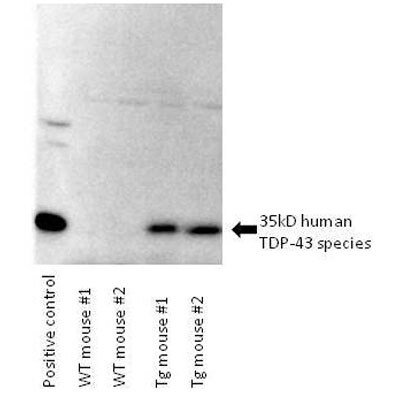
Application: Western BlotSample Tested: Mouse Brain TissueSpecies: MouseVerified Customer | Posted 06/28/2012Western blot showing brain lysate of WT mice and transgenic mice expressing a C-terminal fragment of human TDP-43
There are no reviews that match your criteria.
Protocols
Find general support by application which include: protocols, troubleshooting, illustrated assays, videos and webinars.
- Antigen Retrieval Protocol (PIER)
- Antigen Retrieval for Frozen Sections Protocol
- Appropriate Fixation of IHC/ICC Samples
- Cellular Response to Hypoxia Protocols
- Chromogenic IHC Staining of Formalin-Fixed Paraffin-Embedded (FFPE) Tissue Protocol
- Chromogenic Immunohistochemistry Staining of Frozen Tissue
- ClariTSA™ Fluorophore Kits
- Detection & Visualization of Antibody Binding
- ELISA Sample Preparation & Collection Guide
- ELISA Troubleshooting Guide
- Fluorescent IHC Staining of Frozen Tissue Protocol
- Graphic Protocol for Heat-induced Epitope Retrieval
- Graphic Protocol for the Preparation and Fluorescent IHC Staining of Frozen Tissue Sections
- Graphic Protocol for the Preparation and Fluorescent IHC Staining of Paraffin-embedded Tissue Sections
- Graphic Protocol for the Preparation of Gelatin-coated Slides for Histological Tissue Sections
- How to Run an R&D Systems DuoSet ELISA
- How to Run an R&D Systems Quantikine ELISA
- How to Run an R&D Systems Quantikine™ QuicKit™ ELISA
- ICC Cell Smear Protocol for Suspension Cells
- ICC Immunocytochemistry Protocol Videos
- ICC for Adherent Cells
- IHC Sample Preparation (Frozen sections vs Paraffin)
- Immunocytochemistry (ICC) Protocol
- Immunocytochemistry Troubleshooting
- Immunofluorescence of Organoids Embedded in Cultrex Basement Membrane Extract
- Immunofluorescent IHC Staining of Formalin-Fixed Paraffin-Embedded (FFPE) Tissue Protocol
- Immunohistochemistry (IHC) and Immunocytochemistry (ICC) Protocols
- Immunohistochemistry Frozen Troubleshooting
- Immunohistochemistry Paraffin Troubleshooting
- Immunoprecipitation Protocol
- Preparing Samples for IHC/ICC Experiments
- Preventing Non-Specific Staining (Non-Specific Binding)
- Primary Antibody Selection & Optimization
- Protocol for Heat-Induced Epitope Retrieval (HIER)
- Protocol for Making a 4% Formaldehyde Solution in PBS
- Protocol for VisUCyte™ HRP Polymer Detection Reagent
- Protocol for the Fluorescent ICC Staining of Cell Smears - Graphic
- Protocol for the Fluorescent ICC Staining of Cultured Cells on Coverslips - Graphic
- Protocol for the Preparation & Fixation of Cells on Coverslips
- Protocol for the Preparation and Chromogenic IHC Staining of Frozen Tissue Sections
- Protocol for the Preparation and Chromogenic IHC Staining of Frozen Tissue Sections - Graphic
- Protocol for the Preparation and Chromogenic IHC Staining of Paraffin-embedded Tissue Sections
- Protocol for the Preparation and Chromogenic IHC Staining of Paraffin-embedded Tissue Sections - Graphic
- Protocol for the Preparation and Fluorescent ICC Staining of Cells on Coverslips
- Protocol for the Preparation and Fluorescent ICC Staining of Non-adherent Cells
- Protocol for the Preparation and Fluorescent ICC Staining of Stem Cells on Coverslips
- Protocol for the Preparation and Fluorescent IHC Staining of Frozen Tissue Sections
- Protocol for the Preparation and Fluorescent IHC Staining of Paraffin-embedded Tissue Sections
- Protocol for the Preparation of Gelatin-coated Slides for Histological Tissue Sections
- Protocol for the Preparation of a Cell Smear for Non-adherent Cell ICC - Graphic
- Quantikine HS ELISA Kit Assay Principle, Alkaline Phosphatase
- Quantikine HS ELISA Kit Principle, Streptavidin-HRP Polymer
- R&D Systems Quality Control Western Blot Protocol
- Sandwich ELISA (Colorimetric) – Biotin/Streptavidin Detection Protocol
- Sandwich ELISA (Colorimetric) – Direct Detection Protocol
- TUNEL and Active Caspase-3 Detection by IHC/ICC Protocol
- The Importance of IHC/ICC Controls
- Troubleshooting Guide: ELISA
- Troubleshooting Guide: Immunohistochemistry
- Troubleshooting Guide: Western Blot Figures
- Western Blot Conditions
- Western Blot Protocol
- Western Blot Protocol for Cell Lysates
- Western Blot Troubleshooting
- Western Blot Troubleshooting Guide
- View all Protocols, Troubleshooting, Illustrated assays and Webinars
Loading...