Tenascin C Antibody (4C8MS) - BSA Free
Novus Biologicals | Catalog # NB110-68136
Key Product Details
Validated by
Species Reactivity
Validated:
Cited:
Applications
Validated:
Cited:
Label
Antibody Source
Format
Product Specifications
Immunogen
Reactivity Notes
Localization
Specificity
Clonality
Host
Isotype
Scientific Data Images for Tenascin C Antibody (4C8MS) - BSA Free
![Flow (Intracellular): Tenascin C Antibody (4C8MS) - BSA Free [NB110-68136]](https://resources.rndsystems.com/images/products/Tenascin-C-Antibody-4C8MS-Flow-Intracellular-NB110-68136-img0010.jpg "Flow (Intracellular): Tenascin C Antibody (4C8MS) - BSA Free [NB110-68136]")
Flow (Intracellular): Tenascin C Antibody (4C8MS) - BSA Free [NB110-68136]
Flow (Intracellular): Tenascin C Antibody (4C8MS) [NB110-68136] - Figure A: Intracellular stain performed on U87MG Cells with Tenascin C (4C8MS) antibody NB110-68136 (blue) and a matched isotype control NBP1-97005 (orange). Cells were fixed with 4% paraformaldehyde, following fixation, cells were permeabilized with 0.1% saponin. Cells were incubated in an antibody dilution of 1 ug/mL for 30 minutes at room temperature, followed by mouse F(ab)2 IgG (H+L) APC-conjugated secondary antibody [F0101B, R&D Systems].Figure B: U87MG Cells were either untreated (orange) or treated with 3uM Monensin (blue). An intracellular stain was performed with Tenascin C (4C8MS) antibody NB110-68136. Cells were fixed with 4% paraformaldehyde, following fixation, cells were permeabilized with 0.1% saponin. Cells were incubated in an antibody dilution of 1 ug/mL for 30 minutes at room temperature, followed by mouse F(ab)2 IgG (H+L) APC-conjugated secondary antibody [F0101B, R&D Systems].![Western Blot: Tenascin C Antibody (4C8MS)BSA Free [NB110-68136]](https://resources.rndsystems.com/images/products/Tenascin-C-Antibody-4C8MS-Western-Blot-NB110-68136-img0015.jpg "Western Blot: Tenascin C Antibody (4C8MS)BSA Free [NB110-68136]")
![Immunocytochemistry/ Immunofluorescence: Tenascin C Antibody (4C8MS) - BSA Free [NB110-68136]](https://resources.rndsystems.com/images/products/Tenascin-C-Antibody-4C8MS-Immunocytochemistry-Immunofluorescence-NB110-68136-img0012.jpg "Immunocytochemistry/ Immunofluorescence: Tenascin C Antibody (4C8MS) - BSA Free [NB110-68136]")
Immunocytochemistry/ Immunofluorescence: Tenascin C Antibody (4C8MS) - BSA Free [NB110-68136]
Immunocytochemistry/Immunofluorescence: Tenascin C Antibody (4C8MS) [NB110-68136] - SK-MEL-28 cells were fixed for 10 minutes using 10% formalin and then permeabilized for 5 minutes using 1X PBS + 0.05% Triton-X100. The cells were incubated with anti-Tenascin C at 5 ug/ml overnight at 4C and detected with an anti-Mouse IgG Dylight 488 (Green) at a 1:500 dilution. Nuclei were counterstained with DAPI (Blue). Cells were imaged using a 40X objective.![Immunohistochemistry-Paraffin: Tenascin C Antibody (4C8MS) - BSA Free [NB110-68136]](https://resources.rndsystems.com/images/products/Tenascin-C-Antibody-4C8MS-Immunohistochemistry-Paraffin-NB110-68136-img0009.jpg "Immunohistochemistry-Paraffin: Tenascin C Antibody (4C8MS) - BSA Free [NB110-68136]")
Immunohistochemistry-Paraffin: Tenascin C Antibody (4C8MS) - BSA Free [NB110-68136]
Immunohistochemistry-Paraffin: Tenascin C Antibody (4C8MS) [NB110-68136] - IHC analysis of a formalin fixed paraffin embedded tissue section of mouse bone-tendon using Tenascin C antibody (clone 4C8MS) at 1:25 dilution. The signal was detected using HRP-DAB detection method which followed counterstaining using hematoxylin. The antibody generated a very specific cytoplasmic, membrane and extra-cellular signal in tendon fibroblasts, osteoblasts, osteoclasts, and some bone marrow cells. The mineralized areas were largely negative for the staining.![Flow Cytometry: Tenascin C Antibody (4C8MS) - BSA Free [NB110-68136]](https://resources.rndsystems.com/images/products/Tenascin-C-Antibody-4C8MS-BSA-Free-Flow-Cytometry-NB110-68136-img0016.jpg "Flow Cytometry: Tenascin C Antibody (4C8MS) - BSA Free [NB110-68136]")
Flow Cytometry: Tenascin C Antibody (4C8MS) - BSA Free [NB110-68136]
Flow Cytometry: Tenascin C Antibody (4C8MS) - BSA Free [NB110-68136] - An intracellular stain was performed on MCF7 cells with Tenascin C Antibody (4C8MS) NB110-68136 (blue) and a matched isotype control MAB002 (orange). Cells were fixed with 4% PFA and then permeabilized with 0.1% saponin. Cells were incubated in an antibody dilution of 1 ug/mL for 30 minutes at room temperature, followed by Mouse IgG (H+L) Cross-Adsorbed Secondary Antibody, Dylight 550 (84540, Thermo Fisher).![Immunohistochemistry-Paraffin: Tenascin C Antibody (4C8MS) - BSA Free [NB110-68136]](https://resources.rndsystems.com/images/products/Tenascin-C-Antibody-4C8MS-Immunohistochemistry-Paraffin-NB110-68136-img0008.jpg "Immunohistochemistry-Paraffin: Tenascin C Antibody (4C8MS) - BSA Free [NB110-68136]")
Immunohistochemistry-Paraffin: Tenascin C Antibody (4C8MS) - BSA Free [NB110-68136]
Immunohistochemistry-Paraffin: Tenascin C Antibody (4C8MS) [NB110-68136] - IHC analysis of a formalin fixed paraffin embedded tissue section of mouse bone-tendon using Tenascin C antibody (clone 4C8MS) at 1:25 dilution. The signal was detected using HRP-DAB detection method which followed counterstaining using hematoxylin. The antibody generated a very specific cytoplasmic, membrane and extra-cellular signal in tendon fibroblasts, osteoblasts, osteoclasts, and some bone marrow cells. The mineralized areas were largely negative for the staining.![Flow Cytometry: Tenascin C Antibody (4C8MS) - BSA Free [NB110-68136]](https://resources.rndsystems.com/images/products/Tenascin-C-Antibody-4C8MS-Flow-Cytometry-NB110-68136-img0007.jpg "Flow Cytometry: Tenascin C Antibody (4C8MS) - BSA Free [NB110-68136]")
Flow Cytometry: Tenascin C Antibody (4C8MS) - BSA Free [NB110-68136]
Flow Cytometry: Tenascin C Antibody (4C8MS) [NB110-68136] - Intracellular flow cytometric staining of 1 x 10^6 MCF-7 cells using Tenascin C antibody (dark blue). Isotype control shown in orange. An antibody concentration of 1 ug/1x10^6 cells was used.![Flow Cytometry: Tenascin C Antibody (4C8MS) - BSA Free [NB110-68136]](https://resources.rndsystems.com/images/products/Tenascin-C-Antibody-4C8MS-Flow-Cytometry-NB110-68136-img0011.jpg "Flow Cytometry: Tenascin C Antibody (4C8MS) - BSA Free [NB110-68136]")
Flow Cytometry: Tenascin C Antibody (4C8MS) - BSA Free [NB110-68136]
Flow Cytometry: Tenascin C Antibody (4C8MS) [NB110-68136] - An intracellular stain was performed on SK-MEL-28 cells with Tenascin C Antibody (4C8MS) NB110-68136 and a matched isotype control. Cells were fixed with 4% PFA and then permeablized with 0.1% saponin. Cells were incubated in an antibody dilution of 2.5 ug/mL for 30 minutes at room temperature, followed by mouse F(ab)2 IgG (H+L) APC-conjugated secondary antibody (F0101B, R&D Systems).![Flow Cytometry: Tenascin C Antibody (4C8MS) - BSA Free [NB110-68136]](https://resources.rndsystems.com/images/products/Tenascin-C-Antibody-4C8MS-Flow-Cytometry-NB110-68136-img0013.jpg "Flow Cytometry: Tenascin C Antibody (4C8MS) - BSA Free [NB110-68136]")
Flow Cytometry: Tenascin C Antibody (4C8MS) - BSA Free [NB110-68136]
Flow Cytometry: Tenascin C Antibody (4C8MS) [NB110-68136] - An intracellular stain was performed on SK-MEL-28 cells with Tenascin C Antibody (4C8MS) NB110-68136AF488 (blue) and a matched isotype control (orange). Cells were fixed with 4% PFA and then permeabilized with 0.1% saponin. Cells were incubated in an antibody dilution of 5 ug/mL for 30 minutes at room temperature. Both antibodies were conjugated to Alexa Fluor 488.![Flow Cytometry: Tenascin C Antibody (4C8MS) - BSA Free [NB110-68136]](https://resources.rndsystems.com/images/products/Tenascin-C-Antibody-4C8MS-Flow-Cytometry-NB110-68136-img0014.jpg "Flow Cytometry: Tenascin C Antibody (4C8MS) - BSA Free [NB110-68136]")
Flow Cytometry: Tenascin C Antibody (4C8MS) - BSA Free [NB110-68136]
Flow Cytometry: Tenascin C Antibody (4C8MS) [NB110-68136] - An intracellular stain was performed on SK-MEL-28 cells with Tenascin C Antibody [4C8MS] NB110-68136AF647 (blue) and a matched isotype control (orange). Cells were fixed with 4% PFA and then permeabilized with 0.1% saponin. Cells were incubated in an antibody dilution of 2.5 ug/mL for 30 minutes at room temperature. Both antibodies were conjugated to Alexa Fluor 647.Applications for Tenascin C Antibody (4C8MS) - BSA Free
Flow Cytometry
Immunocytochemistry/ Immunofluorescence
Immunohistochemistry
Immunohistochemistry-Paraffin
Western Blot
Reviewed Applications
Read 2 reviews rated 3.5 using NB110-68136 in the following applications:
Flow Cytometry Panel Builder
Bio-Techne Knows Flow Cytometry
Save time and reduce costly mistakes by quickly finding compatible reagents using the Panel Builder Tool.
Advanced Features
- Spectra Viewer - Custom analysis of spectra from multiple fluorochromes
- Spillover Popups - Visualize the spectra of individual fluorochromes
- Antigen Density Selector - Match fluorochrome brightness with antigen density

Formulation, Preparation, and Storage
Purification
Formulation
Format
Preservative
Concentration
Shipping
Stability & Storage
Background: Tenascin C
Alternate Names
Gene Symbol
UniProt
Additional Tenascin C Products
Product Documents for Tenascin C Antibody (4C8MS) - BSA Free
Certificate of Analysis
To download a Certificate of Analysis, please enter a lot or batch number in the search box below.
Product Specific Notices for Tenascin C Antibody (4C8MS) - BSA Free
This product is for research use only and is not approved for use in humans or in clinical diagnosis. Primary Antibodies are guaranteed for 1 year from date of receipt.
Related Research Areas
Citations for Tenascin C Antibody (4C8MS) - BSA Free
 Powered by Bioz
Customer Reviews for Tenascin C Antibody (4C8MS) - BSA Free (2)
Have you used Tenascin C Antibody (4C8MS) - BSA Free?
Submit a review and receive an Amazon gift card!
$25/€18/£15/$25CAN/¥2500 Yen for a review with an image
$10/€7/£6/$10CAN/¥1110 Yen for a review without an image
Submit a review
Customer Images

-
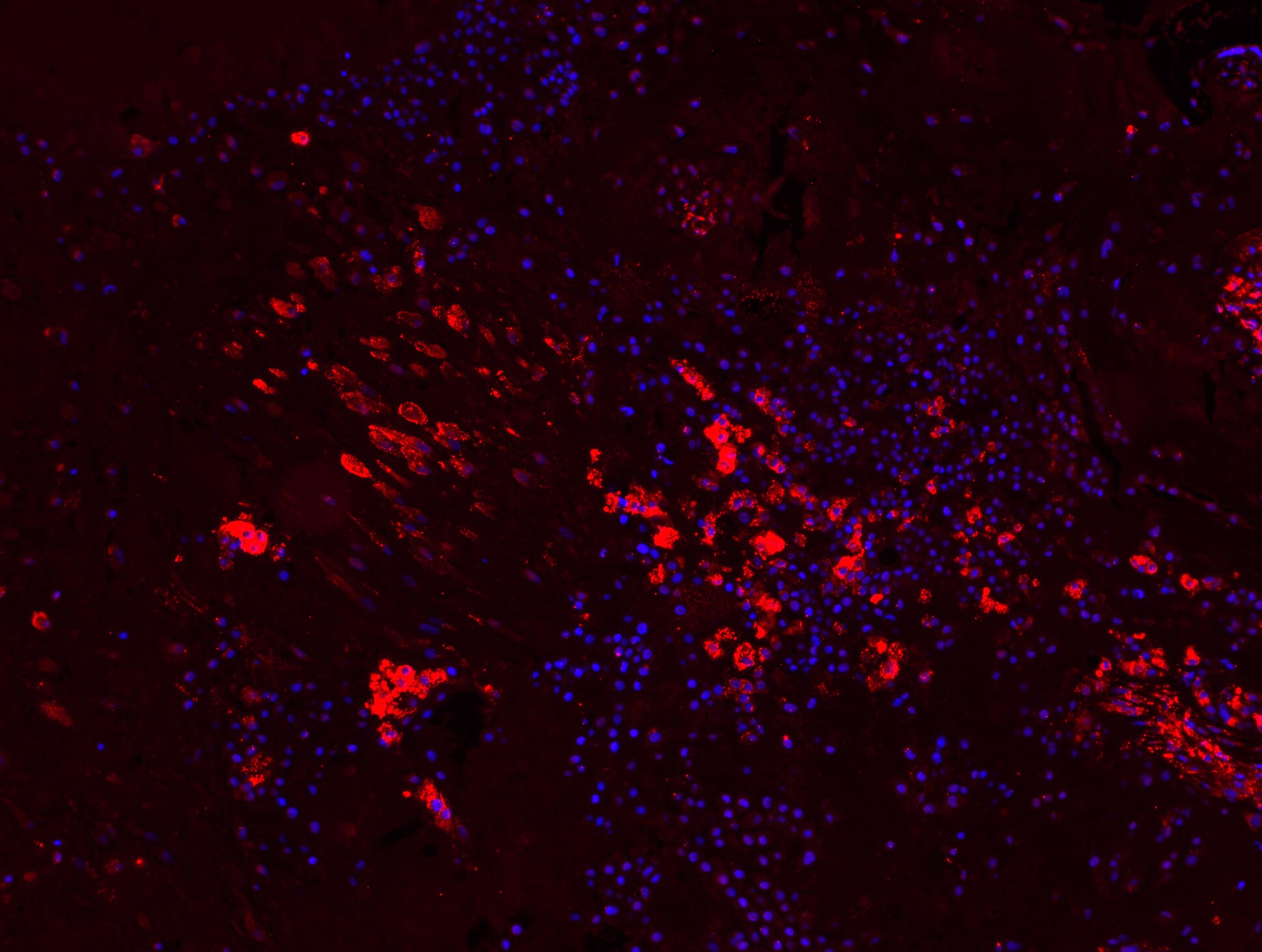
Application: Immunohistochemistry-FrozenSample Tested: ScleraSpecies: FelineVerified Customer | Posted 09/15/2018Tenascin-C antibody was immunolabeled shown as red. DAPI (blue). The image was captured using epifluorescent microscopy.

-
Application: ImmunocytochemistrySample Tested: Uterus tissueSpecies: HumanVerified Customer | Posted 03/22/2017
There are no reviews that match your criteria.
Protocols
View specific protocols for Tenascin C Antibody (4C8MS) - BSA Free (NB110-68136):
Immunohistochemistry-Paraffin Embedded Sections for NB110-68136
Antigen Unmasking:
Bring slides to a boil in 10 mM sodium citrate buffer (pH 6.0) then maintain at a sub-boiling temperature for 10 minutes. Cool slides on bench-top for 30 minutes.
Staining:
1. Wash sections in deionized water three times for 5 minutes each.
2. Wash sections in wash buffer for 5 minutes.
3. Block each section with 100-400 ul blocking solution for 1 hour at room temperature.
4. Remove blocking solution and add 100-400 ul diluted primary antibody. Incubate overnight at 4C.
5. Remove antibody solution and wash sections in wash buffer three times for 5 minutes each.
6. Add 100-400 ul biotinylated diluted secondary antibody. Incubate 30 minutes at room temperature.
7. Remove secondary antibody solution and wash sections three times with wash buffer for 5 minutes each.
8. Add 100-400 ul Streptavidin-HRP reagent to each section and incubate for 30 minutes at room temperature.
9. Wash sections three times in wash buffer for 5 minutes each.
10. Add 100-400 ul DAB substrate to each section and monitor staining closely.
11. As soon as the sections develop, immerse slides in deionized water.
12. Counterstain sections in hematoxylin.
13. Wash sections in deionized water two times for 5 minutes each.
14. Dehydrate sections.
15. Mount coverslips.
*The above information is only intended as a guide. The researcher should determine what protocol best meets their needs. Please follow safe laboratory procedures.
Find general support by application which include: protocols, troubleshooting, illustrated assays, videos and webinars.
- 7-Amino Actinomycin D (7-AAD) Cell Viability Flow Cytometry Protocol
- Antigen Retrieval Protocol (PIER)
- Antigen Retrieval for Frozen Sections Protocol
- Appropriate Fixation of IHC/ICC Samples
- Cellular Response to Hypoxia Protocols
- Chromogenic IHC Staining of Formalin-Fixed Paraffin-Embedded (FFPE) Tissue Protocol
- Chromogenic Immunohistochemistry Staining of Frozen Tissue
- ClariTSA™ Fluorophore Kits
- Detection & Visualization of Antibody Binding
- ELISA Sample Preparation & Collection Guide
- ELISA Troubleshooting Guide
- Extracellular Membrane Flow Cytometry Protocol
- Flow Cytometry Protocol for Cell Surface Markers
- Flow Cytometry Protocol for Staining Membrane Associated Proteins
- Flow Cytometry Staining Protocols
- Flow Cytometry Troubleshooting Guide
- Fluorescent IHC Staining of Frozen Tissue Protocol
- Graphic Protocol for Heat-induced Epitope Retrieval
- Graphic Protocol for the Preparation and Fluorescent IHC Staining of Frozen Tissue Sections
- Graphic Protocol for the Preparation and Fluorescent IHC Staining of Paraffin-embedded Tissue Sections
- Graphic Protocol for the Preparation of Gelatin-coated Slides for Histological Tissue Sections
- How to Run an R&D Systems DuoSet ELISA
- How to Run an R&D Systems Quantikine ELISA
- How to Run an R&D Systems Quantikine™ QuicKit™ ELISA
- ICC Cell Smear Protocol for Suspension Cells
- ICC Immunocytochemistry Protocol Videos
- ICC for Adherent Cells
- IHC Sample Preparation (Frozen sections vs Paraffin)
- Immunocytochemistry (ICC) Protocol
- Immunocytochemistry Troubleshooting
- Immunofluorescence of Organoids Embedded in Cultrex Basement Membrane Extract
- Immunofluorescent IHC Staining of Formalin-Fixed Paraffin-Embedded (FFPE) Tissue Protocol
- Immunohistochemistry (IHC) and Immunocytochemistry (ICC) Protocols
- Immunohistochemistry Frozen Troubleshooting
- Immunohistochemistry Paraffin Troubleshooting
- Intracellular Flow Cytometry Protocol Using Alcohol (Methanol)
- Intracellular Flow Cytometry Protocol Using Detergents
- Intracellular Nuclear Staining Flow Cytometry Protocol Using Detergents
- Intracellular Staining Flow Cytometry Protocol Using Alcohol Permeabilization
- Intracellular Staining Flow Cytometry Protocol Using Detergents to Permeabilize Cells
- Preparing Samples for IHC/ICC Experiments
- Preventing Non-Specific Staining (Non-Specific Binding)
- Primary Antibody Selection & Optimization
- Propidium Iodide Cell Viability Flow Cytometry Protocol
- Protocol for Heat-Induced Epitope Retrieval (HIER)
- Protocol for Liperfluo
- Protocol for Making a 4% Formaldehyde Solution in PBS
- Protocol for VisUCyte™ HRP Polymer Detection Reagent
- Protocol for the Characterization of Human Th22 Cells
- Protocol for the Characterization of Human Th9 Cells
- Protocol for the Fluorescent ICC Staining of Cell Smears - Graphic
- Protocol for the Fluorescent ICC Staining of Cultured Cells on Coverslips - Graphic
- Protocol for the Preparation & Fixation of Cells on Coverslips
- Protocol for the Preparation and Chromogenic IHC Staining of Frozen Tissue Sections
- Protocol for the Preparation and Chromogenic IHC Staining of Frozen Tissue Sections - Graphic
- Protocol for the Preparation and Chromogenic IHC Staining of Paraffin-embedded Tissue Sections
- Protocol for the Preparation and Chromogenic IHC Staining of Paraffin-embedded Tissue Sections - Graphic
- Protocol for the Preparation and Fluorescent ICC Staining of Cells on Coverslips
- Protocol for the Preparation and Fluorescent ICC Staining of Non-adherent Cells
- Protocol for the Preparation and Fluorescent ICC Staining of Stem Cells on Coverslips
- Protocol for the Preparation and Fluorescent IHC Staining of Frozen Tissue Sections
- Protocol for the Preparation and Fluorescent IHC Staining of Paraffin-embedded Tissue Sections
- Protocol for the Preparation of Gelatin-coated Slides for Histological Tissue Sections
- Protocol for the Preparation of a Cell Smear for Non-adherent Cell ICC - Graphic
- Protocol: Annexin V and PI Staining by Flow Cytometry
- Protocol: Annexin V and PI Staining for Apoptosis by Flow Cytometry
- Quantikine HS ELISA Kit Assay Principle, Alkaline Phosphatase
- Quantikine HS ELISA Kit Principle, Streptavidin-HRP Polymer
- R&D Systems Quality Control Western Blot Protocol
- Sandwich ELISA (Colorimetric) – Biotin/Streptavidin Detection Protocol
- Sandwich ELISA (Colorimetric) – Direct Detection Protocol
- TUNEL and Active Caspase-3 Detection by IHC/ICC Protocol
- The Importance of IHC/ICC Controls
- Troubleshooting Guide: ELISA
- Troubleshooting Guide: Fluorokine Flow Cytometry Kits
- Troubleshooting Guide: Immunohistochemistry
- Troubleshooting Guide: Western Blot Figures
- Western Blot Conditions
- Western Blot Protocol
- Western Blot Protocol for Cell Lysates
- Western Blot Troubleshooting
- Western Blot Troubleshooting Guide
- View all Protocols, Troubleshooting, Illustrated assays and Webinars
FAQs for Tenascin C Antibody (4C8MS) - BSA Free
-
Q: I would like to see image of WB to see the isoforms of the tenascin NB110-68136.
A:
I am afraid we do not have notes or images on the westerns for this antibody. There are 2 publications that do have a double band in westerns for this antibody: http://www.ncbi.nlm.nih.gov/pmc/articles/PMC3680762/. http://www.ncbi.nlm.nih.gov/pmc/articles/PMC1868127/. We have 2 Tenascin C antibodies that have been validated in mouse for western blot but none with images.
-
Q: One of our clients is looking for antibodies against tenascin C and fibronectin that could be used for FFPE rat tissues. These antibodies should exclusively recognize tenascin C and fibronectin and do not cross react with each other. Can you inform me if you have these antibodies?
A:
Here is a link to Tenascin C antibodies we have validated in rat tissues for IHC-P Here are antibodies we have validated in fibronectin in rat tissues for IHC-P
-
Q: I would like to see image of WB to see the isoforms of the tenascin NB110-68136.
A:
I am afraid we do not have notes or images on the westerns for this antibody. There are 2 publications that do have a double band in westerns for this antibody: http://www.ncbi.nlm.nih.gov/pmc/articles/PMC3680762/. http://www.ncbi.nlm.nih.gov/pmc/articles/PMC1868127/. We have 2 Tenascin C antibodies that have been validated in mouse for western blot but none with images.
-
Q: One of our clients is looking for antibodies against tenascin C and fibronectin that could be used for FFPE rat tissues. These antibodies should exclusively recognize tenascin C and fibronectin and do not cross react with each other. Can you inform me if you have these antibodies?
A:
Here is a link to Tenascin C antibodies we have validated in rat tissues for IHC-P Here are antibodies we have validated in fibronectin in rat tissues for IHC-P