TFEB Antibody (S1) - Azide and BSA Free
Novus Biologicals | Catalog # H00007942-M01
![Western Blot: TFEB Antibody (S1) [H00007942-M01]](https://resources.rndsystems.com/images/products/TFEB-Antibody-S1-Western-Blot-H00007942-M01-img0009.jpg "Western Blot: TFEB Antibody (S1) [H00007942-M01]")
Loading...
Key Product Details
Species Reactivity
Validated:
Human, Mouse, Rat
Cited:
Mouse
Applications
Validated:
Immunohistochemistry-Paraffin, Western Blot, Immunoblotting, ELISA, Immunocytochemistry/ Immunofluorescence
Cited:
Immunohistochemistry-Paraffin, Western Blot
Label
Unconjugated
Antibody Source
Monoclonal Mouse IgG1 kappa Clone # S1
Format
Azide and BSA Free
Loading...
Product Specifications
Immunogen
TFEB (AAH32448, 1 a.a. ~ 476 a.a) full-length recombinant protein with GST tag. MW of the GST tag alone is 26 KDa. MASRIGLRMQLMREQAQQEEQRERMQQQAVMHYMQQQQQQQQQQLGGPPTPAINTPVHFQSPPPVPGEVLKVQSYLENPTSYHLQQSQHQKVREYLSETYGNKFAAHISPAQGSPKPPPAASPGVRAGHVLSSSAGNSAPNSPMAMLHIGSNPERELDDVIDNIMRLDDVLGYINPEMQMPNTLPLSSSHLNVYSSDPQVTASLVGVTSSSCPADLTQKRELTDAESRALAKERQKKDNHNLIERRRRFNINDRIKELGMLIPKANDLDVRWNKGTILKASVDYIRRMQKDLQKSRELENHSRRLEMTNKQLWLRIQELEMQARVHGLPTTSPSGMNMAELAQQVVKQELPSEEGPGEALMLGAEVPDPEPLPALPPQAPLPLPTQPPSPFHHLDFSHSLSFGGREDEGPPGYPEPLAPGHGSPFPSLSKKDLDLMLLDDSLLPLASDPLLSTMSPEASKASSRRSSFSMEEGDVL
Specificity
TFEB - transcription factor EB
Clonality
Monoclonal
Host
Mouse
Isotype
IgG1 kappa
Description
Quality control test: Antibody Reactive Against Recombinant Protein.
Scientific Data Images for TFEB Antibody (S1) - Azide and BSA Free
Western Blot: TFEB Antibody (S1) [H00007942-M01]
Western Blot: TFEB Antibody (S1) [H00007942-M01] - TFEB monoclonal antibody (M01), clone S1. Analysis of TFEB expression in rat brain.![Immunocytochemistry/ Immunofluorescence: TFEB Antibody (S1) [H00007942-M01]](https://resources.rndsystems.com/images/products/TFEB-Antibody-S1-Immunocytochemistry-Immunofluorescence-H00007942-M01-img0010.jpg "Immunocytochemistry/ Immunofluorescence: TFEB Antibody (S1) [H00007942-M01]")
Immunocytochemistry/ Immunofluorescence: TFEB Antibody (S1) [H00007942-M01]
Immunocytochemistry/Immunofluorescence: TFEB Antibody (S1) [H00007942-M01] - Analysis of monoclonal antibody to TFEB on HeLa cell. Antibody concentration 20 ug/ml.![Western Blot: TFEB Antibody (S1) [H00007942-M01]](https://resources.rndsystems.com/images/products/TFEB-Antibody-S1-Western-Blot-H00007942-M01-img0011.jpg "Western Blot: TFEB Antibody (S1) [H00007942-M01]")
Western Blot: TFEB Antibody (S1) [H00007942-M01]
Western Blot: TFEB Antibody (S1) [H00007942-M01] - TFEB monoclonal antibody (M01), clone 3E1-G6 Western Blot analysis of TFEB expression in Jurkat ( Cat # L017V1 ).![ELISA: TFEB Antibody (S1) [H00007942-M01]](https://resources.rndsystems.com/images/products/TFEB-Antibody-S1-ELISA-H00007942-M01-img0008.jpg "ELISA: TFEB Antibody (S1) [H00007942-M01]")
ELISA: TFEB Antibody (S1) [H00007942-M01]
ELISA: TFEB Antibody (S1) [H00007942-M01] - TFEB Antibody (3E1-G6) [H00007942-M01] - Detection limit for recombinant GST tagged TFEB is approximately 0.03ng/ml as a capture antibody. [H00007942-M01] -")
Western Blot: TFEB Antibody (S1) [H00007942-M01] -
Western Blot: TFEB Antibody (S1) [H00007942-M01] - GDF11 inhibits TFEB activity. (a) AML‐12 cells were cultured for 48 h in presence of GDF11 (100 ng/ml). qRT‐PCR analysis of the mRNA levels of TFEB & its target genes VPS11 & ATP6V1H. (b) AML‐12 cells transient expressing GFP‐TFEB were cultured for 48 h in presence of GDF11 (100 ng/ml). Immunofluorescence confocal microscopy showing TFEB localization with or without GDF11 (original magnification, 400×). (c) Nuclear TFEB fluorescence intensity normalized to vehicle controls. (d) Western blot analysis of total TFEB protein expression. (e) Densitometry analysis of the total TFEB. (f) Western blot analysis of nuclear TFEB protein expression. (g) Densitometry analysis of the nuclear TFEB. The experiment was performed in triplicate with similar results. The data are shown as mean ± SD, *p < 0.05 compared to the vehicle group Image collected & cropped by CiteAb from the following publication (https://pubmed.ncbi.nlm.nih.gov/34905649), licensed under a CC-BY license. Not internally tested by Novus Biologicals. [H00007942-M01] -")
Western Blot: TFEB Antibody (S1) [H00007942-M01] -
Western Blot: TFEB Antibody (S1) [H00007942-M01] - TFEB expression reduces PHF1 levels in the P301S model of tauopathy, & significantly attenuates the loss of neuronal & synaptic markers in the hippocampus. A, Lysates were prepared from the hippocampal regions of WT, P301S, & P301S/flag-TFEB mice, & were subjected to immunoblotting. B, Quantitation by ImageJ revealed PHF1 levels of 436% in the P301S mice compared with WT controls, which were significantly attenuated to 212% in the P301S/flag-TFEB double-transgenic mice. However, TFEB expression did not alter the levels of total tau, which remained similar in the P301S & the double-transgenic mice. NeuN levels were reduced by 25% in the P301S mice, which was significantly reversed to WT levels in the double-transgenic mice. Similarly, spinophilin levels were reversed from 40% reduction to 6% due to TFEB overexpression. TFEB expression also increased LAMP1 levels by 207% & cathepsin D levels by 33% in the P301S/flag-TFEB mice compared with WT controls. Statistical analysis by one-way ANOVA followed by Student–Newman–Keuls post hoc test revealed significant differences. *p < 0.05, **p < 0.01, & ***p < 0.001, compared with WT controls; $$p < 0.01, $$$p < 0.001, compared with P301S mice. The data are reported as the mean ± SEM. n = 4 WT mice; n = 5 P301S & P301S/flag-TFEB mice. Image collected & cropped by CiteAb from the following publication (https://www.eneuro.org/lookup/doi/10.1523/ENEURO.0042-16.2016), licensed under a CC-BY license. Not internally tested by Novus Biologicals. [H00007942-M01] -")
Western Blot: TFEB Antibody (S1) [H00007942-M01] -
Western Blot: TFEB Antibody (S1) [H00007942-M01] - GDF11 inhibits TFEB activity. (a) AML‐12 cells were cultured for 48 h in presence of GDF11 (100 ng/ml). qRT‐PCR analysis of the mRNA levels of TFEB & its target genes VPS11 & ATP6V1H. (b) AML‐12 cells transient expressing GFP‐TFEB were cultured for 48 h in presence of GDF11 (100 ng/ml). Immunofluorescence confocal microscopy showing TFEB localization with or without GDF11 (original magnification, 400×). (c) Nuclear TFEB fluorescence intensity normalized to vehicle controls. (d) Western blot analysis of total TFEB protein expression. (e) Densitometry analysis of the total TFEB. (f) Western blot analysis of nuclear TFEB protein expression. (g) Densitometry analysis of the nuclear TFEB. The experiment was performed in triplicate with similar results. The data are shown as mean ± SD, *p < 0.05 compared to the vehicle group Image collected & cropped by CiteAb from the following publication (https://pubmed.ncbi.nlm.nih.gov/34905649), licensed under a CC-BY license. Not internally tested by Novus Biologicals.Applications for TFEB Antibody (S1) - Azide and BSA Free
Application
Recommended Usage
ELISA
Optimal dilutions of this antibody should be experimentally determined.
Immunoblotting
Optimal dilutions of this antibody should be experimentally determined.
Immunocytochemistry/ Immunofluorescence
Optimal dilutions of this antibody should be experimentally determined.
Immunohistochemistry-Paraffin
Optimal dilutions of this antibody should be experimentally determined.
Western Blot
Optimal dilutions of this antibody should be experimentally determined.
Application Notes
Antibody reactivity against cell lysate and recombinant protein for WB. It has also been used for IF and ELISA. Use in IHC-P reported in scientific literature (PMID:35897713).
Reviewed Applications
Read 1 review rated 3 using H00007942-M01 in the following applications:
Formulation, Preparation, and Storage
Purification
IgG purified
Formulation
In 1x PBS, pH 7.4
Format
Azide and BSA Free
Preservative
No Preservative
Concentration
Concentrations vary lot to lot. See vial label for concentration. If unlisted please contact technical services.
Shipping
The product is shipped with polar packs. Upon receipt, store it immediately at the temperature recommended below.
Stability & Storage
Aliquot and store at -20C or -80C. Avoid freeze-thaw cycles.
Background: TFEB
Long Name
Transcription Factor EB
Alternate Names
ALPHATFEB, BHLHE35, TCFEB
Entrez Gene IDs
7942 (Human)
Gene Symbol
TFEB
OMIM
600744 (Human)
UniProt
Additional TFEB Products
Product Documents for TFEB Antibody (S1) - Azide and BSA Free
Certificate of Analysis
To download a Certificate of Analysis, please enter a lot or batch number in the search box below.
Product Specific Notices for TFEB Antibody (S1) - Azide and BSA Free
This product is produced by and distributed for Abnova, a company based in Taiwan.
This product is for research use only and is not approved for use in humans or in clinical diagnosis. Primary Antibodies are guaranteed for 1 year from date of receipt.
Related Research Areas
Citations for TFEB Antibody (S1) - Azide and BSA Free
 Powered by Bioz
Customer Reviews for TFEB Antibody (S1) - Azide and BSA Free (1)
3 out of 5
1 Customer Rating
Have you used TFEB Antibody (S1) - Azide and BSA Free?
Submit a review and receive an Amazon gift card!
$25/€18/£15/$25CAN/¥2500 Yen for a review with an image
$10/€7/£6/$10CAN/¥1110 Yen for a review without an image
Submit a review
Customer Images
Showing
1
-
1 of
1 review
Showing All
Filter By:
-
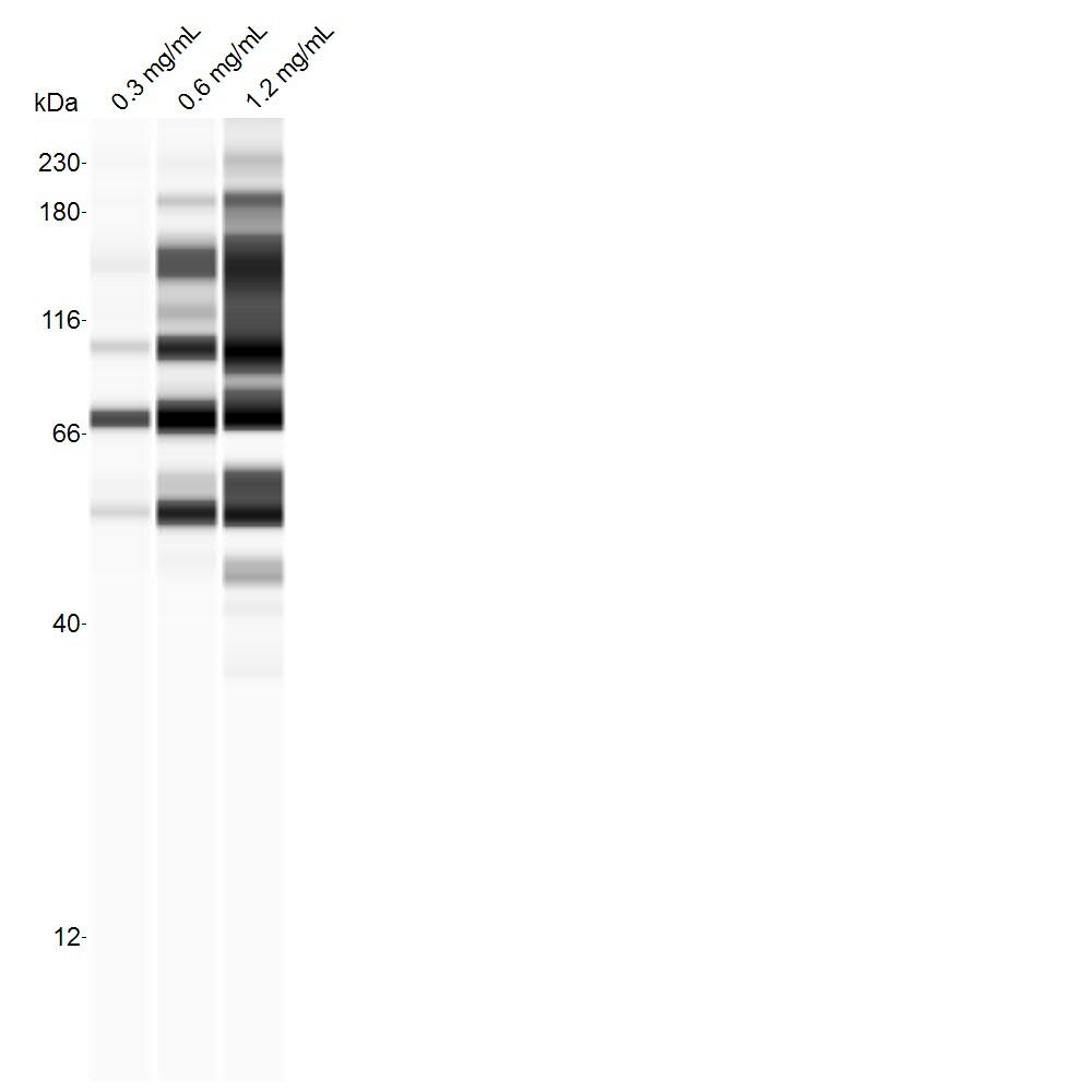
Application: Simple WesternSample Tested: cell line, whole cell lysateSpecies: RatVerified Customer | Posted 08/18/2017B103 whole-cell lysates (concentrations indicated), probed with 1:25 dilution of antibody on the standard separation matrix (12-230 kDa).

There are no reviews that match your criteria.
Protocols
Find general support by application which include: protocols, troubleshooting, illustrated assays, videos and webinars.
- Antigen Retrieval Protocol (PIER)
- Antigen Retrieval for Frozen Sections Protocol
- Appropriate Fixation of IHC/ICC Samples
- Cellular Response to Hypoxia Protocols
- Chromogenic IHC Staining of Formalin-Fixed Paraffin-Embedded (FFPE) Tissue Protocol
- Chromogenic Immunohistochemistry Staining of Frozen Tissue
- ClariTSA™ Fluorophore Kits
- Detection & Visualization of Antibody Binding
- ELISA Sample Preparation & Collection Guide
- ELISA Troubleshooting Guide
- Fluorescent IHC Staining of Frozen Tissue Protocol
- Graphic Protocol for Heat-induced Epitope Retrieval
- Graphic Protocol for the Preparation and Fluorescent IHC Staining of Frozen Tissue Sections
- Graphic Protocol for the Preparation and Fluorescent IHC Staining of Paraffin-embedded Tissue Sections
- Graphic Protocol for the Preparation of Gelatin-coated Slides for Histological Tissue Sections
- How to Run an R&D Systems DuoSet ELISA
- How to Run an R&D Systems Quantikine ELISA
- How to Run an R&D Systems Quantikine™ QuicKit™ ELISA
- ICC Cell Smear Protocol for Suspension Cells
- ICC Immunocytochemistry Protocol Videos
- ICC for Adherent Cells
- IHC Sample Preparation (Frozen sections vs Paraffin)
- Immunocytochemistry (ICC) Protocol
- Immunocytochemistry Troubleshooting
- Immunofluorescence of Organoids Embedded in Cultrex Basement Membrane Extract
- Immunofluorescent IHC Staining of Formalin-Fixed Paraffin-Embedded (FFPE) Tissue Protocol
- Immunohistochemistry (IHC) and Immunocytochemistry (ICC) Protocols
- Immunohistochemistry Frozen Troubleshooting
- Immunohistochemistry Paraffin Troubleshooting
- Preparing Samples for IHC/ICC Experiments
- Preventing Non-Specific Staining (Non-Specific Binding)
- Primary Antibody Selection & Optimization
- Protocol for Heat-Induced Epitope Retrieval (HIER)
- Protocol for Making a 4% Formaldehyde Solution in PBS
- Protocol for VisUCyte™ HRP Polymer Detection Reagent
- Protocol for the Fluorescent ICC Staining of Cell Smears - Graphic
- Protocol for the Fluorescent ICC Staining of Cultured Cells on Coverslips - Graphic
- Protocol for the Preparation & Fixation of Cells on Coverslips
- Protocol for the Preparation and Chromogenic IHC Staining of Frozen Tissue Sections
- Protocol for the Preparation and Chromogenic IHC Staining of Frozen Tissue Sections - Graphic
- Protocol for the Preparation and Chromogenic IHC Staining of Paraffin-embedded Tissue Sections
- Protocol for the Preparation and Chromogenic IHC Staining of Paraffin-embedded Tissue Sections - Graphic
- Protocol for the Preparation and Fluorescent ICC Staining of Cells on Coverslips
- Protocol for the Preparation and Fluorescent ICC Staining of Non-adherent Cells
- Protocol for the Preparation and Fluorescent ICC Staining of Stem Cells on Coverslips
- Protocol for the Preparation and Fluorescent IHC Staining of Frozen Tissue Sections
- Protocol for the Preparation and Fluorescent IHC Staining of Paraffin-embedded Tissue Sections
- Protocol for the Preparation of Gelatin-coated Slides for Histological Tissue Sections
- Protocol for the Preparation of a Cell Smear for Non-adherent Cell ICC - Graphic
- Quantikine HS ELISA Kit Assay Principle, Alkaline Phosphatase
- Quantikine HS ELISA Kit Principle, Streptavidin-HRP Polymer
- R&D Systems Quality Control Western Blot Protocol
- Sandwich ELISA (Colorimetric) – Biotin/Streptavidin Detection Protocol
- Sandwich ELISA (Colorimetric) – Direct Detection Protocol
- TUNEL and Active Caspase-3 Detection by IHC/ICC Protocol
- The Importance of IHC/ICC Controls
- Troubleshooting Guide: ELISA
- Troubleshooting Guide: Immunohistochemistry
- Troubleshooting Guide: Western Blot Figures
- Western Blot Conditions
- Western Blot Protocol
- Western Blot Protocol for Cell Lysates
- Western Blot Troubleshooting
- Western Blot Troubleshooting Guide
- View all Protocols, Troubleshooting, Illustrated assays and Webinars
Loading...