TRF-2 Antibody (4A794.15) - BSA Free
Novus Biologicals | Catalog # NB100-56506
Key Product Details
Species Reactivity
Validated:
Human, Mouse, Rat, Deer, Marsupial
Cited:
Human, Mouse, Rat, Marsupial, Muntjac (deer)
Applications
Validated:
Immunohistochemistry, Immunohistochemistry-Paraffin, Immunohistochemistry-Frozen, Western Blot, Immunoblotting, ELISA, Flow Cytometry, Flow (Intracellular), Immunocytochemistry/ Immunofluorescence, Simple Western, Immunoprecipitation, Chromatin Immunoprecipitation (ChIP), Proximity Ligation Assay, CyTOF-ready
Cited:
Immunohistochemistry-Paraffin, Western Blot, Immunofluorescence, Immunocytochemistry, Immunocytochemistry/ Immunofluorescence, Immunoprecipitation, Chemotaxis, Proximity Ligation Assay
Label
Unconjugated
Antibody Source
Monoclonal Mouse IgG1 kappa Clone # 4A794.15
Format
BSA Free
Loading...
Product Specifications
Immunogen
This TRF-2 Antibody (4A794.15) was developed against Baculovirus expressed whole length TRF2 protein, used for immunizing mice and splenocytes used to generate the hybridoma clone (NP_005643).
Marker
Telomeres marker
Specificity
The TRF2 antibody recognizes full-length TRF2 as well as TRF2 forms lacking both the N-terminal basic domain (B) and the telobox Myb-like C-terminal DNA-binding domain (M). TRF2 forms missing the B and M domains are often referred to as mutant TRF2. Although the exact epitope recognized by the TRF2 antibody has not been mapped, the scientific literature indicates it is in the D or L domain, but not in the B or M domain.
Clonality
Monoclonal
Host
Mouse
Isotype
IgG1 kappa
Theoretical MW
59.6 kDa.
Disclaimer note: The observed molecular weight of the protein may vary from the listed predicted molecular weight due to post translational modifications, post translation cleavages, relative charges, and other experimental factors.
Disclaimer note: The observed molecular weight of the protein may vary from the listed predicted molecular weight due to post translational modifications, post translation cleavages, relative charges, and other experimental factors.
Description
The TRF2 antibody is referred to as both clone 4A794.15 and 4A794 in the published literature. The TRF2 antibody recognizes full-length TRF2 as well as TRF2 forms lacking both the N-terminal basic domain (B) and the telobox Myb-like C-terminal DNA-binding domain (M). TRF2 forms missing the B and M domains are often referred to as mutant TRF2. Although the exact epitope recognized by the TRF2 antibody has not been mapped, the scientific literature indicates it is in the D or L domain, but not in the B or M domain.
Scientific Data Images for TRF-2 Antibody (4A794.15) - BSA Free
![Simple Western: TRF-2 Antibody (4A794.15)BSA Free [NB100-56506]](https://resources.rndsystems.com/images/products/TRF-2-Antibody-4A794-15-Simple-Western-NB100-56506-img0010.jpg "Simple Western: TRF-2 Antibody (4A794.15)BSA Free [NB100-56506]")
Simple Western: TRF-2 Antibody (4A794.15)BSA Free [NB100-56506]
Simple Western: TRF-2 Antibody (4A794.15) [NB100-56506] - Image shows a specific band for TRF2 in 1.0 mg/mL of HeLa lysate. This experiment was performed under reducing conditions using the 12-230 kDa separation system. Non-specific interaction with the 230 kDa Simple Western standard may be seen with this antibody.![Western Blot: TRF-2 Antibody (4A794.15)BSA Free [NB100-56506]](https://resources.rndsystems.com/images/products/TRF-2-Antibody-4A794-15-Western-Blot-NB100-56506-img0008.jpg "Western Blot: TRF-2 Antibody (4A794.15)BSA Free [NB100-56506]")
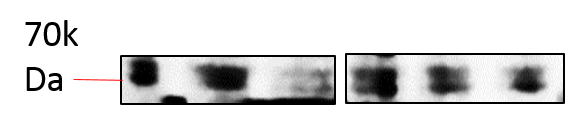
Western Blot: TRF-2 Antibody (4A794.15)BSA Free [NB100-56506]
Western Blot: TRF-2 Antibody (4A794.15) [NB100-56506] - Analysis in human Jurkat cell lysate at 2 ug/mL. Goat anti-mouse Ig HRP secondary antibody and PicoTect ECL substrate solution were used.![Immunohistochemistry-Paraffin: TRF-2 Antibody (4A794.15) - BSA Free [NB100-56506]](https://resources.rndsystems.com/images/products/TRF-2-Antibody-4A794-15-Immunohistochemistry-Paraffin-NB100-56506-img0012.jpg "Immunohistochemistry-Paraffin: TRF-2 Antibody (4A794.15) - BSA Free [NB100-56506]")
Immunohistochemistry-Paraffin: TRF-2 Antibody (4A794.15) - BSA Free [NB100-56506]
Immunohistochemistry-Paraffin: TRF-2 Antibody (4A794.15) [NB100-56506] - Transitional cell carcinoma, urinary bladder, stained with TRF2 antibody (4 ug/mL), peroxidase-conjugate and DAB chromogen. Note specific nuclear staining. Tumor/normal adjacent tissue array slide was used for this test. Staining of formalin-fixed tissues is enhanced by boiling tissue sections in 10 mM sodium citrate buffer, pH 6.0 for 10-20 min followed by cooling at RT for 20 min.![Immunocytochemistry/ Immunofluorescence: TRF-2 Antibody (4A794.15) - BSA Free [NB100-56506]](https://resources.rndsystems.com/images/products/TRF-2-Antibody-4A794-15-Immunocytochemistry-Immunofluorescence-NB100-56506-img0018.jpg "Immunocytochemistry/ Immunofluorescence: TRF-2 Antibody (4A794.15) - BSA Free [NB100-56506]")
Immunocytochemistry/ Immunofluorescence: TRF-2 Antibody (4A794.15) - BSA Free [NB100-56506]
TRF-2-Antibody-4A794-15-Immunocytochemistry-Immunofluorescence-NB100-56506-img0018.jpg![Flow Cytometry: TRF-2 Antibody (4A794.15) - BSA Free [NB100-56506]](https://resources.rndsystems.com/images/products/TRF-2-Antibody-4A794-15-Flow-Cytometry-NB100-56506-img0001.jpg "Flow Cytometry: TRF-2 Antibody (4A794.15) - BSA Free [NB100-56506]")
Flow Cytometry: TRF-2 Antibody (4A794.15) - BSA Free [NB100-56506]
Flow Cytometry: TRF-2 Antibody (4A794.15) [NB100-56506] - Intracellular flow cytometric analysis of TRF2 in 10^6 human Jurkat cells using 0.1 ug of NB100-56506. The shaded histogram represents cells alone, blue represents isotype control and red represents NB100-56506, anti-TRF2.![Western Blot: TRF-2 Antibody (4A794.15)BSA Free [NB100-56506]](https://resources.rndsystems.com/images/products/TRF-2-Antibody-4A794-15-Western-Blot-NB100-56506-img0016.jpg "Western Blot: TRF-2 Antibody (4A794.15)BSA Free [NB100-56506]")
Western Blot: TRF-2 Antibody (4A794.15)BSA Free [NB100-56506]
Western Blot: TRF-2 Antibody (4A794.15) [NB100-56506] - Total protein from mouse 3T3 cells was separated on a 12% gel by SDS-PAGE, transferred to PVDF membrane and blocked in 5% non-fat milk in TBST. The membrane was probed with 2.0 ug/mL anti-TRF2 in 1% non-fat milk in TBST and detected with an anti-mouse HRP secondary antibody using chemiluminescence.![Immunocytochemistry/ Immunofluorescence: TRF-2 Antibody (4A794.15) - BSA Free [NB100-56506]](https://resources.rndsystems.com/images/products/TRF-2-Antibody-4A794-15-Immunocytochemistry-Immunofluorescence-NB100-56506-img0019.jpg "Immunocytochemistry/ Immunofluorescence: TRF-2 Antibody (4A794.15) - BSA Free [NB100-56506]")
Immunocytochemistry/ Immunofluorescence: TRF-2 Antibody (4A794.15) - BSA Free [NB100-56506]
TRF-2-Antibody-4A794-15-Immunocytochemistry-Immunofluorescence-NB100-56506-img0019.jpg![Flow Cytometry: TRF-2 Antibody (4A794.15) - BSA Free [NB100-56506]](https://resources.rndsystems.com/images/products/TRF-2-Antibody-4A794-15-Flow-Cytometry-NB100-56506-img0020.jpg "Flow Cytometry: TRF-2 Antibody (4A794.15) - BSA Free [NB100-56506]")
Flow Cytometry: TRF-2 Antibody (4A794.15) - BSA Free [NB100-56506]
Flow Cytometry: TRF-2 Antibody (4A794.15) [NB100-56506] - An intracellular stain was performed on A431 cells with TRF2 Antibody [4A794.15] NB100-56506 (blue) and a matched isotype control (orange). Cells were fixed with 4% PFA and then permeabilized with 0.1% saponin. Cells were incubated in an antibody dilution of 1.0 ug/mL for 30 minutes at room temperature, followed by Mouse IgG (H+L) Cross-Adsorbed Secondary Antibody, Dylight 550 (35503, Thermo Fisher).![Immunocytochemistry/ Immunofluorescence: TRF-2 Antibody (4A794.15) - BSA Free [NB100-56506]](https://resources.rndsystems.com/images/products/TRF-2-Antibody-4A794-15-Immunocytochemistry-Immunofluorescence-NB100-56506-img0005.jpg "Immunocytochemistry/ Immunofluorescence: TRF-2 Antibody (4A794.15) - BSA Free [NB100-56506]")
Immunocytochemistry/ Immunofluorescence: TRF-2 Antibody (4A794.15) - BSA Free [NB100-56506]
Immunocytochemistry/Immunofluorescence: TRF-2 Antibody (4A794.15) [NB100-56506] - Staining of TRF2-bound telomeres in human HeLa cells (Courtesy of Fotiadou, et al, 2004).![Immunocytochemistry/ Immunofluorescence: TRF-2 Antibody (4A794.15) - BSA Free [NB100-56506]](https://resources.rndsystems.com/images/products/TRF-2-Antibody-4A794-15-Immunocytochemistry-Immunofluorescence-NB100-56506-img0017.jpg "Immunocytochemistry/ Immunofluorescence: TRF-2 Antibody (4A794.15) - BSA Free [NB100-56506]")
Immunocytochemistry/ Immunofluorescence: TRF-2 Antibody (4A794.15) - BSA Free [NB100-56506]
Immunocytochemistry/Immunofluorescence: TRF-2 Antibody (4A794.15) [NB100-56506] - HeLa cells were fixed for 10 minutes using 10% formalin and then permeabilized for 5 minutes using 1X PBS + 0.5% Triton-X100. The cells were incubated with anti-TRF-2 (4A794.15) conjugated to FITC [NB100-56506F] at 5 ug/mL for 1 hour at room temperature. Nuclei were counterstained with DAPI (Blue). Cells were imaged using a 40X objective. - BSA Free [NB100-56506] -")
Immunocytochemistry/ Immunofluorescence: TRF-2 Antibody (4A794.15) - BSA Free [NB100-56506] -
Immunocytochemistry/ Immunofluorescence: TRF-2 Antibody (4A794.15) - BSA Free [NB100-56506] - ZBTB48‐dependent loss of MTFP1 phenocopies MTFP1 depletionFluorescence microscopy analysis of the structure & localization of the mitochondrial network in HeLa WT & ZBTB48 KO clones. Mitochondria are marked with the MitoTracker dye (red), & nuclei are counterstained with DAPI (blue). Scale bars represent 20 μm.The same analysis as in (A) for U2OS WT & ZBTB48 KO clones. Image collected & cropped by CiteAb from the following publication (https://pubmed.ncbi.nlm.nih.gov/28500257), licensed under a CC-BY license. Not internally tested by Novus Biologicals. - BSA Free [NB100-56506] -")
Western Blot: TRF-2 Antibody (4A794.15) - BSA Free [NB100-56506] -
Western Blot: TRF-2 Antibody (4A794.15) - BSA Free [NB100-56506] - TRF2 delta B delta M expression induces chromosome end-to-end fusions in all inducible cell lines(A) Immunoblots of MCF-10A, TO, SH-TO & SV-TO cell lines with & without DOX & HEK 293T. After 96 h of DOX treatment, the inducible cell lines expressed the truncated TRF2 delta B delta M protein (50 kDa); in contrast, uninduced cell lines, MCF-10A parental cell line & HEK 293T cells displayed only the endogen TRF2 protein (66 kDa). Lamin B1 was used as loading control. (B) After sustained expression of TRF2 delta B delta M for 96 h there was a significant increase in aberrant metaphases only in TO cells when compared to uninduced matched cells. (C) Nevertheless, the efficacy of the inducible system was validated by the statistical increase of cells with end-to-end fusions in all inducible cell lines. (D) Moreover, a high incidence of fusions per cell was found in all modified cell lines after sustained TRF2 depletion. Data was presented as mean + SEM. (E) Example of TO, SH-TO & SV-TO karyotypes after 96 h of TRF2 delta B delta M expression. Open arrows indicate clonal aberrations in the parental MCF-10A cell line. Insets in the karyotype show rearranged chromosomes stained with centromeric (green) & telomeric (red) PNA probes. Note the presence of telomere FISH signals at the fusion point of chromatid- or chromosome-type end-to-end fusions. Image collected & cropped by CiteAb from the following publication (https://www.oncotarget.com/lookup/doi/10.18632/oncotarget.25502), licensed under a CC-BY license. Not internally tested by Novus Biologicals. - BSA Free [NB100-56506] -")
Immunocytochemistry/ Immunofluorescence: TRF-2 Antibody (4A794.15) - BSA Free [NB100-56506] -
Immunocytochemistry/ Immunofluorescence: TRF-2 Antibody (4A794.15) - BSA Free [NB100-56506] - ZBTB48 ZnF11 necessary to bind to telomeres. Sequence‐specific DNA pull‐downs w/ either telomeric (TTAGGG)/a control sequence (GTGAGT) for FLAG‐ZBTB48 WT, domain deletion constructs for different zinc finger & combinations of deletion constructs w/ ZnF10/11 point mutants. Domain structures indicated - right.Sequence‐specific DNA pull‐downs for FLAG‐ZBTB48 WT & ZnF11 point mutant for telomeric repeat sequences of different phyla (green) & their respective scrambled controls (blue).Protein expression analysis of ZBTB48 by WB for cell lines used. GAPDH serves as a loading control.IF stainings for exogenous FLAG‐ZBTB48 WT & point mutants for ZnF10 & ZnF11 in U2OS cells. The same analysis as in Fig 1E performed & average co‐localization frequencies shown (n = 24–37 cells).Co‐localization analysis of endogenous ZBTB48/exogenous FLAG‐ZBTB48 WT w/ TRF2 in HeLa cells by IF (IF) staining. Image illustrating co‐localization between ZBTB48/FLAG‐ZBTB48 WT (green) & TRF2 (red) as a marker for telomeres is shown w/ DAPI (blue) used as nuclear counterstain. Co‐localization events indicated by white arrows. Quantification of frequency of co‐localization events (right) done after 3D reconstruction of acquired z‐stacks (n = 30 cells).Co‐localization analysis of endogenous ZBTB48/exogenous FLAG‐ZBTB48 WT w/ TRF2 in HeLa 1.3 cells by IF (IF) staining analogous to (E) (n = 30 cells).Co‐localization analysis of endogenous ZBTB48/exogenous FLAG‐ZBTB48 WT w/ TRF2 in HT1080 super‐telomerase cells by IF (IF) staining analogous to (E) (n = 30 cells).Data information: (D–G) Scale bars = 5 μm. Error bars indicate standard deviations, & P‐values based on Student's t‐test.Source data available online for this figure. Image collected & cropped by CiteAb from following publication (https://pubmed.ncbi.nlm.nih.gov/28500257), licensed under a CC-BY license. Not internally tested by Novus Biologicals.Applications for TRF-2 Antibody (4A794.15) - BSA Free
Application
Recommended Usage
Chromatin Immunoprecipitation (ChIP)
1:10-1:500
ELISA
2 ug/ml
Flow Cytometry
0.1 ug/10^6 cells
Immunoblotting
reported in scientific literature (PMID 23708666)
Immunocytochemistry/ Immunofluorescence
1:10 - 1:500
Immunohistochemistry
1:10 - 1:500
Immunohistochemistry-Frozen
5 ug/ml
Immunohistochemistry-Paraffin
1:200
Immunoprecipitation
2 ug/10^6 cells
Proximity Ligation Assay
reported in scientific literature (PMID 27366950)
Simple Western
1:50
Western Blot
2-4 ug/ml
Application Notes
TRF-2 may be detected as a single band or as a doublet in Western blot. Okabe (2000) described the doublet as 65 and 69 kDa using clone 4A794.15. Observed molecular weights could vary depending on molecular weight standards used and gel conditions.
In Simple Western only 10 - 15 uL of the recommended dilution is used per data point.
See Simple Western Antibody Database for Simple Western validation: Tested in HeLa lysate 1.0 mg/mL, separated by Size, antibody dilution of 1:50, apparent MW was 80 kDa. Separated by Size-Wes, Sally Sue/Peggy Sue.
The observed molecular weight of the protein may vary from the listed predicted molecular weight due to post translational modifications, post translation cleavages, relative charges, and other experimental factors.
Reviewed Applications
Read 1 review rated 4 using NB100-56506 in the following applications:
Flow Cytometry Panel Builder
Bio-Techne Knows Flow Cytometry
Save time and reduce costly mistakes by quickly finding compatible reagents using the Panel Builder Tool.
Advanced Features
- Spectra Viewer - Custom analysis of spectra from multiple fluorochromes
- Spillover Popups - Visualize the spectra of individual fluorochromes
- Antigen Density Selector - Match fluorochrome brightness with antigen density

Formulation, Preparation, and Storage
Purification
Protein G purified
Formulation
PBS
Format
BSA Free
Preservative
0.02% Sodium Azide
Concentration
1 mg/ml
Shipping
The product is shipped with polar packs. Upon receipt, store it immediately at the temperature recommended below.
Stability & Storage
Store at 4C short term. Aliquot and store at -20C long term. Avoid freeze-thaw cycles.
Background: TRF-2
Both TRF2 and TRF1 bind to telomeric double stranded 5'-TTAGGG-3' DNA repeats, then recruit RAP1, TIN2, TPP1, and POT1 for the assembly of the shelterin complex. The telomeric association of TRF2 is greatly increased in the S phase of the cell cycle (2). Loss of TRF2 leads to telomere shortening, the DNA damage response, chromosomal instability, and replicative senescence. Interestingly, the contribution of TRF2 to telomere shortening via a telomerase-independent mechanism has also been reported (3). In conjunction with the exonuclease, Apollo, TRF2 protects telomeres during replication and negatively regulates the accumulation of DNA topoisomerase (TOP1, TOP2A and TOP2B).
TRF2 has been implicated in cancer, shown to be a major oncogene in telomerase-deficient mice. A link to Werner syndrome, a premature aging disease caused by the loss of WRN, has been reported based on TRF2 recruitment of WRN for processing of telomeric DNA (4). TRF2 expression is increased during human embryonic stem cell differentiation and has been shown to interact with Repressor Element-1 Silencing Transcription Factor (REST), protecting it from proteasomal degradation (5).
References
1. Grammatikakis, I., Zhang, P., Mattson, M. P., & Gorospe, M. (2016). The long and the short of TRF2 in neurogenesis. Cell cycle (Georgetown, Tex.), 15(22), 3026-3032. PMID: 27565210
2. Li, F., Kim, H., Ji, Z., Zhang, T., Chen, B., Ge, Y., Hu, Y., Feng, X., Han, X., Xu, H., Zhang, Y., Yu, H., Liu, D., Ma, W., & Songyang, Z. (2018). The BUB3-BUB1 Complex Promotes Telomere DNA Replication. Molecular cell, 70(3), 395-407. PMID: 29727616
3. Ancelin, K., Brunori, M., Bauwens, S., Koering, C. E., Brun, C., Ricoul, M., Pommier, J. P., Sabatier, L., & Gilson, E. (2002). Targeting assay to study the cis functions of human telomeric proteins: evidence for inhibition of telomerase by TRF1 and for activation of telomere degradation by TRF2. Molecular and cellular biology, 22(10), 3474-3487. PMID: 11971978
4. Machwe A, Xiao L, & Orren DK. (2004) TRF2 recruits the Werner syndrome (WRN) exonuclease for processing of telomeric DNA. Oncogene. 23(1):149-56. PMID: 14712220.
5. Diotti, R., & Loayza, D. (2011). Shelterin complex and associated factors at human telomeres. Nucleus (Austin, Tex.), 2(2), 119-135. PMID: 21738835
Long Name
Telomeric Repeat Binding Factor 2
Alternate Names
TERF2, TRBF2, TRF2, 4A794, 4A794 trf2, 4A794.15, 4A794.15 trf2, anti-trf2 4A794, anti-trf2 4A794.15, clone 4A794, clone 4A794.15, Telomeric repeat binding protein 2
Gene Symbol
TERF2
UniProt
Additional TRF-2 Products
Product Documents for TRF-2 Antibody (4A794.15) - BSA Free
Certificate of Analysis
To download a Certificate of Analysis, please enter a lot or batch number in the search box below.
Product Specific Notices for TRF-2 Antibody (4A794.15) - BSA Free
This product is for research use only and is not approved for use in humans or in clinical diagnosis. Primary Antibodies are guaranteed for 1 year from date of receipt.
Related Research Areas
Citations for TRF-2 Antibody (4A794.15) - BSA Free
 Powered by Bioz
Customer Reviews for TRF-2 Antibody (4A794.15) - BSA Free (1)
4 out of 5
1 Customer Rating
Have you used TRF-2 Antibody (4A794.15) - BSA Free?
Submit a review and receive an Amazon gift card!
$25/€18/£15/$25CAN/¥2500 Yen for a review with an image
$10/€7/£6/$10CAN/¥1110 Yen for a review without an image
Submit a review
Customer Images
Showing
1
-
1 of
1 review
Showing All
Filter By:
-
Application: Western BlotSample Tested: 293T lysateSpecies: HumanVerified Customer | Posted 09/08/2017

There are no reviews that match your criteria.
Protocols
View specific protocols for TRF-2 Antibody (4A794.15) - BSA Free (NB100-56506):
Protocol for Flow Cytometry Intracellular Staining
Sample Preparation.
1. Grow cells to 60-85% confluency. Flow cytometry requires between 2 x 10⁵ and 1 x 10⁶ cells for optimal performance.
2. If cells are adherent, harvest gently by washing once with staining buffer and then scraping. Avoid using trypsin as this can disrupt certain epitopes of interest. If enzymatic harvest is required, use Accutase, Collagenase, or TrypLE Express for a less damaging option.
3. Reserve 100 uL for counting, then transfer cell volume into a 50 mL conical tube and centrifuge for 8 minutes at 400 RCF.
a. Count cells using a hemocytometer and a 1:1 trypan blue exclusion stain to determine cell viability before starting the flow protocol. If cells appear blue, do not proceed.
4. Re-suspend cells to a concentration of 1 x 10⁶ cells/mL in staining buffer (NBP2-26247).
5. Aliquot out 100 uL samples in accordance with your experimental samples.
Tip: When cell surface and intracellular staining are required in the same sample, it is advisable that the cell surface staining be performed first since the fixation and permeabilization steps might reduce the availability of surface antigens.
Intracellular Staining.
Tip: When performing intracellular staining, it is important to use appropriate fixation and permeabilization reagents based upon the target and its subcellular location. Generally, our Intracellular Flow Assay Kit (NBP2-29450) is a good place to start as it contains an optimized combination of reagents for intracellular staining as well as an inhibitor of intracellular protein transport (necessary if staining secreted proteins). Certain targets may require more gentle or transient permeabilization protocols such as the commonly employed methanol or saponin-based methods.
Protocol for Cytoplasmic Targets:
1. Fix the cells by adding 100 uL fixation solution (such as 4% PFA) to each sample for 10-15 minutes.
2. Permeabilize cells by adding 100 uL of a permeabilization buffer to every 1 x 10⁶ cells present in the sample. Mix well and incubate at room temperature for 15 minutes.
a. For cytoplasmic targets, use a gentle permeabilization solution such as 1X PBS + 0.5% Saponin or 1X PBS + 0.5% Tween-20.
b. To maintain the permeabilized state throughout your experiment, use staining buffer + 0.1% of the permeabilization reagent (i.e. 0.1% Tween-20 or 0.1% Saponin).
3. Following the 15 minute incubation, add 2 mL of the staining buffer + 0.1% permeabilizer to each sample.
4. Centrifuge for 1 minute at 400 RCF.
5. Discard supernatant and re-suspend in 100 uL of staining buffer + 0.1% permeabilizer.
6. Add appropriate amount of each antibody (eg. 1 test or 1 ug per sample, as experimentally determined).
7. Mix well and incubate at room temperature for 30 minutes- 1 hour. Gently mix samples every 10-15 minutes.
8. Following the primary/conjugate incubation, add 1-2 mL/sample of staining buffer +0.1% permeabilizer and centrifuge for 1 minute at 400 RCF.
9. Wash twice by re-suspending cells in staining buffer (2 mL for tubes or 200 uL for wells) and centrifuging at 400 RCF for 5 minutes. Discard supernatant.
10. Add appropriate amount of secondary antibody (as experimentally determined) to each sample.
11. Incubate at room temperature in dark for 20 minutes.
12. Add 1-2 mL of staining buffer and centrifuge at 400 RCF for 1 minute and discard supernatant.
13. Wash twice by re-suspending cells in staining buffer (2 mL for tubes or 200 uL for wells) and centrifuging at 400 RCF for 5 minutes. Discard supernatant.
14. Resuspend in an appropriate volume of staining buffer (usually 500 uL per sample) and proceed with analysis on your flow cytometer.
Immunocytochemistry Protocol
Culture cells to appropriate density in 35 mm culture dishes or 6-well plates.
1. Remove culture medium and wash the cells briefly in PBS. Add 10% formalin to the dish and fix at room temperature for 10 minutes.
2. Remove the formalin and wash the cells in PBS.
3. Permeablize the cells with 0.1% Triton X100 or other suitable detergent for 10 min.
4. Remove the permeablization buffer and wash three times for 10 minutes each in PBS. Be sure to not let the specimen dry out.
5. To block nonspecific antibody binding, incubate in 10% normal goat serum from 1 hour to overnight at room temperature.
6. Add primary antibody at appropriate dilution and incubate overnight at 4C.
7. Remove primary antibody and replace with PBS. Wash three times for 10 minutes each.
8. Add secondary antibody at appropriate dilution. Incubate for 1 hour at room temperature.
9. Remove secondary antibody and replace with PBS. Wash three times for 10 minutes each.
10. Counter stain DNA with DAPi if required.
Immunohistochemistry-Paraffin Embedded Sections
Antigen Unmasking:
Bring slides to a boil in 10 mM sodium citrate buffer (pH 6.0) then maintain at a sub-boiling temperature for 10 minutes. Cool slides on bench-top for 30 minutes (keep slides in the sodium citrate buffer at all times).
Staining:
1. Wash sections in deionized water three times for 5 minutes each.
2. Wash sections in PBS for 5 minutes.
3. Block each section with 100-400 ul blocking solution (1% BSA in PBS) for 1 hour at room temperature.
4. Remove blocking solution and add 100-400 ul diluted primary antibody. Incubate overnight at 4 C.
5. Remove antibody solution and wash sections in wash buffer three times for 5 minutes each.
6. Add 100-400 ul HRP polymer conjugated secondary antibody. Incubate 30 minutes at room temperature.
7. Wash sections three times in wash buffer for 5 minutes each.
8. Add 100-400 ul DAB substrate to each section and monitor staining closely.
9. As soon as the sections develop, immerse slides in deionized water.
10. Counterstain sections in hematoxylin.
11. Wash sections in deionized water two times for 5 minutes each.
12. Dehydrate sections.
13. Mount coverslips.
Find general support by application which include: protocols, troubleshooting, illustrated assays, videos and webinars.
- 7-Amino Actinomycin D (7-AAD) Cell Viability Flow Cytometry Protocol
- Antigen Retrieval Protocol (PIER)
- Antigen Retrieval for Frozen Sections Protocol
- Appropriate Fixation of IHC/ICC Samples
- Cellular Response to Hypoxia Protocols
- ChIP Protocol Video
- Chromatin Immunoprecipitation (ChIP) Protocol
- Chromatin Immunoprecipitation Protocol
- Chromogenic IHC Staining of Formalin-Fixed Paraffin-Embedded (FFPE) Tissue Protocol
- Chromogenic Immunohistochemistry Staining of Frozen Tissue
- ClariTSA™ Fluorophore Kits
- Detection & Visualization of Antibody Binding
- ELISA Sample Preparation & Collection Guide
- ELISA Troubleshooting Guide
- Extracellular Membrane Flow Cytometry Protocol
- Flow Cytometry Protocol for Cell Surface Markers
- Flow Cytometry Protocol for Staining Membrane Associated Proteins
- Flow Cytometry Staining Protocols
- Flow Cytometry Troubleshooting Guide
- Fluorescent IHC Staining of Frozen Tissue Protocol
- Graphic Protocol for Heat-induced Epitope Retrieval
- Graphic Protocol for the Preparation and Fluorescent IHC Staining of Frozen Tissue Sections
- Graphic Protocol for the Preparation and Fluorescent IHC Staining of Paraffin-embedded Tissue Sections
- Graphic Protocol for the Preparation of Gelatin-coated Slides for Histological Tissue Sections
- How to Run an R&D Systems DuoSet ELISA
- How to Run an R&D Systems Quantikine ELISA
- How to Run an R&D Systems Quantikine™ QuicKit™ ELISA
- ICC Cell Smear Protocol for Suspension Cells
- ICC Immunocytochemistry Protocol Videos
- ICC for Adherent Cells
- IHC Sample Preparation (Frozen sections vs Paraffin)
- Immunocytochemistry (ICC) Protocol
- Immunocytochemistry Troubleshooting
- Immunofluorescence of Organoids Embedded in Cultrex Basement Membrane Extract
- Immunofluorescent IHC Staining of Formalin-Fixed Paraffin-Embedded (FFPE) Tissue Protocol
- Immunohistochemistry (IHC) and Immunocytochemistry (ICC) Protocols
- Immunohistochemistry Frozen Troubleshooting
- Immunohistochemistry Paraffin Troubleshooting
- Immunoprecipitation Protocol
- Intracellular Flow Cytometry Protocol Using Alcohol (Methanol)
- Intracellular Flow Cytometry Protocol Using Detergents
- Intracellular Nuclear Staining Flow Cytometry Protocol Using Detergents
- Intracellular Staining Flow Cytometry Protocol Using Alcohol Permeabilization
- Intracellular Staining Flow Cytometry Protocol Using Detergents to Permeabilize Cells
- Preparing Samples for IHC/ICC Experiments
- Preventing Non-Specific Staining (Non-Specific Binding)
- Primary Antibody Selection & Optimization
- Propidium Iodide Cell Viability Flow Cytometry Protocol
- Protocol for Heat-Induced Epitope Retrieval (HIER)
- Protocol for Liperfluo
- Protocol for Making a 4% Formaldehyde Solution in PBS
- Protocol for VisUCyte™ HRP Polymer Detection Reagent
- Protocol for the Characterization of Human Th22 Cells
- Protocol for the Characterization of Human Th9 Cells
- Protocol for the Fluorescent ICC Staining of Cell Smears - Graphic
- Protocol for the Fluorescent ICC Staining of Cultured Cells on Coverslips - Graphic
- Protocol for the Preparation & Fixation of Cells on Coverslips
- Protocol for the Preparation and Chromogenic IHC Staining of Frozen Tissue Sections
- Protocol for the Preparation and Chromogenic IHC Staining of Frozen Tissue Sections - Graphic
- Protocol for the Preparation and Chromogenic IHC Staining of Paraffin-embedded Tissue Sections
- Protocol for the Preparation and Chromogenic IHC Staining of Paraffin-embedded Tissue Sections - Graphic
- Protocol for the Preparation and Fluorescent ICC Staining of Cells on Coverslips
- Protocol for the Preparation and Fluorescent ICC Staining of Non-adherent Cells
- Protocol for the Preparation and Fluorescent ICC Staining of Stem Cells on Coverslips
- Protocol for the Preparation and Fluorescent IHC Staining of Frozen Tissue Sections
- Protocol for the Preparation and Fluorescent IHC Staining of Paraffin-embedded Tissue Sections
- Protocol for the Preparation of Gelatin-coated Slides for Histological Tissue Sections
- Protocol for the Preparation of a Cell Smear for Non-adherent Cell ICC - Graphic
- Protocol: Annexin V and PI Staining by Flow Cytometry
- Protocol: Annexin V and PI Staining for Apoptosis by Flow Cytometry
- Quantikine HS ELISA Kit Assay Principle, Alkaline Phosphatase
- Quantikine HS ELISA Kit Principle, Streptavidin-HRP Polymer
- R&D Systems Quality Control Western Blot Protocol
- Sandwich ELISA (Colorimetric) – Biotin/Streptavidin Detection Protocol
- Sandwich ELISA (Colorimetric) – Direct Detection Protocol
- TUNEL and Active Caspase-3 Detection by IHC/ICC Protocol
- The Importance of IHC/ICC Controls
- Troubleshooting Guide: ELISA
- Troubleshooting Guide: Fluorokine Flow Cytometry Kits
- Troubleshooting Guide: Immunohistochemistry
- Troubleshooting Guide: Western Blot Figures
- Western Blot Conditions
- Western Blot Protocol
- Western Blot Protocol for Cell Lysates
- Western Blot Troubleshooting
- Western Blot Troubleshooting Guide
- View all Protocols, Troubleshooting, Illustrated assays and Webinars
FAQs for TRF-2 Antibody (4A794.15) - BSA Free
Showing
1
-
4 of
4 FAQs
Showing All
-
Q: Are the TRF-2 antibodies validated in Simple Western?
A: Yes, we offer a 2 TRF-2 antibodies that have been tested in Simple Western: NB100-56506 and NB110-57130.
-
Q: Does TRF-2 antibodies comes in lyophilized form?
A: we carry 2 TERF2/TRBF2 antibodies in lyophilized form: AF5635, MAB5635.
-
Q: What is the immunogen sequence of this TRF-2 antibody?
A: The whole length of Baculovirus expressed TRF-2 protein.
-
Q: What the theoretical molecular weight for TRF-2 antibodies?
A: The TMW of TRF-2 antibodies is approximately 55 - 56 kDa.
-
Q: Are the TRF-2 antibodies validated in Simple Western?
A: Yes, we offer a 2 TRF-2 antibodies that have been tested in Simple Western: NB100-56506 and NB110-57130.
-
Q: Does TRF-2 antibodies comes in lyophilized form?
A: we carry 2 TERF2/TRBF2 antibodies in lyophilized form: AF5635, MAB5635.
-
Q: What is the immunogen sequence of this TRF-2 antibody?
A: The whole length of Baculovirus expressed TRF-2 protein.
-
Q: What the theoretical molecular weight for TRF-2 antibodies?
A: The TMW of TRF-2 antibodies is approximately 55 - 56 kDa.
Loading...