Tryptophan hydroxylase 2 Antibody - BSA Free
Novus Biologicals | Catalog # NB100-74555
![Immunohistochemistry-Paraffin: Tryptophan hydroxylase 2 Antibody - BSA Free [NB100-74555]](https://resources.rndsystems.com/images/products/Tryptophan-hydroxylase-2-Antibody-Immunohistochemistry-Paraffin-NB100-74555-img0009.jpg "Immunohistochemistry-Paraffin: Tryptophan hydroxylase 2 Antibody - BSA Free [NB100-74555]")
Key Product Details
Validated by
Biological Validation
Species Reactivity
Validated:
Human, Mouse, Rat, Primate, Rabbit
Cited:
Human, Mouse, Rat, Primate
Applications
Validated:
Immunohistochemistry, Immunohistochemistry-Paraffin, Immunohistochemistry-Frozen, Immunohistochemistry Free-Floating, Western Blot, Flow Cytometry, Immunocytochemistry/ Immunofluorescence, Immunoprecipitation
Cited:
Immunohistochemistry, Immunohistochemistry-Frozen, Immunohistochemistry Free-Floating, Western Blot, Flow Cytometry, Immunocytochemistry/ Immunofluorescence, IF/IHC
Label
Unconjugated
Antibody Source
Polyclonal Rabbit IgG
Format
BSA Free
Loading...
Product Specifications
Immunogen
Synthetic peptide make to an internal portion of the mouse Tryptophan hydroxylase 2 protein (between amino acids 10-75) [UniProt Q8CGV2]
Reactivity Notes
Human reactivity is weak. Mouse reactivity reported in scientific literature (PMID: 16581041). Rat reactivity reported in scientific literature (PMID: 24066056).
Clonality
Polyclonal
Host
Rabbit
Isotype
IgG
Scientific Data Images for Tryptophan hydroxylase 2 Antibody - BSA Free
Immunohistochemistry-Paraffin: Tryptophan hydroxylase 2 Antibody - BSA Free [NB100-74555]
Immunohistochemistry-Paraffin: Tryptophan hydroxylase 2 Antibody [NB100-74555] - IHC analysis of a formalin fixed paraffin embedded tissue section of mouse brain using Tryptophan hydroxylase 2 antibody at 1:25 dilution. The staining was developed using HRP-DAB detection method and the nuclei were counterstained with hematoxylin. The antibody generated staining of the neurons.![Western Blot: Tryptophan hydroxylase 2 AntibodyBSA Free [NB100-74555]](https://resources.rndsystems.com/images/products/Tryptophan-hydroxylase-2-Antibody-Western-Blot-NB100-74555-img0010.jpg "Western Blot: Tryptophan hydroxylase 2 AntibodyBSA Free [NB100-74555]")
Western Blot: Tryptophan hydroxylase 2 AntibodyBSA Free [NB100-74555]
Western Blot: Tryptophan hydroxylase 2 Antibody [NB100-74555] - Total protein from mouse brain, stomach and human brain was separated on a 7.5% gel by SDS-PAGE, transferred to PVDF membrane and blocked in 5% non-fat milk in TBST. The membrane was probed with 2.0 ug/ml anti-Tryptophan Hydroxylase 2 in 1% non-fat milk in TBST and detected with an anti-rabbit HRP secondary antibody using chemiluminescence.![Immunocytochemistry/ Immunofluorescence: Tryptophan hydroxylase 2 Antibody - BSA Free [NB100-74555]](https://resources.rndsystems.com/images/products/Tryptophan-hydroxylase-2-Antibody-Immunocytochemistry-Immunofluorescence-NB100-74555-img0011.jpg "Immunocytochemistry/ Immunofluorescence: Tryptophan hydroxylase 2 Antibody - BSA Free [NB100-74555]")
![Immunocytochemistry/ Immunofluorescence: Tryptophan hydroxylase 2 Antibody - BSA Free [NB100-74555]](https://resources.rndsystems.com/images/products/Tryptophan-hydroxylase-2-Antibody-Immunocytochemistry-Immunofluorescence-NB100-74555-img0006.jpg "Immunocytochemistry/ Immunofluorescence: Tryptophan hydroxylase 2 Antibody - BSA Free [NB100-74555]")
Immunocytochemistry/ Immunofluorescence: Tryptophan hydroxylase 2 Antibody - BSA Free [NB100-74555]
Immunocytochemistry/Immunofluorescence: Tryptophan hydroxylase 2 Antibody [NB100-74555] - Analysis in mouse brain cells. Image courtesy of product review by Assaf Vestin.![Immunohistochemistry Free-Floating: Tryptophan hydroxylase 2 Antibody - BSA Free [NB100-74555]](https://resources.rndsystems.com/images/products/Tryptophan-hydroxylase-2-Antibody-Immunohistochemistry-Free-Floating-NB100-74555-img0008.jpg "Immunohistochemistry Free-Floating: Tryptophan hydroxylase 2 Antibody - BSA Free [NB100-74555]")
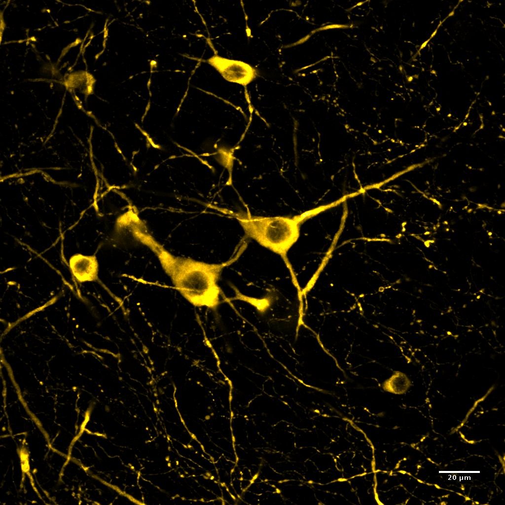
Immunohistochemistry Free-Floating: Tryptophan hydroxylase 2 Antibody - BSA Free [NB100-74555]
Immunohistochemistry Free-Floating: Tryptophan hydroxylase 2 Antibody [NB100-74555] - PFA-perfused mouse midbrain serotoninergic neurons. AlexaFluor488 used as secondary antibody. Neurons pseudocolored in yellow hot LUT. Image from verified customer review.![Immunoprecipitation: Tryptophan hydroxylase 2 Antibody - BSA Free [NB100-74555]](https://resources.rndsystems.com/images/products/Tryptophan-hydroxylase-2-Antibody-Immunoprecipitation-NB100-74555-img0007.jpg "Immunoprecipitation: Tryptophan hydroxylase 2 Antibody - BSA Free [NB100-74555]")
Immunoprecipitation: Tryptophan hydroxylase 2 Antibody - BSA Free [NB100-74555]
Immunoprecipitation: Tryptophan hydroxylase 2 Antibody [NB100-74555] - Analysis of raphe nuclei isolated from Sprague-Dawley rat brains using TPH2 Polyclonal Antibody. PA1-778 was coupled to magnetic beads and added to raphe nuclei whole cell extracts for 15 minutes at RT. Immunoprecipitated proteins were assessed by Western blotting (WB), using either mouse anti-TPH2 (Ms TPH2, Lane 2) or sheep anti-TPH2 antibodies (Sh TPH2, Lane 3). As a positive control for the Western blot, non-immunoprecipitated brain lysates were probed with the mouse anti-TPH2 antibody (Lane 1). As a specificity control, a blot containing immunoprecipitated proteins was probed with an antibody specific for Tyrosine Hydroxylase (TH, Lane 4). Data courtesy of the Innovators Program.
Immunohistochemistry: Tryptophan hydroxylase 2 Antibody - BSA Free [NB100-74555] -
The 5-HT and TPH2 in the raphe nuclei of the brain. The 5-HT- and TPH2-positive double stained cells in the dorsal raphe nuclei (A). Representative photomicrographs were taken at magnifications of 100×. Quantification of 5-HT and TPH2 (B). Data are expressed as the means ± SD (n = 3). *, p < 0.05; **, p < 0.01 compared with the vehicle group. 5-HT; 5-hydroxytryptamine, TPH2; tryptophan hydroxylase, DAPI; 4′,6-diamidine-2-phenylindole dihydrochloride.
Western Blot: Tryptophan hydroxylase 2 Antibody - BSA Free [NB100-74555] -
Enhanced serotonergic phenotypes of differentiated HT22 cells. (A) Immunofluorescence staining of SERT (green), 5-HT1aR (orange) and TPH2 (magenta) in hippocampal primary culture. DAPI staining (blue) indicates the nucleus. Scale bar = 50 μm. (B,C) SERT (green), 5-HT1aR (orange) and TPH2 (magenta) immunofluorescence staining (B) and western blotting (C) of HT22 cells grown in normal growth medium and differentiation medium for 3 d. DAPI staining (blue) indicates the nucleus. Scale bar = 25 μm. GAPDH was used as loading control. Data are representative of at least three independent experiments and are quantified using densitometric analysis. Statistical significance: *p < 0.05, **p < 0.01, ***p < 0.001 and ****p < 0.0001. Image collected and cropped by CiteAb from the following open publication (https://pubmed.ncbi.nlm.nih.gov/36703746), licensed under a CC-BY license. Not internally tested by Novus Biologicals.
Western Blot: Tryptophan hydroxylase 2 Antibody - BSA Free [NB100-74555] -
Pet-1 enhances the serotonergic phenotype in differentiated HT22 cells. (A) Immunofluorescence staining of Pet-1 (red) in hippocampal primary culture. DAPI staining (blue) indicates the nucleus. Scale bar = 50 μm. (B,C) Pet-1 (red) immunofluorescence staining (B) and western blotting (C) of HT22 cells grown in normal growth medium and differentiation medium for 3 d. DAPI staining (blue) indicates the nucleus. Scale bar = 25 μm. GAPDH is used as loading control. (D,E) Control siRNA and Pet-1 siRNA were transiently transfected in HT22 cells, changed with normal growth medium and differentiation medium for 3 d. Western blotting for analyzing Pet-1 (D) and SERT, 5-HT1aR, and TPH2 (E) expression. GAPDH, vinculin, and beta -actin were used as loading controls. Data are representative of at least three independent experiments and are quantified using densitometric analysis. Statistical significance: *p <.05, and ***p <.001. Image collected and cropped by CiteAb from the following open publication (https://pubmed.ncbi.nlm.nih.gov/36703746), licensed under a CC-BY license. Not internally tested by Novus Biologicals.
Western Blot: Tryptophan hydroxylase 2 Antibody - BSA Free [NB100-74555] -
Functional loss of 5-HT1Areceptor on the corticosterone and thermoregulatory responses. After fluoxetine injection for 4 weeks, the protein expression of TPH2 and c-Fos following 15 min of 8-OH-DPAT challenge was determined by western blot analysis in RN (A and B). Plasma corticosterone levels (C) or rectal temperature measurement (D) following 0, 15, 30, or 45 min of challenge with 8-OH-DPAT were analyzed. In addition, the plasma corticosterone levels were determined both pre- and post-exercise (E). The data are expressed as the mean +/- SD (n = 4 or 8/group). *p < 0.05 and **p < 0.01 compared to the saline group, #p < 0.05 and ##p < 0.01 compared to the saline-injected group without 8-OH-DPAT challenge or exercise. Serum cortisol levels of patients with ME/CFS were determined by EIA (F). The data are expressed as the mean +/- SD (n = 10 and 17, respectively). *p < 0.05 compared to the healthy controls Image collected and cropped by CiteAb from the following open publication (https://pubmed.ncbi.nlm.nih.gov/38191373), licensed under a CC-BY license. Not internally tested by Novus Biologicals.
Immunocytochemistry/ Immunofluorescence: Tryptophan hydroxylase 2 Antibody - BSA Free [NB100-74555] -
The 5-HT and TPH2 in the raphe nuclei of the brain. The 5-HT- and TPH2-positive double stained cells in the dorsal raphe nuclei (A). Representative photomicrographs were taken at magnifications of 100×. Quantification of 5-HT and TPH2 (B). Data are expressed as the means +/- SD (n = 3). *, p < 0.05; **, p < 0.01 compared with the vehicle group. 5-HT; 5-hydroxytryptamine, TPH2; tryptophan hydroxylase, DAPI; 4′,6-diamidine-2-phenylindole dihydrochloride. Image collected and cropped by CiteAb from the following open publication (https://pubmed.ncbi.nlm.nih.gov/31906307), licensed under a CC-BY license. Not internally tested by Novus Biologicals.Applications for Tryptophan hydroxylase 2 Antibody - BSA Free
Application
Recommended Usage
Flow Cytometry
reported in scientific literature (PMID 30581079)
Immunocytochemistry/ Immunofluorescence
1:500
Immunohistochemistry
1:10-1:500
Immunohistochemistry Free-Floating
reported in scientific literature (PMID 26428905)
Immunohistochemistry-Frozen
reported in scientific literature (PMID 30350781)
Immunohistochemistry-Paraffin
1:100
Immunoprecipitation
1:1000
Western Blot
1:1000
Application Notes
IF usage was reported in scientific literature (see Soiza-Reillly M et al, 2011).
Reviewed Applications
Read 3 reviews rated 4.7 using NB100-74555 in the following applications:
Flow Cytometry Panel Builder
Bio-Techne Knows Flow Cytometry
Save time and reduce costly mistakes by quickly finding compatible reagents using the Panel Builder Tool.
Advanced Features
- Spectra Viewer - Custom analysis of spectra from multiple fluorochromes
- Spillover Popups - Visualize the spectra of individual fluorochromes
- Antigen Density Selector - Match fluorochrome brightness with antigen density

Formulation, Preparation, and Storage
Purification
Immunogen affinity purified
Formulation
PBS
Format
BSA Free
Preservative
0.02% Sodium Azide
Concentration
1.0 mg/ml
Shipping
The product is shipped with polar packs. Upon receipt, store it immediately at the temperature recommended below.
Stability & Storage
Store at 4C short term. Aliquot and store at -20C long term. Avoid freeze-thaw cycles.
Background: Tryptophan Hydroxylase 2
Alternate Names
NTPH, TPH2
Gene Symbol
TPH2
UniProt
Additional Tryptophan Hydroxylase 2 Products
Product Documents for Tryptophan hydroxylase 2 Antibody - BSA Free
Certificate of Analysis
To download a Certificate of Analysis, please enter a lot or batch number in the search box below.
Product Specific Notices for Tryptophan hydroxylase 2 Antibody - BSA Free
This product is for research use only and is not approved for use in humans or in clinical diagnosis. Primary Antibodies are guaranteed for 1 year from date of receipt.
Citations for Tryptophan hydroxylase 2 Antibody - BSA Free
 Powered by Bioz
Customer Reviews for Tryptophan hydroxylase 2 Antibody - BSA Free (3)
4.7 out of 5
3 Customer Ratings
Have you used Tryptophan hydroxylase 2 Antibody - BSA Free?
Submit a review and receive an Amazon gift card!
$25/€18/£15/$25CAN/¥2500 Yen for a review with an image
$10/€7/£6/$10CAN/¥1110 Yen for a review without an image
Submit a review
Customer Images
Showing
1
-
3 of
3 reviews
Showing All
Filter By:
-
Application: Immunohistochemistry-FrozenSample Tested: midbrainSpecies: MouseVerified Customer | Posted 07/19/2018The image shows midbrain serotoninergic neurons from mice. Neurons are pseudocolored in yellow hot LUTThis is a fantastic antibody to detect TPH2 in free floating slices from PFA-perfused mice. I use it with Alexa Fluor 488 2nd antibody.

-
Application: ImmunocytochemistrySample Tested: MouseSpecies: MouseVerified Customer | Posted 12/27/2011
-
Application: Immunohistochemistry-ParaffinSample Tested: BrainSpecies: MouseVerified Customer | Posted 10/25/2010
There are no reviews that match your criteria.
Protocols
View specific protocols for Tryptophan hydroxylase 2 Antibody - BSA Free (NB100-74555):
Immunohistochemistry-Paraffin Embedded Sections
Antigen Unmasking:
Bring slides to a boil in 10 mM sodium citrate buffer (pH 6.0) then maintain at a sub-boiling temperature for 10 minutes. Cool slides on bench-top for 30 minutes (keep slides in the sodium citrate buffer all the time).
Staining:
1. Wash sections in deionized water three times for 5 minutes each.
2. Wash sections in PBS for 5 minutes.
3. Block each section with 100-400 ul blocking solution (1% BSA in PBS) for 1 hour at room temperature.
4. Remove blocking solution and add 100-400 ul diluted primary antibody. Incubate overnight at 4 C.
5. Remove antibody solution and wash sections in wash buffer three times for 5 minutes each.
6. Add 100-400 ul HRP polymer conjugated secondary antibody. Incubate 30 minutes at room temperature.
7. Wash sections three times in wash buffer for 5 minutes each.
8. Add 100-400 ul DAB substrate to each section and monitor staining closely.
9. As soon as the sections develop, immerse slides in deionized water.
10. Counterstain sections in hematoxylin.
11. Wash sections in deionized water two times for 5 minutes each.
12. Dehydrate sections.
13. Mount coverslips.
Western Blot Protocol
1. Perform SDS-PAGE on samples to be analyzed, loading 10-25 ug of total protein per lane.
2. Transfer proteins to PVDF membrane according to the instructions provided by the manufacturer of the membrane and transfer apparatus.
3. Stain the membrane with Ponceau S (or similar product) to assess transfer success, and mark molecular weight standards where appropriate.
4. Rinse the blot TBS -0.05% Tween 20 (TBST).
5. Block the membrane in 5% Non-fat milk in TBST (blocking buffer) for at least 1 hour.
6. Wash the membrane in TBST three times for 10 minutes each.
7. Dilute primary antibody in 5% BSA and incubate overnight at 4C with gentle rocking.
8. Wash the membrane in TBST three times for 10 minutes each.
9. Incubate the membrane in diluted HRP conjugated secondary antibody in blocking buffer (as per manufacturer's instructions) for 1 hour at room temperature.
10. Wash the blot in TBST three times for 10 minutes each (this step can be repeated as required to reduce background).
11. Apply the detection reagent of choice in accordance with the manufacturers instructions.
Find general support by application which include: protocols, troubleshooting, illustrated assays, videos and webinars.
- 7-Amino Actinomycin D (7-AAD) Cell Viability Flow Cytometry Protocol
- Antigen Retrieval Protocol (PIER)
- Antigen Retrieval for Frozen Sections Protocol
- Appropriate Fixation of IHC/ICC Samples
- Cellular Response to Hypoxia Protocols
- Chromogenic IHC Staining of Formalin-Fixed Paraffin-Embedded (FFPE) Tissue Protocol
- Chromogenic Immunohistochemistry Staining of Frozen Tissue
- ClariTSA™ Fluorophore Kits
- Detection & Visualization of Antibody Binding
- Extracellular Membrane Flow Cytometry Protocol
- Flow Cytometry Protocol for Cell Surface Markers
- Flow Cytometry Protocol for Staining Membrane Associated Proteins
- Flow Cytometry Staining Protocols
- Flow Cytometry Troubleshooting Guide
- Fluorescent IHC Staining of Frozen Tissue Protocol
- Graphic Protocol for Heat-induced Epitope Retrieval
- Graphic Protocol for the Preparation and Fluorescent IHC Staining of Frozen Tissue Sections
- Graphic Protocol for the Preparation and Fluorescent IHC Staining of Paraffin-embedded Tissue Sections
- Graphic Protocol for the Preparation of Gelatin-coated Slides for Histological Tissue Sections
- ICC Cell Smear Protocol for Suspension Cells
- ICC Immunocytochemistry Protocol Videos
- ICC for Adherent Cells
- IHC Sample Preparation (Frozen sections vs Paraffin)
- Immunocytochemistry (ICC) Protocol
- Immunocytochemistry Troubleshooting
- Immunofluorescence of Organoids Embedded in Cultrex Basement Membrane Extract
- Immunofluorescent IHC Staining of Formalin-Fixed Paraffin-Embedded (FFPE) Tissue Protocol
- Immunohistochemistry (IHC) and Immunocytochemistry (ICC) Protocols
- Immunohistochemistry Frozen Troubleshooting
- Immunohistochemistry Paraffin Troubleshooting
- Immunoprecipitation Protocol
- Intracellular Flow Cytometry Protocol Using Alcohol (Methanol)
- Intracellular Flow Cytometry Protocol Using Detergents
- Intracellular Nuclear Staining Flow Cytometry Protocol Using Detergents
- Intracellular Staining Flow Cytometry Protocol Using Alcohol Permeabilization
- Intracellular Staining Flow Cytometry Protocol Using Detergents to Permeabilize Cells
- Preparing Samples for IHC/ICC Experiments
- Preventing Non-Specific Staining (Non-Specific Binding)
- Primary Antibody Selection & Optimization
- Propidium Iodide Cell Viability Flow Cytometry Protocol
- Protocol for Heat-Induced Epitope Retrieval (HIER)
- Protocol for Liperfluo
- Protocol for Making a 4% Formaldehyde Solution in PBS
- Protocol for VisUCyte™ HRP Polymer Detection Reagent
- Protocol for the Characterization of Human Th22 Cells
- Protocol for the Characterization of Human Th9 Cells
- Protocol for the Fluorescent ICC Staining of Cell Smears - Graphic
- Protocol for the Fluorescent ICC Staining of Cultured Cells on Coverslips - Graphic
- Protocol for the Preparation & Fixation of Cells on Coverslips
- Protocol for the Preparation and Chromogenic IHC Staining of Frozen Tissue Sections
- Protocol for the Preparation and Chromogenic IHC Staining of Frozen Tissue Sections - Graphic
- Protocol for the Preparation and Chromogenic IHC Staining of Paraffin-embedded Tissue Sections
- Protocol for the Preparation and Chromogenic IHC Staining of Paraffin-embedded Tissue Sections - Graphic
- Protocol for the Preparation and Fluorescent ICC Staining of Cells on Coverslips
- Protocol for the Preparation and Fluorescent ICC Staining of Non-adherent Cells
- Protocol for the Preparation and Fluorescent ICC Staining of Stem Cells on Coverslips
- Protocol for the Preparation and Fluorescent IHC Staining of Frozen Tissue Sections
- Protocol for the Preparation and Fluorescent IHC Staining of Paraffin-embedded Tissue Sections
- Protocol for the Preparation of Gelatin-coated Slides for Histological Tissue Sections
- Protocol for the Preparation of a Cell Smear for Non-adherent Cell ICC - Graphic
- Protocol: Annexin V and PI Staining by Flow Cytometry
- Protocol: Annexin V and PI Staining for Apoptosis by Flow Cytometry
- R&D Systems Quality Control Western Blot Protocol
- TUNEL and Active Caspase-3 Detection by IHC/ICC Protocol
- The Importance of IHC/ICC Controls
- Troubleshooting Guide: Fluorokine Flow Cytometry Kits
- Troubleshooting Guide: Immunohistochemistry
- Troubleshooting Guide: Western Blot Figures
- Western Blot Conditions
- Western Blot Protocol
- Western Blot Protocol for Cell Lysates
- Western Blot Troubleshooting
- Western Blot Troubleshooting Guide
- View all Protocols, Troubleshooting, Illustrated assays and Webinars
Loading...