TSG101 Antibody (4A10)
Novus Biologicals | Catalog # NB200-112
![Western Blot: TSG101 Antibody (4A10) [NB200-112]](https://resources.rndsystems.com/images/products/TSG101-Antibody-4A10-Western-Blot-NB200-112-img0035.jpg "Western Blot: TSG101 Antibody (4A10) [NB200-112]")
Loading...
Key Product Details
Validated by
Knockout/Knockdown
Species Reactivity
Validated:
Human, Mouse, Rat, Porcine, Canine, Hamster, Monkey, Zebrafish
Cited:
Human, Mouse, Rat, Hamster, Primate, Primate - Macaca mulatta (Rhesus Macaque)
Applications
Validated:
Immunohistochemistry, Immunohistochemistry-Paraffin, Immunohistochemistry Free-Floating, Western Blot, ELISA, Flow Cytometry, Immunocytochemistry/ Immunofluorescence, Immunoprecipitation, Electron Microscopy, Knockdown Validated
Cited:
Western Blot, ELISA, Flow Cytometry, Immunocytochemistry/ Immunofluorescence, Immunoprecipitation, Functional Assay, IF/IHC
Label
Unconjugated
Antibody Source
Monoclonal Mouse IgG1 Clone # 4A10
Loading...
Product Specifications
Immunogen
Amino acids 167-374 of TSG101 protein expressed in E. coli.
Reactivity Notes
Please note that this antibody is reactive to Mouse and derived from the same host, Mouse. Mouse-On-Mouse blocking reagent may be needed for IHC and ICC experiments to reduce high background signal. You can find these reagents under catalog numbers PK-2200-NB and MP-2400-NB. Please contact Technical Support if you have any questions.
Marker
Exosome Marker
Clonality
Monoclonal
Host
Mouse
Isotype
IgG1
Theoretical MW
44 kDa.
Disclaimer note: The observed molecular weight of the protein may vary from the listed predicted molecular weight due to post translational modifications, post translation cleavages, relative charges, and other experimental factors.
Disclaimer note: The observed molecular weight of the protein may vary from the listed predicted molecular weight due to post translational modifications, post translation cleavages, relative charges, and other experimental factors.
Scientific Data Images for TSG101 Antibody (4A10)
Western Blot: TSG101 Antibody (4A10) [NB200-112]
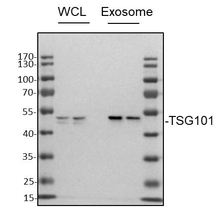
Western Blot: TSG101 Antibody (4A10) [NB200-112] - Whole cell lysates (WCL) or exosome sample from MDA-MB-231 cells was loaded with 10 ug/lane. 10% SDS-PAGE. TSG101 Antibody (NB200-112) was used for primary antibody: 1:1000, 4C, overnight. Image from verified customer review.![Immunohistochemistry-Paraffin: TSG101 Antibody (4A10) [NB200-112]](https://resources.rndsystems.com/images/products/TSG101-Antibody-4A10-Immunohistochemistry-Paraffin-NB200-112-img0026.jpg "Immunohistochemistry-Paraffin: TSG101 Antibody (4A10) [NB200-112]")
Immunohistochemistry-Paraffin: TSG101 Antibody (4A10) [NB200-112]
Immunohistochemistry-Paraffin: TSG101 Antibody (4A10) [NB200-112] - Human ovarian cancer. TSG101 stained by TSG101 antibody [4A10] (NB200-112).Antigen Retrieval: Citrate buffer, pH 6.0, 15 min![Flow Cytometry: TSG101 Antibody (4A10) [NB200-112]](https://resources.rndsystems.com/images/products/TSG101-Antibody-4A10-Flow-Cytometry-NB200-112-img0027.jpg "Flow Cytometry: TSG101 Antibody (4A10) [NB200-112]")
Flow Cytometry: TSG101 Antibody (4A10) [NB200-112]
Flow Cytometry: TSG101 Antibody (4A10) [NB200-112] - TSG101 antibody [4A10] (NB200-112) detects TSG101 protein by flow cytometry analysis.Sample: THP-1 cell.Black: Unlabelled sample was used as a control.Red: TSG101 antibody [4A10] (NB200-112).Acquisition of 20,000 events were collected using a Dylight 488-conjugated secondary antibody for FACS analysis.![Western Blot: TSG101 Antibody (4A10) [NB200-112]](https://resources.rndsystems.com/images/products/TSG101-Antibody-4A10-Western-Blot-NB200-112-img0009.jpg "Western Blot: TSG101 Antibody (4A10) [NB200-112]")
Western Blot: TSG101 Antibody (4A10) [NB200-112]
Western Blot: TSG101 Antibody (4A10) [NB200-112] - A. 30 ug NIH-3T3 whole cell lysate/extract. B. 30 ug JC whole cell lysate/extract. C. 30 ug BCL-1 whole cell lysate/extract.![Western Blot: TSG101 Antibody (4A10) [NB200-112]](https://resources.rndsystems.com/images/products/TSG101-Antibody-4A10-Western-Blot-NB200-112-img0018.jpg "Western Blot: TSG101 Antibody (4A10) [NB200-112]")
Western Blot: TSG101 Antibody (4A10) [NB200-112]
Western Blot: TSG101 Antibody (4A10) [NB200-112] - Various whole cell extracts (30 ug) were separated by 10% SDS-PAGE, and the membrane was blotted with TSG101 antibody [4A10] diluted at 1:500. The HRP-conjugated anti-mouse IgG antibody (NBP2-19382) was used to detect the primary antibody.![Western Blot: TSG101 Antibody (4A10) [NB200-112]](https://resources.rndsystems.com/images/products/TSG101-Antibody-4A10-Western-Blot-NB200-112-img0020.jpg "Western Blot: TSG101 Antibody (4A10) [NB200-112]")
Western Blot: TSG101 Antibody (4A10) [NB200-112]
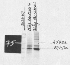
Western Blot: TSG101 Antibody (4A10) [NB200-112] - Lane 1: MWM, Lane 2: TSG101 in RAW 264.7 Whole Cell Lysate, Lane 3: Exosome Lysate. Used primary antibody at 1:500 and Donkey Anti-Mouse at 0.5 ug/mL (this could be titrated down to reduce background). Used picogram sensitivity ECL reagents. Image from verified customer review.![Western Blot: TSG101 Antibody (4A10) [NB200-112]](https://resources.rndsystems.com/images/products/TSG101-Antibody-4A10-Western-Blot-NB200-112-img0033.jpg "Western Blot: TSG101 Antibody (4A10) [NB200-112]")
Western Blot: TSG101 Antibody (4A10) [NB200-112]
Western Blot: TSG101 Antibody (4A10) [NB200-112] - Various tissue extracts (50 ug) were separated by 10% SDS-PAGE, and the membrane was blotted with TSG101 antibody [4A10] diluted at 1:500. The HRP-conjugated anti-mouse IgG antibody (NBP2-19382) was used to detect the primary antibody.![Western Blot: TSG101 Antibody (4A10) [NB200-112]](https://resources.rndsystems.com/images/products/TSG101-Antibody-4A10-Western-Blot-NB200-112-img0034.jpg "Western Blot: TSG101 Antibody (4A10) [NB200-112]")
Western Blot: TSG101 Antibody (4A10) [NB200-112]
TSG101-Antibody-4A10-Western-Blot-NB200-112-img0034.jpg![Immunohistochemistry-Paraffin: TSG101 Antibody (4A10) [NB200-112]](https://resources.rndsystems.com/images/products/TSG101-Antibody-4A10-Immunohistochemistry-Paraffin-NB200-112-img0024.jpg "Immunohistochemistry-Paraffin: TSG101 Antibody (4A10) [NB200-112]")
Immunohistochemistry-Paraffin: TSG101 Antibody (4A10) [NB200-112]
Immunohistochemistry-Paraffin: TSG101 Antibody (4A10) [NB200-112] - Human breast carcinoma. TSG101 stained by TSG101 antibody [4A10] (NB200-112).Antigen Retrieval: Citrate buffer, pH 6.0, 15 min![ELISA: TSG101 Antibody (4A10) [NB200-112]](https://resources.rndsystems.com/images/products/TSG101-Antibody-4A10-ELISA-NB200-112-img0011.jpg "ELISA: TSG101 Antibody (4A10) [NB200-112]")
ELISA: TSG101 Antibody (4A10) [NB200-112]
ELISA: TSG101 Antibody (4A10) [NB200-112] - ELISA detection of TSG101 for capture at a concentration of 5 ug/mL and TSG101 for detection at a concentration of 1.5 ug/mL.![Knockdown Validated: TSG101 Antibody (4A10) [NB200-112]](https://resources.rndsystems.com/images/products/TSG101-Antibody-4A10-Knockdown-Validated-NB200-112-img0014.jpg "Knockdown Validated: TSG101 Antibody (4A10) [NB200-112]")
 [NB200-112] -")
Western Blot: TSG101 Antibody (4A10) [NB200-112] -
Western Blot: TSG101 Antibody (4A10) [NB200-112] - Characterization of pECs-EVs from different women in secretory & proliferative phase of cycle. Representative TEM images of pECs-EVs from secretory (A,B) & proliferative phase (C,D). Scale bars: 50 nm (A,C); 100 nm (B,D). Representative nanoparticle tracking analysis plots of pECs-EVs from secretory (E) & proliferative phase (F). Western blot showing the presence of different canonical EV markers (CD63, CD9, Alix & TSG101) in secretory & proliferative phase pECs-EVs (n = 4) (G). Negative EV markers used were Calnexin (ER), beta -tubulin & the cytosolic form of DCXR (H). The same markers were evaluated in pECs protein extract as controls for EV enrichment of these markers. Image collected & cropped by CiteAb from the following publication (https://pubmed.ncbi.nlm.nih.gov/32483153), licensed under a CC-BY license. Not internally tested by Novus Biologicals. [NB200-112] -")
Western Blot: TSG101 Antibody (4A10) [NB200-112] -
Western Blot: TSG101 Antibody (4A10) [NB200-112] - Characterization of exosomes. (A) Representative TEM images of exosomes at 22000x magnification (Left) HCT116-R exosomes & (Right) HCT116-P exosomes. (B) Enlarged planar view of a cup-shaped structure for single exosome in our samples at 87000x magnification. (C) Western blot analysis of exosomes revealed differential expression of TSG101, CD9, & CD63 between HCT116-R exosomes & HCT116-P exosomes. (D) Average size distribution & zeta potential of exosomes measured in Zetasizer. Image collected & cropped by CiteAb from the following publication (https://pubmed.ncbi.nlm.nih.gov/31712601), licensed under a CC-BY license. Not internally tested by Novus Biologicals. [NB200-112] -")
Western Blot: TSG101 Antibody (4A10) [NB200-112] -
Western Blot: TSG101 Antibody (4A10) [NB200-112] - EPS15 depletion affects late endosomal maturation.(A) Co-depletion of SPOPL an EPS15 has an additive effect on influenza A virus infection. (B) EPS15 depletion stabilizes ESCRT components HRS, STAM & TSG101. (C) EPS15 depletion affects LDL uptake in cells resulting in an accumulation of LDL in enlarged vacuoles (upper panel). Late endosomes, visualized by life-cell microscopy of GFP-RAB7, are enlarged in cells depleted of EPS15 (lower panel).DOI:http://dx.doi.org/10.7554/eLife.13841.016 Image collected & cropped by CiteAb from the following publication (https://elifesciences.org/articles/13841), licensed under a CC-BY license. Not internally tested by Novus Biologicals. [NB200-112] -")
Immunohistochemistry-Paraffin: TSG101 Antibody (4A10) [NB200-112] -
Immunohistochemistry-Paraffin: TSG101 Antibody (4A10) [NB200-112] - TSG101 antibody [4A10] detects TSG101 protein at cytoplasm by immunohistochemical analysis.Sample: Paraffin-embedded human lung cancer.
TSG101 stained by TSG101 antibody [4A10] (NB200-112) diluted at 1:50.
Antigen Retrieval: Citrate buffer, pH 6.0, 15 min
 [NB200-112] -")
Western Blot: TSG101 Antibody (4A10) [NB200-112] -
Western Blot: TSG101 Antibody (4A10) [NB200-112] - Various whole cell extracts (30 ug) were separated by 10% SDS-PAGE, and the membrane was blotted with TSG101 antibody [4A10] (NB200-112) diluted at 1:500. The HRP-conjugated anti-mouse IgG antibody was used to detect the primary antibody. [NB200-112] -")
Western Blot: TSG101 Antibody (4A10) [NB200-112] -
Western Blot: TSG101 Antibody (4A10) [NB200-112] - Various whole cell extracts (30 ug) were separated by 10% SDS-PAGE, and the membrane was blotted with TSG101 antibody [4A10] (NB200-112) diluted at 1:500. The HRP-conjugated anti-mouset IgG antibody was used to detect the primary antibody. [NB200-112] -")
Western Blot: TSG101 Antibody (4A10) [NB200-112] -
(A) Schematic representation of the tetraspanin-based recombinant proteins composition. CD9, CD63 and CD81 tetraspanins are fused to RFP reporter protein, carrying on its 5’terminus a cathepsin B-specific active cleavage site (CS) or non-active cleavage site (NACS). (B) Schematic representation showing the expected tetraspanin-based recombinant proteins orientation on EV membrane and cathepsin B-dependent RFP release. (C) Western blot analysis of cathepsin B from 5637, HEK293 and HeLa cells and respective EVs, representative of three independent experiments; detection of beta-actin for cell extracts and TSG101 for EV extracts was used as loading controls. Image collected and cropped by CiteAb from the following open publication (https://pubmed.ncbi.nlm.nih.gov/36579638), licensed under a CC-BY license. Not internally tested by Novus Biologicals. [NB200-112] -")
Western Blot: TSG101 Antibody (4A10) [NB200-112] -
Exogenous CD9/CD81-RFP CS/NACS protein expression in HEK293 cell lysates and EV fraction. A Fluorescence microscopy images of HEK293 cells stably expressing CD9/CD81-RFP CS/NACS recombinant proteins. Representative western blot analysis comparison between CD9 (B) or CD81 (D) from CD9/CD81-RFP CS/NACS HEK293 cells lysates versus EVs. C, Representative western blot analysis comparison of CD81 and CD63 from CD9-RFP CS/NACS HEK293 derived EVs (C) versus CD9 and CD63 from CD81-RFP CS/NACS HEK293 derived EVs (E). The CD9/CD81-RFP fusion protein was detected also with anti-RFP antibody (CD9-RFP in C e CD81-RFP in E). Alix and TSG101 analysis was used for quantitative protein normalization and RFP detection to confirm the presence of recombinant proteins. Image collected and cropped by CiteAb from the following open publication (https://pubmed.ncbi.nlm.nih.gov/36579638), licensed under a CC-BY license. Not internally tested by Novus Biologicals. [NB200-112] -")
Western Blot: TSG101 Antibody (4A10) [NB200-112] -
EPS15 depletion affects late endosomal maturation.(A) Co-depletion of SPOPL an EPS15 has an additive effect on influenza A virus infection. (B) EPS15 depletion stabilizes ESCRT components HRS, STAM and TSG101. (C) EPS15 depletion affects LDL uptake in cells resulting in an accumulation of LDL in enlarged vacuoles (upper panel). Late endosomes, visualized by life-cell microscopy of GFP-RAB7, are enlarged in cells depleted of EPS15 (lower panel).DOI:http://dx.doi.org/10.7554/eLife.13841.016 Image collected and cropped by CiteAb from the following open publication (https://pubmed.ncbi.nlm.nih.gov/27008177), licensed under a CC-BY license. Not internally tested by Novus Biologicals. [NB200-112] -")
Western Blot: TSG101 Antibody (4A10) [NB200-112] -
Characterization of urine and urinary exosomes isolated by BEST of PE and HC. (A) Representative nanoparticle tracking analysis for untreated urine and isolated urine from PE and HC, depicting particle diameter and the number of particles. (B) and (C) Comparisons of particle concentration and particle size between PE and HC. (D) Western blots of TSG101 and CD63 protein extracted from isolated exosomes. (E) and (F) Quantitative analysis of exosome marker TSG101 and CD63 protein. All data are presented as mean \documentclass[12pt]{minimal}\usepackage{amsmath}\usepackage{wasysym} \usepackage{amsfonts} \usepackage{amssymb} \usepackage{amsbsy}\usepackage{mathrsfs}\usepackage{upgreek}\setlength{\oddsidemargin}{-69pt}\begin{document}$$\:\pm\:$$\end{document} standard error and analyzed by paired t-test and independent two-tailed t-test. BEST, biologically intact exosome separation technology; PE, patients with preeclampsia; HC, healthy controls. Image collected and cropped by CiteAb from the following open publication (https://pubmed.ncbi.nlm.nih.gov/39406891), licensed under a CC-BY license. Not internally tested by Novus Biologicals. [NB200-112] -")
Western Blot: TSG101 Antibody (4A10) [NB200-112] -
Characterization of exosomes. (A) Representative TEM images of exosomes at 22000x magnification (Left) HCT116-R exosomes and (Right) HCT116-P exosomes. (B) Enlarged planar view of a cup-shaped structure for single exosome in our samples at 87000x magnification. (C) Western blot analysis of exosomes revealed differential expression of TSG101, CD9, and CD63 between HCT116-R exosomes and HCT116-P exosomes. (D) Average size distribution and zeta potential of exosomes measured in Zetasizer. Image collected and cropped by CiteAb from the following open publication (https://pubmed.ncbi.nlm.nih.gov/31712601), licensed under a CC-BY license. Not internally tested by Novus Biologicals. [NB200-112] -")
Western Blot: TSG101 Antibody (4A10) [NB200-112] -
Characterization of pECs-EVs from different women in secretory and proliferative phase of cycle. Representative TEM images of pECs-EVs from secretory (A,B) and proliferative phase (C,D). Scale bars: 50 nm (A,C); 100 nm (B,D). Representative nanoparticle tracking analysis plots of pECs-EVs from secretory (E) and proliferative phase (F). Western blot showing the presence of different canonical EV markers (CD63, CD9, Alix and TSG101) in secretory and proliferative phase pECs-EVs (n = 4) (G). Negative EV markers used were Calnexin (ER), beta -tubulin and the cytosolic form of DCXR (H). The same markers were evaluated in pECs protein extract as controls for EV enrichment of these markers. Image collected and cropped by CiteAb from the following open publication (https://pubmed.ncbi.nlm.nih.gov/32483153), licensed under a CC-BY license. Not internally tested by Novus Biologicals.Applications for TSG101 Antibody (4A10)
Application
Recommended Usage
ELISA
1:100 - 1:2000
Electron Microscopy
Assay dependent
Flow Cytometry
1:25-1:200
Immunocytochemistry/ Immunofluorescence
1:500-1:1000
Immunohistochemistry
1:100-1:1000
Immunohistochemistry Free-Floating
Assay dependent
Immunohistochemistry-Paraffin
1:100-1:1000
Immunoprecipitation
Assay dependent
Western Blot
1:500-1:3000
Reviewed Applications
Read 3 reviews rated 4.3 using NB200-112 in the following applications:
Flow Cytometry Panel Builder
Bio-Techne Knows Flow Cytometry
Save time and reduce costly mistakes by quickly finding compatible reagents using the Panel Builder Tool.
Advanced Features
- Spectra Viewer - Custom analysis of spectra from multiple fluorochromes
- Spillover Popups - Visualize the spectra of individual fluorochromes
- Antigen Density Selector - Match fluorochrome brightness with antigen density

Formulation, Preparation, and Storage
Purification
Antigen Affinity-purified
Formulation
PBS
Preservative
No Preservative
Concentration
Concentrations vary lot to lot. See vial label for concentration. If unlisted please contact technical services.
Shipping
The product is shipped with polar packs. Upon receipt, store it immediately at the temperature recommended below.
Stability & Storage
Store at 4C short term. Aliquot and store at -20C long term. Avoid freeze-thaw cycles.
Background: TSG101
Upon its initial discovery, TSG101 was recognized as a tumor suppressor protein due to the identification of deletions within the TSG101 gene in human breast carcinomas. However, re-examination of the initial findings argued against this function and supported that TSG101 promotes tumorigenesis (2). In agreement with this role, TSG101 expression is upregulated in several types of cancer including breast, ovarian, and colorectal carcinoma.
References
1. Schmidt, O., & Teis, D. (2012). The ESCRT machinery. Current Biology. https://doi.org/10.1016/j.cub.2012.01.028
2. Jiang, Y., Ou, Y., & Cheng, X. (2013). Role of TSG101 in cancer. Frontiers in Bioscience. https://doi.org/10.2741/4099
3. Balut, C. M., Gao, Y., Murray, S. A., Thibodeau, P. H., & Devor, D. C. (2010). ESCRT-dependent targeting of plasma membrane localized KCa3.1 to the lysosomes. American Journal of Physiology - Cell Physiology. https://doi.org/10.1152/ajpcell.00120.2010
4. Su, V., & Lau, A. F. (2014). Connexins: Mechanisms regulating protein levels and intercellular communication. FEBS Letters. https://doi.org/10.1016/j.febslet.2014.01.013
Long Name
Tumor Susceptibility 101
Alternate Names
TSG10, VPS23
Gene Symbol
TSG101
UniProt
Additional TSG101 Products
Product Documents for TSG101 Antibody (4A10)
Certificate of Analysis
To download a Certificate of Analysis, please enter a lot or batch number in the search box below.
Product Specific Notices for TSG101 Antibody (4A10)
This product is for research use only and is not approved for use in humans or in clinical diagnosis. Primary Antibodies are guaranteed for 1 year from date of receipt.
Citations for TSG101 Antibody (4A10)
 Powered by Bioz
Customer Reviews for TSG101 Antibody (4A10) (3)
4.3 out of 5
3 Customer Ratings
Have you used TSG101 Antibody (4A10)?
Submit a review and receive an Amazon gift card!
$25/€18/£15/$25CAN/¥2500 Yen for a review with an image
$10/€7/£6/$10CAN/¥1110 Yen for a review without an image
Submit a review
Customer Images
Showing
1
-
3 of
3 reviews
Showing All
Filter By:
-
Application: Western BlotSample Tested: MDA-MB-231Species: HumanVerified Customer | Posted 10/20/2022Western Blot: whole cell lysates (WCL) or exosome sample from MDA-MB-231 cells was loaded with 10 ug/lane. 10% SDS-PAGE. TSG101 Antibody (NB200-112) was used for primary antibody: 1:1000, 4℃, overnight.

-
Application: Western BlotSample Tested: RAW 264.7 mouse monocyte/macrophage cell lineSpecies: MouseVerified Customer | Posted 06/05/2018TSG101 in RAW 264.7 Whole Cell Lysate and Exosome LysateUsed primary @ 1:500 and Donkey Anti-Mouse @ 0.5 ug/mL (this could be titrated down to reduce background). Used picogram sensitivity ECL reagents.
-
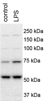
Application: Western BlotSample Tested: Primary mouse hepatocytesSpecies: MouseVerified Customer | Posted 12/08/2017mouse hepatocytes were treated with 100 ng/ml LPS for 4 hoursmouse hepatocytes were treated with 100 ng/ml LPS for 4 hours
There are no reviews that match your criteria.
Protocols
Find general support by application which include: protocols, troubleshooting, illustrated assays, videos and webinars.
- 7-Amino Actinomycin D (7-AAD) Cell Viability Flow Cytometry Protocol
- Antigen Retrieval Protocol (PIER)
- Antigen Retrieval for Frozen Sections Protocol
- Appropriate Fixation of IHC/ICC Samples
- Cellular Response to Hypoxia Protocols
- Chromogenic IHC Staining of Formalin-Fixed Paraffin-Embedded (FFPE) Tissue Protocol
- Chromogenic Immunohistochemistry Staining of Frozen Tissue
- ClariTSA™ Fluorophore Kits
- Detection & Visualization of Antibody Binding
- ELISA Sample Preparation & Collection Guide
- ELISA Troubleshooting Guide
- Extracellular Membrane Flow Cytometry Protocol
- Flow Cytometry Protocol for Cell Surface Markers
- Flow Cytometry Protocol for Staining Membrane Associated Proteins
- Flow Cytometry Staining Protocols
- Flow Cytometry Troubleshooting Guide
- Fluorescent IHC Staining of Frozen Tissue Protocol
- Graphic Protocol for Heat-induced Epitope Retrieval
- Graphic Protocol for the Preparation and Fluorescent IHC Staining of Frozen Tissue Sections
- Graphic Protocol for the Preparation and Fluorescent IHC Staining of Paraffin-embedded Tissue Sections
- Graphic Protocol for the Preparation of Gelatin-coated Slides for Histological Tissue Sections
- How to Run an R&D Systems DuoSet ELISA
- How to Run an R&D Systems Quantikine ELISA
- How to Run an R&D Systems Quantikine™ QuicKit™ ELISA
- ICC Cell Smear Protocol for Suspension Cells
- ICC Immunocytochemistry Protocol Videos
- ICC for Adherent Cells
- IHC Sample Preparation (Frozen sections vs Paraffin)
- Immunocytochemistry (ICC) Protocol
- Immunocytochemistry Troubleshooting
- Immunofluorescence of Organoids Embedded in Cultrex Basement Membrane Extract
- Immunofluorescent IHC Staining of Formalin-Fixed Paraffin-Embedded (FFPE) Tissue Protocol
- Immunohistochemistry (IHC) and Immunocytochemistry (ICC) Protocols
- Immunohistochemistry Frozen Troubleshooting
- Immunohistochemistry Paraffin Troubleshooting
- Immunoprecipitation Protocol
- Intracellular Flow Cytometry Protocol Using Alcohol (Methanol)
- Intracellular Flow Cytometry Protocol Using Detergents
- Intracellular Nuclear Staining Flow Cytometry Protocol Using Detergents
- Intracellular Staining Flow Cytometry Protocol Using Alcohol Permeabilization
- Intracellular Staining Flow Cytometry Protocol Using Detergents to Permeabilize Cells
- Preparing Samples for IHC/ICC Experiments
- Preventing Non-Specific Staining (Non-Specific Binding)
- Primary Antibody Selection & Optimization
- Propidium Iodide Cell Viability Flow Cytometry Protocol
- Protocol for Heat-Induced Epitope Retrieval (HIER)
- Protocol for Liperfluo
- Protocol for Making a 4% Formaldehyde Solution in PBS
- Protocol for VisUCyte™ HRP Polymer Detection Reagent
- Protocol for the Characterization of Human Th22 Cells
- Protocol for the Characterization of Human Th9 Cells
- Protocol for the Fluorescent ICC Staining of Cell Smears - Graphic
- Protocol for the Fluorescent ICC Staining of Cultured Cells on Coverslips - Graphic
- Protocol for the Preparation & Fixation of Cells on Coverslips
- Protocol for the Preparation and Chromogenic IHC Staining of Frozen Tissue Sections
- Protocol for the Preparation and Chromogenic IHC Staining of Frozen Tissue Sections - Graphic
- Protocol for the Preparation and Chromogenic IHC Staining of Paraffin-embedded Tissue Sections
- Protocol for the Preparation and Chromogenic IHC Staining of Paraffin-embedded Tissue Sections - Graphic
- Protocol for the Preparation and Fluorescent ICC Staining of Cells on Coverslips
- Protocol for the Preparation and Fluorescent ICC Staining of Non-adherent Cells
- Protocol for the Preparation and Fluorescent ICC Staining of Stem Cells on Coverslips
- Protocol for the Preparation and Fluorescent IHC Staining of Frozen Tissue Sections
- Protocol for the Preparation and Fluorescent IHC Staining of Paraffin-embedded Tissue Sections
- Protocol for the Preparation of Gelatin-coated Slides for Histological Tissue Sections
- Protocol for the Preparation of a Cell Smear for Non-adherent Cell ICC - Graphic
- Protocol: Annexin V and PI Staining by Flow Cytometry
- Protocol: Annexin V and PI Staining for Apoptosis by Flow Cytometry
- Quantikine HS ELISA Kit Assay Principle, Alkaline Phosphatase
- Quantikine HS ELISA Kit Principle, Streptavidin-HRP Polymer
- R&D Systems Quality Control Western Blot Protocol
- Sandwich ELISA (Colorimetric) – Biotin/Streptavidin Detection Protocol
- Sandwich ELISA (Colorimetric) – Direct Detection Protocol
- TUNEL and Active Caspase-3 Detection by IHC/ICC Protocol
- The Importance of IHC/ICC Controls
- Troubleshooting Guide: ELISA
- Troubleshooting Guide: Fluorokine Flow Cytometry Kits
- Troubleshooting Guide: Immunohistochemistry
- Troubleshooting Guide: Western Blot Figures
- Western Blot Conditions
- Western Blot Protocol
- Western Blot Protocol for Cell Lysates
- Western Blot Troubleshooting
- Western Blot Troubleshooting Guide
- View all Protocols, Troubleshooting, Illustrated assays and Webinars
Loading...