UNC13D/Munc 13-4 Antibody
Novus Biologicals | Catalog # NB100-41385
![Western Blot: UNC13D/Munc 13-4 Antibody [NB100-41385]](https://resources.rndsystems.com/images/products/UNC13D-Munc-13-4-Antibody-Western-Blot-NB100-41385-img0008.jpg "Western Blot: UNC13D/Munc 13-4 Antibody [NB100-41385]")
Key Product Details
Validated by
Species Reactivity
Validated:
Cited:
Predicted:
Applications
Validated:
Cited:
Label
Antibody Source
Product Specifications
Immunogen
Reactivity Notes
Clonality
Host
Isotype
Scientific Data Images for UNC13D/Munc 13-4 Antibody
Western Blot: UNC13D/Munc 13-4 Antibody [NB100-41385]
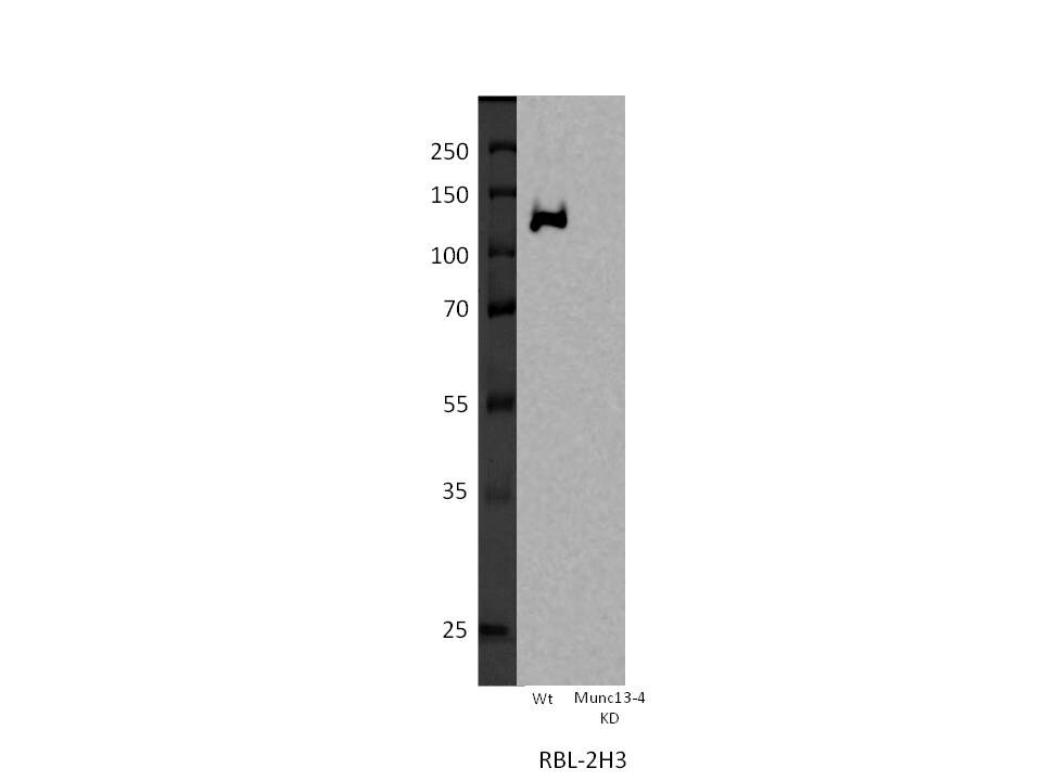
Western Blot: UNC13D/Munc 13-4 Antibody [NB100-41385] - RBL-2H3 from Wt or shRNA Munc13-4 lentiviral stable expression whole cell lysate. 50 ug total protein. WB image submitted by a verified customer review.![Immunohistochemistry-Paraffin: UNC13D/Munc 13-4 Antibody [NB100-41385]](https://resources.rndsystems.com/images/products/UNC13D-Munc-13-4-Antibody-Immunohistochemistry-Paraffin-NB100-41385-img0006.jpg "Immunohistochemistry-Paraffin: UNC13D/Munc 13-4 Antibody [NB100-41385]")
Immunohistochemistry-Paraffin: UNC13D/Munc 13-4 Antibody [NB100-41385]
Immunohistochemistry-Paraffin: UNC13D/Munc 13-4 Antibody [NB100-41385] - Staining of paraffin embedded Mouse Lung with antibody at 0.5 ug/mL (wt in A and KO in B).![Western Blot: UNC13D/Munc 13-4 Antibody [NB100-41385]](https://resources.rndsystems.com/images/products/UNC13D-Munc-13-4-Antibody-Western-Blot-NB100-41385-img0007.jpg "Western Blot: UNC13D/Munc 13-4 Antibody [NB100-41385]")
Western Blot: UNC13D/Munc 13-4 Antibody [NB100-41385]
Western Blot: UNC13D/Munc 13-4 Antibody [NB100-41385] - Staining of Human T-lymphocyte lysate with antibody at 2 ug/mL (35 ug protein in RIPA buffer). Detected by chemiluminescence.![Immunohistochemistry-Paraffin: UNC13D/Munc 13-4 Antibody [NB100-41385]](https://resources.rndsystems.com/images/products/UNC13D-Munc-13-4-Antibody-Immunohistochemistry-Paraffin-NB100-41385-img0005.jpg "Immunohistochemistry-Paraffin: UNC13D/Munc 13-4 Antibody [NB100-41385]")
Immunohistochemistry-Paraffin: UNC13D/Munc 13-4 Antibody [NB100-41385]
Immunohistochemistry-Paraffin: UNC13D/Munc 13-4 Antibody [NB100-41385] - Staining of paraffin embedded Human Thymus. Antibody at 2.5 ug/mL. Steamed antigen retrieval with citrate buffer pH 6, AP-staining.![Knockdown Validated: UNC13D/Munc 13-4 Antibody [NB100-41385]](https://resources.rndsystems.com/images/products/UNC13D-Munc-13-4-Antibody-Knockdown-Validated-NB100-41385-img0009.jpg "Western Blot: UNC13D/Munc 13-4 Antibody [NB100-41385]")
![Knockdown Validated: UNC13D/Munc 13-4 Antibody [NB100-41385]](https://resources.rndsystems.com/images/products/UNC13D-Munc-13-4-Antibody-Knockdown-Validated-NB100-41385-img0010.jpg "Western Blot: UNC13D/Munc 13-4 Antibody [NB100-41385]")

Western Blot: UNC13D/Munc 13-4 Antibody [NB100-41385] -
Western Blot: UNC13D/Munc 13-4 Antibody [NB100-41385] - Munc13-4 KD strongly impairs exosome release. (A) SDS-PAGE Western blot of indicated proteins in MDA-MB-231 cells after stable expression of shRNA for Munc13-4 or Rab27a or a scrambled control (Ctrl). (B) Culture medium from MDA-MB-231 cells either untreated or stimulated w/ 1.25 µM ionomycin for 30 min centrifuged at 1,000 g to remove cellular debris & 10,000 g to remove large extracellular vesicles. (C) The resulting 10,000-g supernatant filtered onto a nitrocellulose membrane & analyzed for CD63, CD9, ALIX, & GM130 content by antibody blotting. (D) Quantification of CD63, CD9, & ALIX blots in C are shown as exosome release as a percentage of total cellular material w/ mean values ± standard error (SE) for n ≥ 3. *, P < 0.05 for comparison w/ corresponding control samples. (E) Panc-1 or A549 cells left untreated or treated w/ TGF beta -1 for 24 h. Indicated proteins detected by SDS-PAGE Western blot. (F) Panc-1 cells left untreated or treated w/ TGF beta -1 for 24 h, & Munc13-4 levels determined by immunofluorescence. TGF beta -1–treated cells exhibited a mesenchymal morphology. Bars, 5 μm. (G) A549 cells stably expressing control shRNA (Ctrl) or Munc13-4 shRNA left untreated (Untr) or treated w/ TGF beta -1 for 24 h, & SDS-PAGE Western blotting for indicated proteins conducted. (H) Culture media supernatants (as in B) from A549 cells that either untreated or stimulated w/ 1.25 µM ionomycin for 30 min filtered onto nitrocellulose membrane & analyzed for CD63 & GM130. (I) Quantification of CD63+ exosome release shown as a percentage of total cellular material w/ mean values ± SE for n = 5. *, P < 0.05; **, P < 0.01 for comparison w/ corresponding control samples. Image collected & cropped by CiteAb from the following publication (https://pubmed.ncbi.nlm.nih.gov/29930202), licensed under a CC-BY license. Not internally tested by Novus Biologicals.
Immunocytochemistry/ Immunofluorescence: UNC13D/Munc 13-4 Antibody [NB100-41385] -
Munc13-4 translocation to membrane is Ca2+ dependent. (A) Live-cell epifluorescence imaging of GFP-Munc13-4 in MDA-MB-231 cells at indicated times after ionomycin stimulation. See Video 1. (B) MDA-MB-231 cells expressing wild-type GFP-Munc13-4, GFP-Munc13-4 C2A*, or GFP-Munc13-4 C2B* either left untreated or stimulated with 1.25 uM ionomycin for 5 min were fixed and imaged by confocal microscopy. (C) Indicated proteins were detected by SDS-PAGE and Western blotting of lysates with Munc13-4 antibody from MDA-MB-231 cells stably expressing control shRNA (Ctrl) or shRNA targeting Munc13-4 (KD), or Munc13-4 KD cells rescued with shRNA-resistant wild-type Munc13-4, Munc13-4 C2A*, or Munc13-4 C2B*. (D) Culture media supernatants (as in Fig. 1 B) from MDA-MB-231 cells as in C either untreated or stimulated with 1.25 uM ionomycin for 30 min were filtered onto membrane and analyzed for CD63 or GM130. (E) Quantification of CD63+ exosome release (from Fig 1 D) shown as mean values +/- SE for n = 3. *, P < 0.05 for comparison between ionomycin-treated and basal. Bars, 5 μm. Image collected and cropped by CiteAb from the following open publication (https://pubmed.ncbi.nlm.nih.gov/29930202), licensed under a CC-BY license. Not internally tested by Novus Biologicals.Applications for UNC13D/Munc 13-4 Antibody
Immunohistochemistry
Immunohistochemistry-Paraffin
Peptide ELISA
Western Blot
Reviewed Applications
Read 1 review rated 5 using NB100-41385 in the following applications:
Formulation, Preparation, and Storage
Purification
Formulation
Preservative
Concentration
Shipping
Stability & Storage
Background: UNC13D
Long Name
Alternate Names
Gene Symbol
UniProt
Additional UNC13D Products
Product Documents for UNC13D/Munc 13-4 Antibody
Certificate of Analysis
To download a Certificate of Analysis, please enter a lot or batch number in the search box below.
Product Specific Notices for UNC13D/Munc 13-4 Antibody
This product is for research use only and is not approved for use in humans or in clinical diagnosis. Primary Antibodies are guaranteed for 1 year from date of receipt.
Related Research Areas
Citations for UNC13D/Munc 13-4 Antibody
 Powered by Bioz
Customer Reviews for UNC13D/Munc 13-4 Antibody (1)
Have you used UNC13D/Munc 13-4 Antibody?
Submit a review and receive an Amazon gift card!
$25/€18/£15/$25CAN/¥2500 Yen for a review with an image
$10/€7/£6/$10CAN/¥1110 Yen for a review without an image
Submit a review
Customer Images
-
Application: Western BlotSample Tested: RBL-2H3 rat basophilic leukemia cell lineSpecies: RatVerified Customer | Posted 04/14/2017RBL-2H3 from Wt or shRNA Munc13-4 lentiviral stable expression whole cell lysate. 50ug protein

There are no reviews that match your criteria.
Protocols
Find general support by application which include: protocols, troubleshooting, illustrated assays, videos and webinars.
- Antigen Retrieval Protocol (PIER)
- Antigen Retrieval for Frozen Sections Protocol
- Appropriate Fixation of IHC/ICC Samples
- Cellular Response to Hypoxia Protocols
- Chromogenic IHC Staining of Formalin-Fixed Paraffin-Embedded (FFPE) Tissue Protocol
- Chromogenic Immunohistochemistry Staining of Frozen Tissue
- ClariTSA™ Fluorophore Kits
- Detection & Visualization of Antibody Binding
- ELISA Sample Preparation & Collection Guide
- ELISA Troubleshooting Guide
- Fluorescent IHC Staining of Frozen Tissue Protocol
- Graphic Protocol for Heat-induced Epitope Retrieval
- Graphic Protocol for the Preparation and Fluorescent IHC Staining of Frozen Tissue Sections
- Graphic Protocol for the Preparation and Fluorescent IHC Staining of Paraffin-embedded Tissue Sections
- Graphic Protocol for the Preparation of Gelatin-coated Slides for Histological Tissue Sections
- How to Run an R&D Systems DuoSet ELISA
- How to Run an R&D Systems Quantikine ELISA
- How to Run an R&D Systems Quantikine™ QuicKit™ ELISA
- ICC Cell Smear Protocol for Suspension Cells
- ICC Immunocytochemistry Protocol Videos
- ICC for Adherent Cells
- IHC Sample Preparation (Frozen sections vs Paraffin)
- Immunocytochemistry (ICC) Protocol
- Immunocytochemistry Troubleshooting
- Immunofluorescence of Organoids Embedded in Cultrex Basement Membrane Extract
- Immunofluorescent IHC Staining of Formalin-Fixed Paraffin-Embedded (FFPE) Tissue Protocol
- Immunohistochemistry (IHC) and Immunocytochemistry (ICC) Protocols
- Immunohistochemistry Frozen Troubleshooting
- Immunohistochemistry Paraffin Troubleshooting
- Preparing Samples for IHC/ICC Experiments
- Preventing Non-Specific Staining (Non-Specific Binding)
- Primary Antibody Selection & Optimization
- Protocol for Heat-Induced Epitope Retrieval (HIER)
- Protocol for Making a 4% Formaldehyde Solution in PBS
- Protocol for VisUCyte™ HRP Polymer Detection Reagent
- Protocol for the Fluorescent ICC Staining of Cell Smears - Graphic
- Protocol for the Fluorescent ICC Staining of Cultured Cells on Coverslips - Graphic
- Protocol for the Preparation & Fixation of Cells on Coverslips
- Protocol for the Preparation and Chromogenic IHC Staining of Frozen Tissue Sections
- Protocol for the Preparation and Chromogenic IHC Staining of Frozen Tissue Sections - Graphic
- Protocol for the Preparation and Chromogenic IHC Staining of Paraffin-embedded Tissue Sections
- Protocol for the Preparation and Chromogenic IHC Staining of Paraffin-embedded Tissue Sections - Graphic
- Protocol for the Preparation and Fluorescent ICC Staining of Cells on Coverslips
- Protocol for the Preparation and Fluorescent ICC Staining of Non-adherent Cells
- Protocol for the Preparation and Fluorescent ICC Staining of Stem Cells on Coverslips
- Protocol for the Preparation and Fluorescent IHC Staining of Frozen Tissue Sections
- Protocol for the Preparation and Fluorescent IHC Staining of Paraffin-embedded Tissue Sections
- Protocol for the Preparation of Gelatin-coated Slides for Histological Tissue Sections
- Protocol for the Preparation of a Cell Smear for Non-adherent Cell ICC - Graphic
- Quantikine HS ELISA Kit Assay Principle, Alkaline Phosphatase
- Quantikine HS ELISA Kit Principle, Streptavidin-HRP Polymer
- R&D Systems Quality Control Western Blot Protocol
- Sandwich ELISA (Colorimetric) – Biotin/Streptavidin Detection Protocol
- Sandwich ELISA (Colorimetric) – Direct Detection Protocol
- TUNEL and Active Caspase-3 Detection by IHC/ICC Protocol
- The Importance of IHC/ICC Controls
- Troubleshooting Guide: ELISA
- Troubleshooting Guide: Immunohistochemistry
- Troubleshooting Guide: Western Blot Figures
- Western Blot Conditions
- Western Blot Protocol
- Western Blot Protocol for Cell Lysates
- Western Blot Troubleshooting
- Western Blot Troubleshooting Guide
- View all Protocols, Troubleshooting, Illustrated assays and Webinars
FAQs for UNC13D/Munc 13-4 Antibody
-
Q: I'm looking Munc13-4 N terminal antibody with related epitope sequence.
A:
Unfortunately we do not epitope map our antibodies, but we do have a whole list of antibodies against your target depending on species reactivity and application you want to use them for. Please see this link to our MUNC 13-4 antibodies. All of the immunogen information that is not proprietary, or known should be presented on the datasheet. Often times a range will be provided where the peptide falls within, or if it was raised against a larger recombinant protein as is the case for the one you inquired about we do not know exactly where the binding occurs.