V5 Epitope Tag Antibody - BSA Free
Novus Biologicals | Catalog # NB600-381
![Western Blot: V5 Epitope Tag AntibodyBSA Free [NB600-381]](https://resources.rndsystems.com/images/products/V5-Epitope-Tag-Antibody-Western-Blot-NB600-381-img0013.jpg "Western Blot: V5 Epitope Tag AntibodyBSA Free [NB600-381]")
Key Product Details
Species Reactivity
Validated:
Cited:
Applications
Validated:
Cited:
Label
Antibody Source
Format
Product Specifications
Immunogen
Clonality
Host
Isotype
Scientific Data Images for V5 Epitope Tag Antibody - BSA Free
Western Blot: V5 Epitope Tag AntibodyBSA Free [NB600-381]
V5-Epitope-Tag-Antibody-Western-Blot-NB600-381-img0013.jpg![Immunocytochemistry/ Immunofluorescence: V5 Epitope Tag Antibody - BSA Free [NB600-381]](https://resources.rndsystems.com/images/products/V5-Epitope-Tag-Antibody-Immunocytochemistry-Immunofluorescence-NB600-381-img0009.jpg "Immunocytochemistry/ Immunofluorescence: V5 Epitope Tag Antibody - BSA Free [NB600-381]")
Immunocytochemistry/ Immunofluorescence: V5 Epitope Tag Antibody - BSA Free [NB600-381]
Immunocytochemistry/Immunofluorescence: V5 Epitope Tag Antibody [NB600-381] - ATG2A-V5 overexpression reduces intracellular neutral lipids in THP-1 cells. Scale bars, 10 um. Regulation of phagocyte triglyceride by a STAT-ATG2 pathway controls mycobacterial infection. Nat Commun (2017)![Western Blot: V5 Epitope Tag AntibodyBSA Free [NB600-381]](https://resources.rndsystems.com/images/products/V5-Epitope-Tag-Antibody-Western-Blot-NB600-381-img0002.jpg "Western Blot: V5 Epitope Tag AntibodyBSA Free [NB600-381]")
Western Blot: V5 Epitope Tag AntibodyBSA Free [NB600-381]
Western Blot: V5 Epitope Tag Antibody [NB600-381] - 200, 100, or 50 ng of E. coli whole cell lysate expressing a multi-tag fusion protein. Antibody used at 0.04 ug/mL (1:25,000).![Western Blot: V5 Epitope Tag AntibodyBSA Free [NB600-381]](https://resources.rndsystems.com/images/products/V5-Epitope-Tag-Antibody-Western-Blot-NB600-381-img0001.jpg "Western Blot: V5 Epitope Tag AntibodyBSA Free [NB600-381]")
Western Blot: V5 Epitope Tag AntibodyBSA Free [NB600-381]
Western Blot: V5 Epitope Tag Antibody [NB600-381] - NP-40 whole cell lysate (10 ug) from wild-type HEK293 cells (lane 1) or HEK293 cells expressing V5 tagged Nicastrin (lane 2). NB600-381 was used at 0.2 ug/mL (1:5000).![Western Blot: V5 Epitope Tag AntibodyBSA Free [NB600-381]](https://resources.rndsystems.com/images/products/V5-Epitope-Tag-Antibody-Western-Blot-NB600-381-img0005.jpg "Western Blot: V5 Epitope Tag AntibodyBSA Free [NB600-381]")
Western Blot: V5 Epitope Tag AntibodyBSA Free [NB600-381]
Western Blot: V5 Epitope Tag Antibody [NB600-381] - Analysis using the HRP conjugate of NB600-381. Detection of 200, 100, or 50 ng of E. coli whole cell lysate expressing a multi-tag fusion protein. Antibody used at 0.2 ug/mL (1:5,000).![Immunocytochemistry/ Immunofluorescence: V5 Epitope Tag Antibody - BSA Free [NB600-381]](https://resources.rndsystems.com/images/products/V5-Epitope-Tag-Antibody-Immunocytochemistry-Immunofluorescence-NB600-381-img0007.jpg "Immunocytochemistry/ Immunofluorescence: V5 Epitope Tag Antibody - BSA Free [NB600-381]")
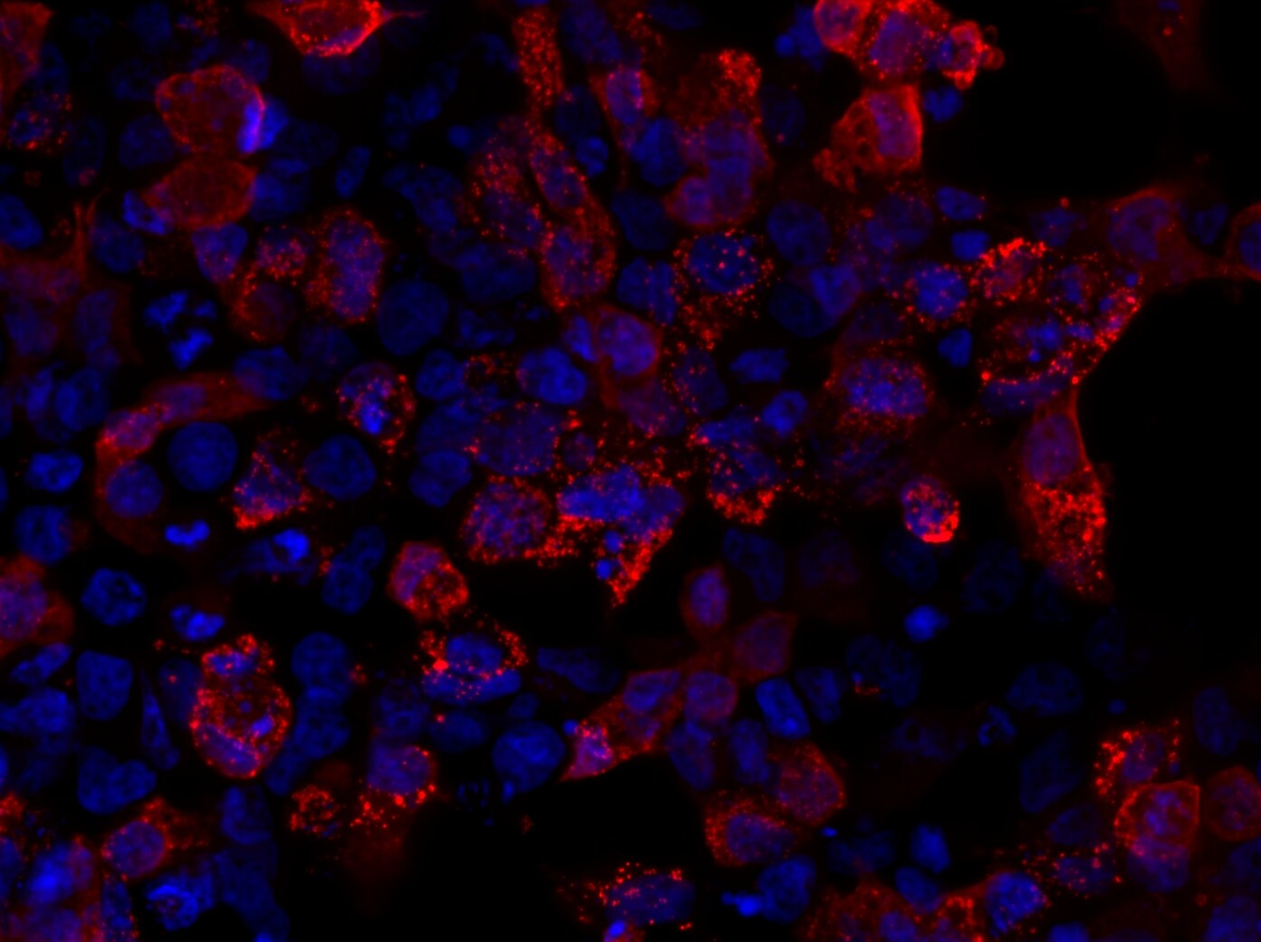
Immunocytochemistry/ Immunofluorescence: V5 Epitope Tag Antibody - BSA Free [NB600-381]
Immunocytochemistry/Immunofluorescence: V5 Epitope Tag Antibody [NB600-381] - HEK293 cells transiently expressing V5-tagged mutant PFN1 stained with V5 Epitope Tag antibody (red). ICC/IF image submitted by a verified customer review.![Immunocytochemistry/ Immunofluorescence: V5 Epitope Tag Antibody - BSA Free [NB600-381]](https://resources.rndsystems.com/images/products/V5-Epitope-Tag-Antibody-Immunohistochemistry-NB600-381-img0008.jpg "Immunocytochemistry/ Immunofluorescence: V5 Epitope Tag Antibody - BSA Free [NB600-381]")
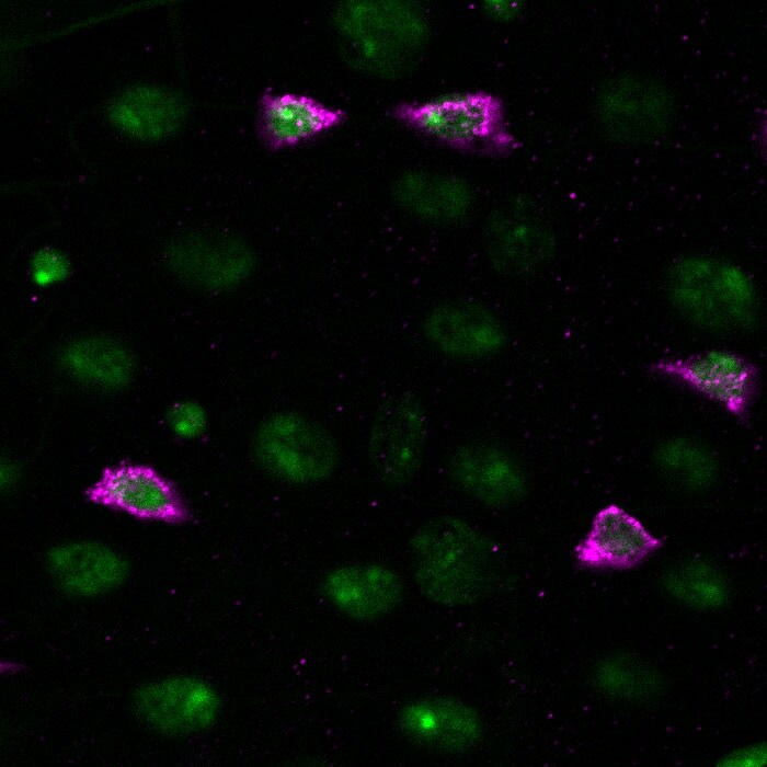
Immunocytochemistry/ Immunofluorescence: V5 Epitope Tag Antibody - BSA Free [NB600-381]
Immunocytochemistry/Immunofluorescence: V5 Epitope Tag Antibody [NB600-381] - Drosophila adult midgut expressing a V5-tagged protein (magenta) using the GAL4/UAS system. Nuclei are shown in green. pAb dilution of 1:500. ICC/IF image submitted by a verified customer review.![Immunoprecipitation: V5 Epitope Tag Antibody - BSA Free [NB600-381]](https://resources.rndsystems.com/images/products/V5-Epitope-Tag-Antibody-Immunoprecipitation-NB600-381-img0012.jpg "Immunoprecipitation: V5 Epitope Tag Antibody - BSA Free [NB600-381]")
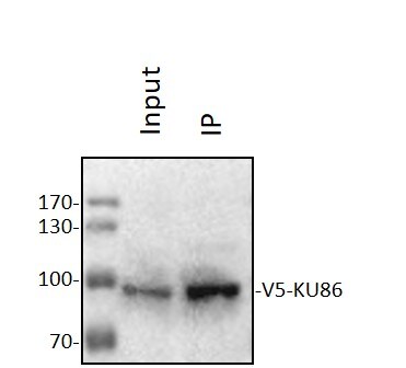
Immunoprecipitation: V5 Epitope Tag Antibody - BSA Free [NB600-381]
Immunoprecipitation: V5 Epitope Tag Antibody [NB600-381] - HEK293T were transfected with V5-KU86 plasmid, followed by V5 antibody IP then Western blot with v5 antibody (NB600-381). Primary antibody at 1:1000, 4C, overnight. IP/WB image submitted by a verified customer review.
Western Blot: V5 Epitope Tag Antibody - BSA Free [NB600-381] -
PRDX proteins bind to HIF-1 alpha & HIF-2 alpha A. HeLa cells were transfected with an expression vector encoding V5-epitope-tagged PRDX2 (PRDX2-V5) & exposed to 1% O2 for 24 h. Whole cell lysate (WCL) was subject to immunoprecipitation (IP) using anti-HIF-1 alpha antibody or control IgG, followed by immunoblot assays with antibody against V5 epitope or HIF-1 alpha. B. HeLa cells were transfected with PRDX2-V5 vector & exposed to 1% O2 for 24 h. The WCL was subject to IP using anti-V5 antibody or control IgG, followed by immunoblot assays with antibody against V5 or HIF-1 alpha. Light IgG: immunoglobulin light chain from the secondary antibody. C. HeLa cells were transfected with vector encoding a V5-tagged PRDX family member & exposed to 1% O2 for 24 h. WCL was subject to IP using anti-HIF-1 alpha antibody, followed by immunoblot assays with antibody against V5 or HIF-1 alpha. D. HeLa cells were transfected with empty vector (EV) or vector encoding a V5-tagged PRDX family member & exposed to 1% O2 for 24 h. WCL was subject to IP using anti-HIF-2 alpha antibody, followed by immunoblot assays with antibody against V5 or HIF-2 alpha. Image collected & cropped by CiteAb from the following publication (https://www.oncotarget.com/lookup/doi/10.18632/oncotarget.7142), licensed under a CC-BY license. Not internally tested by Novus Biologicals.
Western Blot: V5 Epitope Tag Antibody - BSA Free [NB600-381] -
Hypoxia induces the nuclear translocation of PRDX2 & PRDX4HeLa cells were transfected with vector encoding PRDX2-V5 (P2) or PRDX4-V5 (P4), or empty vector (EV), & exposed to 20% or 1% O2 for 48 h. Nuclear & cytosolic fractions were isolated & subject to immunoblot assays with antibodies against HIF-1 alpha, HIF-2 alpha, V5, alpha -tubulin, & histone H3. Image collected & cropped by CiteAb from the following publication (https://www.oncotarget.com/lookup/doi/10.18632/oncotarget.7142), licensed under a CC-BY license. Not internally tested by Novus Biologicals.
Western Blot: V5 Epitope Tag Antibody - BSA Free [NB600-381] -
PRDX proteins bind to HIF-1 alpha & HIF-2 alpha A. HeLa cells were transfected with an expression vector encoding V5-epitope-tagged PRDX2 (PRDX2-V5) & exposed to 1% O2 for 24 h. Whole cell lysate (WCL) was subject to immunoprecipitation (IP) using anti-HIF-1 alpha antibody or control IgG, followed by immunoblot assays with antibody against V5 epitope or HIF-1 alpha. B. HeLa cells were transfected with PRDX2-V5 vector & exposed to 1% O2 for 24 h. The WCL was subject to IP using anti-V5 antibody or control IgG, followed by immunoblot assays with antibody against V5 or HIF-1 alpha. Light IgG: immunoglobulin light chain from the secondary antibody. C. HeLa cells were transfected with vector encoding a V5-tagged PRDX family member & exposed to 1% O2 for 24 h. WCL was subject to IP using anti-HIF-1 alpha antibody, followed by immunoblot assays with antibody against V5 or HIF-1 alpha. D. HeLa cells were transfected with empty vector (EV) or vector encoding a V5-tagged PRDX family member & exposed to 1% O2 for 24 h. WCL was subject to IP using anti-HIF-2 alpha antibody, followed by immunoblot assays with antibody against V5 or HIF-2 alpha. Image collected & cropped by CiteAb from the following publication (https://www.oncotarget.com/lookup/doi/10.18632/oncotarget.7142), licensed under a CC-BY license. Not internally tested by Novus Biologicals.
Western Blot: V5 Epitope Tag Antibody - BSA Free [NB600-381] -
Western Blot: V5 Epitope Tag Antibody - BSA Free [NB600-381] - (a) Light & fluorescent micrographs of stable HN33.11 cells expressing (A)-GFP or (B)-GFP, respectively. Results from two stable clones of each transfection pool are shown. Cells are treated with vehicle (Dox−) or 1 μg/mL Dox (Dox+). (b) Western blot of homogenates of stable clones showing endogenous production of full-length p75NTR (FLp75), Dox-induced, but “leaky” production of (A) & (B) & of the V5 tag. Control values for each clone do not differ significantly between GFP(−) & GFP(+) cells (Student's t-test). ***P < 0.001 relative to GFP(−) cells. Image collected & cropped by CiteAb from the following publication (https://pubmed.ncbi.nlm.nih.gov/21904642), licensed under a CC-BY license. Not internally tested by Novus Biologicals.
Western Blot: V5 Epitope Tag Antibody - BSA Free [NB600-381] -
Western Blot: V5 Epitope Tag Antibody - BSA Free [NB600-381] - Mapping the PRDX2 & PRDX4 binding domains of HIF-1 alpha A. & B. HeLa cells were transfected with PRDX2-V5 (A) or PRDX4-V5 (B) vector & WCL was incubated with purified GST or GST-HIF-1 alpha fusion protein in the presence of glutathione-Sepharose beads, followed by immunoblot assays with anti-V5 antibody (upper panels) or Ponceau S staining (lower panels). Image collected & cropped by CiteAb from the following publication (https://www.oncotarget.com/lookup/doi/10.18632/oncotarget.7142), licensed under a CC-BY license. Not internally tested by Novus Biologicals.Applications for V5 Epitope Tag Antibody - BSA Free
ELISA
Immunocytochemistry/ Immunofluorescence
Immunohistochemistry
Immunoprecipitation
Western Blot
Reviewed Applications
Read 3 reviews rated 5 using NB600-381 in the following applications:
Flow Cytometry Panel Builder
Bio-Techne Knows Flow Cytometry
Save time and reduce costly mistakes by quickly finding compatible reagents using the Panel Builder Tool.
Advanced Features
- Spectra Viewer - Custom analysis of spectra from multiple fluorochromes
- Spillover Popups - Visualize the spectra of individual fluorochromes
- Antigen Density Selector - Match fluorochrome brightness with antigen density

Formulation, Preparation, and Storage
Purification
Formulation
Format
Preservative
Concentration
Shipping
Stability & Storage
Background: V5 Epitope Tag
Alternate Names
Additional V5 Epitope Tag Products
Product Documents for V5 Epitope Tag Antibody - BSA Free
Certificate of Analysis
To download a Certificate of Analysis, please enter a lot or batch number in the search box below.
Product Specific Notices for V5 Epitope Tag Antibody - BSA Free
This product is for research use only and is not approved for use in humans or in clinical diagnosis. Primary Antibodies are guaranteed for 1 year from date of receipt.
Citations for V5 Epitope Tag Antibody - BSA Free
 Powered by Bioz
Customer Reviews for V5 Epitope Tag Antibody - BSA Free (3)
Have you used V5 Epitope Tag Antibody - BSA Free?
Submit a review and receive an Amazon gift card!
$25/€18/£15/$25CAN/¥2500 Yen for a review with an image
$10/€7/£6/$10CAN/¥1110 Yen for a review without an image
Submit a review
Customer Images
-
Application: ImmunoprecipitationSample Tested: 293 cell extractSpecies: HumanVerified Customer | Posted 11/21/2020Western Blot: HEK293T were transfected with V5-KU86 plasmid, followed by V5 antibody IP then blot with v5 antibody (NB600-381). Primary antibody: 1:1000, 4℃, overnight.

-
Application: ImmunohistochemistrySample Tested: Adult intestineSpecies: DrosophilaVerified Customer | Posted 06/10/2017Drosophila adult midgut expressing a V5-tagged protein (magenta) using the GAL4/UAS system. Nuclei are shown in green.Dilution 1:500
-
Application: ImmunofluorescenceSample Tested: HEK293 cellsSpecies: HumanVerified Customer | Posted 07/06/2016HEK293 cells transiently expressing V5-tagged mutant PFN1
There are no reviews that match your criteria.
Protocols
Find general support by application which include: protocols, troubleshooting, illustrated assays, videos and webinars.
- 7-Amino Actinomycin D (7-AAD) Cell Viability Flow Cytometry Protocol
- Antigen Retrieval Protocol (PIER)
- Antigen Retrieval for Frozen Sections Protocol
- Appropriate Fixation of IHC/ICC Samples
- Cellular Response to Hypoxia Protocols
- Chromogenic IHC Staining of Formalin-Fixed Paraffin-Embedded (FFPE) Tissue Protocol
- Chromogenic Immunohistochemistry Staining of Frozen Tissue
- ClariTSA™ Fluorophore Kits
- Detection & Visualization of Antibody Binding
- ELISA Sample Preparation & Collection Guide
- ELISA Troubleshooting Guide
- Extracellular Membrane Flow Cytometry Protocol
- Flow Cytometry Protocol for Cell Surface Markers
- Flow Cytometry Protocol for Staining Membrane Associated Proteins
- Flow Cytometry Staining Protocols
- Flow Cytometry Troubleshooting Guide
- Fluorescent IHC Staining of Frozen Tissue Protocol
- Graphic Protocol for Heat-induced Epitope Retrieval
- Graphic Protocol for the Preparation and Fluorescent IHC Staining of Frozen Tissue Sections
- Graphic Protocol for the Preparation and Fluorescent IHC Staining of Paraffin-embedded Tissue Sections
- Graphic Protocol for the Preparation of Gelatin-coated Slides for Histological Tissue Sections
- How to Run an R&D Systems DuoSet ELISA
- How to Run an R&D Systems Quantikine ELISA
- How to Run an R&D Systems Quantikine™ QuicKit™ ELISA
- ICC Cell Smear Protocol for Suspension Cells
- ICC Immunocytochemistry Protocol Videos
- ICC for Adherent Cells
- IHC Sample Preparation (Frozen sections vs Paraffin)
- Immunocytochemistry (ICC) Protocol
- Immunocytochemistry Troubleshooting
- Immunofluorescence of Organoids Embedded in Cultrex Basement Membrane Extract
- Immunofluorescent IHC Staining of Formalin-Fixed Paraffin-Embedded (FFPE) Tissue Protocol
- Immunohistochemistry (IHC) and Immunocytochemistry (ICC) Protocols
- Immunohistochemistry Frozen Troubleshooting
- Immunohistochemistry Paraffin Troubleshooting
- Immunoprecipitation Protocol
- Intracellular Flow Cytometry Protocol Using Alcohol (Methanol)
- Intracellular Flow Cytometry Protocol Using Detergents
- Intracellular Nuclear Staining Flow Cytometry Protocol Using Detergents
- Intracellular Staining Flow Cytometry Protocol Using Alcohol Permeabilization
- Intracellular Staining Flow Cytometry Protocol Using Detergents to Permeabilize Cells
- Preparing Samples for IHC/ICC Experiments
- Preventing Non-Specific Staining (Non-Specific Binding)
- Primary Antibody Selection & Optimization
- Propidium Iodide Cell Viability Flow Cytometry Protocol
- Protocol for Heat-Induced Epitope Retrieval (HIER)
- Protocol for Liperfluo
- Protocol for Making a 4% Formaldehyde Solution in PBS
- Protocol for VisUCyte™ HRP Polymer Detection Reagent
- Protocol for the Characterization of Human Th22 Cells
- Protocol for the Characterization of Human Th9 Cells
- Protocol for the Fluorescent ICC Staining of Cell Smears - Graphic
- Protocol for the Fluorescent ICC Staining of Cultured Cells on Coverslips - Graphic
- Protocol for the Preparation & Fixation of Cells on Coverslips
- Protocol for the Preparation and Chromogenic IHC Staining of Frozen Tissue Sections
- Protocol for the Preparation and Chromogenic IHC Staining of Frozen Tissue Sections - Graphic
- Protocol for the Preparation and Chromogenic IHC Staining of Paraffin-embedded Tissue Sections
- Protocol for the Preparation and Chromogenic IHC Staining of Paraffin-embedded Tissue Sections - Graphic
- Protocol for the Preparation and Fluorescent ICC Staining of Cells on Coverslips
- Protocol for the Preparation and Fluorescent ICC Staining of Non-adherent Cells
- Protocol for the Preparation and Fluorescent ICC Staining of Stem Cells on Coverslips
- Protocol for the Preparation and Fluorescent IHC Staining of Frozen Tissue Sections
- Protocol for the Preparation and Fluorescent IHC Staining of Paraffin-embedded Tissue Sections
- Protocol for the Preparation of Gelatin-coated Slides for Histological Tissue Sections
- Protocol for the Preparation of a Cell Smear for Non-adherent Cell ICC - Graphic
- Protocol: Annexin V and PI Staining by Flow Cytometry
- Protocol: Annexin V and PI Staining for Apoptosis by Flow Cytometry
- Quantikine HS ELISA Kit Assay Principle, Alkaline Phosphatase
- Quantikine HS ELISA Kit Principle, Streptavidin-HRP Polymer
- R&D Systems Quality Control Western Blot Protocol
- Sandwich ELISA (Colorimetric) – Biotin/Streptavidin Detection Protocol
- Sandwich ELISA (Colorimetric) – Direct Detection Protocol
- TUNEL and Active Caspase-3 Detection by IHC/ICC Protocol
- The Importance of IHC/ICC Controls
- Troubleshooting Guide: ELISA
- Troubleshooting Guide: Fluorokine Flow Cytometry Kits
- Troubleshooting Guide: Immunohistochemistry
- Troubleshooting Guide: Western Blot Figures
- Western Blot Conditions
- Western Blot Protocol
- Western Blot Protocol for Cell Lysates
- Western Blot Troubleshooting
- Western Blot Troubleshooting Guide
- View all Protocols, Troubleshooting, Illustrated assays and Webinars
FAQs for V5 Epitope Tag Antibody - BSA Free
-
Q: I can't find any images or the antibody used in publications for this application, but the description says it works in this particular application. Could the company provide additional data?
A: This antibody has been validated in IHC on both frozen and paraffin-embedded tissues and is 100% guaranteed for this use. Validation came from customer feedback, so unfortunately, these images are not available. I apologize for the inconvenience. If you are concerned about images, you may want to consider NB600-381 or NB100-62264 as these two products both have over 20 publications.