Wnt-5a Antibody - BSA Free
Novus Biologicals | Catalog # NBP2-24752
![Western Blot: Wnt-5a AntibodyBSA Free [NBP2-24752]](https://resources.rndsystems.com/images/products/Wnt-5a-Antibody-Western-Blot-NBP2-24752-img0004.jpg "Western Blot: Wnt-5a AntibodyBSA Free [NBP2-24752]")
Key Product Details
Species Reactivity
Validated:
Human, Mouse, Rat, Bovine, Monkey
Cited:
Human, Mouse
Predicted:
Amphibian (90%), Guinea Pig (100%). Backed by our 100% Guarantee.
Applications
Validated:
Immunohistochemistry, Immunohistochemistry-Paraffin, Immunohistochemistry-Frozen, Western Blot
Cited:
Immunohistochemistry-Paraffin, Western Blot, IF/IHC
Label
Unconjugated
Antibody Source
Polyclonal Rabbit IgG
Format
BSA Free
Loading...
Product Specifications
Immunogen
A synthetic peptide corresponding to amino acids 190-230 of human Wnt-5a was used as the immunogen for this antibody, GenBank no NP_003383.2.
Reactivity Notes
The amino acid sequence used as immunogen is 90% homologous in newt (Pleurodeles waltl), and 88% in Xenopus and axolotl (Ambystoma mexicanum). Reactivity to Petarus breviceps reported in verified customer review.
Clonality
Polyclonal
Host
Rabbit
Isotype
IgG
Scientific Data Images for Wnt-5a Antibody - BSA Free
Western Blot: Wnt-5a AntibodyBSA Free [NBP2-24752]
Western Blot: Wnt-5a Antibody [NBP2-24752] - Analysis of Wnt-5a in human heart lysate in the 1) absence, 2) presence of immunizing peptide, 3) mouse and 4) rat heart![Immunohistochemistry-Frozen: Wnt-5a Antibody - BSA Free [NBP2-24752]](https://resources.rndsystems.com/images/products/Wnt-5a-Antibody-Immunohistochemistry-Frozen-NBP2-24752-img0005.jpg "Immunohistochemistry-Frozen: Wnt-5a Antibody - BSA Free [NBP2-24752]")
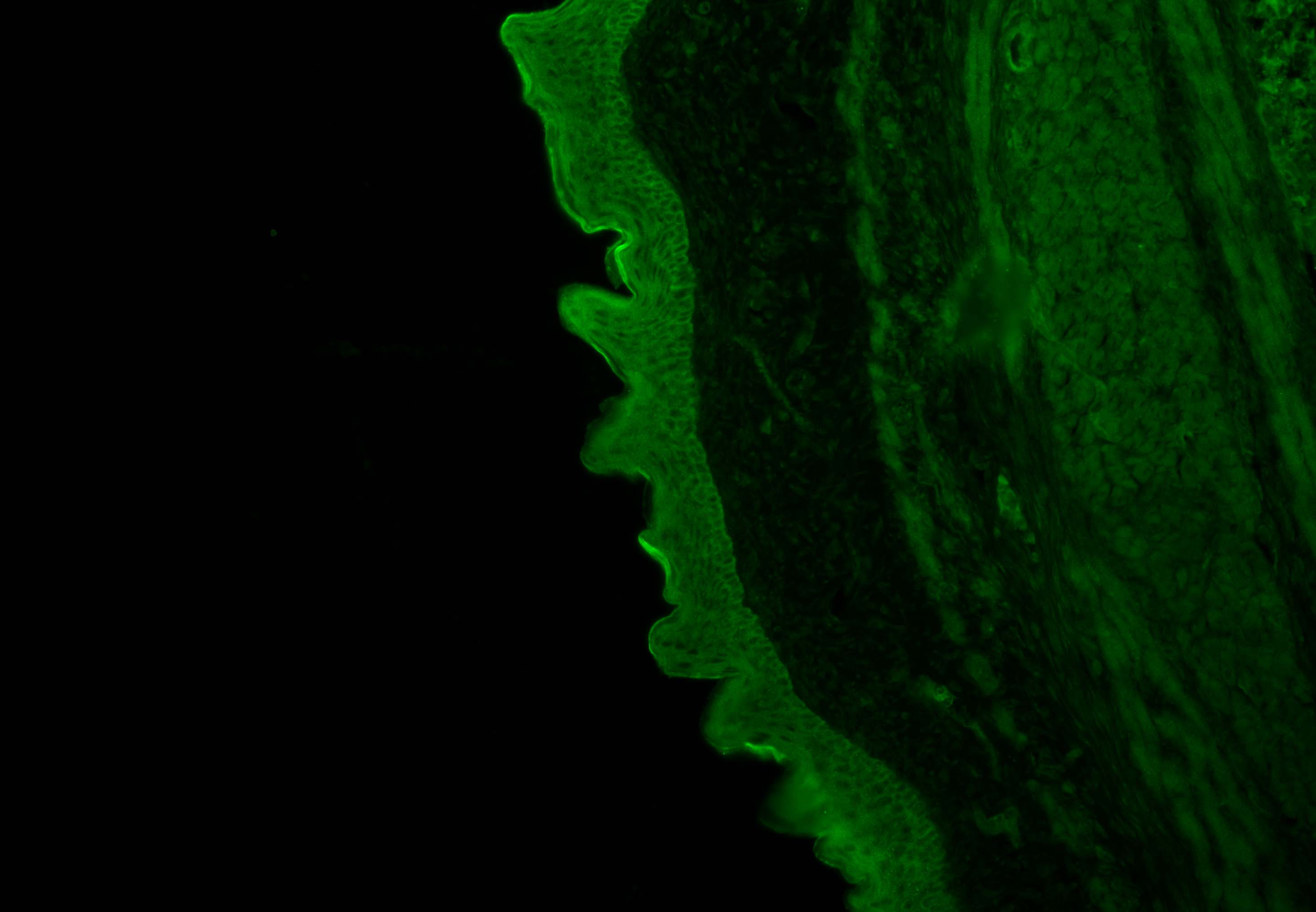
Immunohistochemistry-Frozen: Wnt-5a Antibody - BSA Free [NBP2-24752]
Immunohistochemistry-Frozen: Wnt-5a Antibody [NBP2-24752] - P. breviceps epithelial expression of Wnt5a at 1:100 dilution. Tissue samples Cryo-section fixed (12um slices) in OCT, washed for 10min in PBT (PBS + 0.1% tween), blocked for 1hr in 3%BSA/PBS, and then incubated with the antibody overnight at 4C. The next day washed 3x for 10min in PBT and incubated for one hr in Alexa 488 (1:500), washed 3x10min in PBS, and coverslipped. Image from verified customer review.![Immunohistochemistry-Paraffin: Wnt-5a Antibody - BSA Free [NBP2-24752]](https://resources.rndsystems.com/images/products/Wnt-5a-Antibody-Immunohistochemistry-Paraffin-NBP2-24752-img0003.jpg "Immunohistochemistry-Paraffin: Wnt-5a Antibody - BSA Free [NBP2-24752]")
Immunohistochemistry-Paraffin: Wnt-5a Antibody - BSA Free [NBP2-24752]
Immunohistochemistry-Paraffin: Wnt-5a Antibody [NBP2-24752] - Analysis of Wnt-5a in FFPE human breast tumor using an isotype control (left) and Wnt-5a antibody (right)![Wnt-5a Antibody - BSA Free Immunohistochemistry-Paraffin: Wnt-5a Antibody - BSA Free [NBP2-24752]](https://resources.rndsystems.com/images/products/antibody/nbp2-24752_rabbit-polyclonal-wnt-5a-antibody-imgenex-img-6075a-immunohistochemistry-paraffin-162202682014.png "Immunohistochemistry-Paraffin: Wnt-5a Antibody - BSA Free [NBP2-24752]")
Immunohistochemistry-Paraffin: Wnt-5a Antibody - BSA Free [NBP2-24752]
Analysis of a FFPE tissue section of human placenta using 1:200 dilution of Wnt-5a antibody. The staining was developed using HRP labeled anti-rabbit secondary antibody and DAB reagent, and nuclei of cells were counter-stained with hematoxylin.Applications for Wnt-5a Antibody - BSA Free
Application
Recommended Usage
Immunohistochemistry-Frozen
reported by customer review
Immunohistochemistry-Paraffin
5-10 ug/ml
Western Blot
0.5-2 ug/ml
Reviewed Applications
Read 1 review rated 4 using NBP2-24752 in the following applications:
Formulation, Preparation, and Storage
Purification
Affinity purified
Formulation
PBS
Format
BSA Free
Preservative
0.05% Sodium Azide
Concentration
1.0 mg/ml
Shipping
The product is shipped with polar packs. Upon receipt, store it immediately at the temperature recommended below.
Stability & Storage
Store at 4C short term. Aliquot and store at -20C long term. Avoid freeze-thaw cycles.
Background: Wnt-5a
Long Name
Wingless-type MMTV Integration Site Family, Member 5a
Alternate Names
Wnt5a
Gene Symbol
WNT5A
UniProt
Additional Wnt-5a Products
Product Documents for Wnt-5a Antibody - BSA Free
Certificate of Analysis
To download a Certificate of Analysis, please enter a lot or batch number in the search box below.
Product Specific Notices for Wnt-5a Antibody - BSA Free
This product is for research use only and is not approved for use in humans or in clinical diagnosis. Primary Antibodies are guaranteed for 1 year from date of receipt.
Related Research Areas
Citations for Wnt-5a Antibody - BSA Free
 Powered by Bioz
Customer Reviews for Wnt-5a Antibody - BSA Free (1)
4 out of 5
1 Customer Rating
Have you used Wnt-5a Antibody - BSA Free?
Submit a review and receive an Amazon gift card!
$25/€18/£15/$25CAN/¥2500 Yen for a review with an image
$10/€7/£6/$10CAN/¥1110 Yen for a review without an image
Submit a review
Customer Images
Showing
1
-
1 of
1 review
Showing All
Filter By:
-
Application: Immunohistochemistry-FrozenSample Tested: Skin tissue and Skin frozen tissueSpecies: Petaurus breviceps (marsupial)Verified Customer | Posted 02/25/2019Epithelial expression of Wnt5a1:100 dilution. We cryo-sectioned fixed tissue (12um slices) in OCT, washed samples for 10min in PBT (PBS + 0.1% tween), blocked the sample for 1hr in 3%BSA/PBS and then incubated with the antibody overnight at 4C. The next day we washed 3x for 10min in PBT and incubated for one hr in Alexa488 (1:500), washed 3x10min in PBS and coverslipped.

There are no reviews that match your criteria.
Protocols
View specific protocols for Wnt-5a Antibody - BSA Free (NBP2-24752):
Immunohistochemistry-Paraffin Embedded Sections
Antigen Unmasking:
Bring slides to a boil in 10 mM sodium citrate buffer (pH 6.0) then maintain at a sub-boiling temperature for 10 minutes. Cool slides on bench-top for 30 minutes (keep slides in the sodium citrate buffer at all times).
Staining:
1. Wash sections in deionized water three times for 5 minutes each.
2. Wash sections in PBS for 5 minutes.
3. Block each section with 100-400 ul blocking solution (1% BSA in PBS) for 1 hour at room temperature.
4. Remove blocking solution and add 100-400 ul diluted primary antibody. Incubate overnight at 4 C.
5. Remove antibody solution and wash sections in wash buffer three times for 5 minutes each.
6. Add 100-400 ul HRP polymer conjugated secondary antibody. Incubate 30 minutes at room temperature.
7. Wash sections three times in wash buffer for 5 minutes each.
8. Add 100-400 ul DAB substrate to each section and monitor staining closely.
9. As soon as the sections develop, immerse slides in deionized water.
10. Counterstain sections in hematoxylin.
11. Wash sections in deionized water two times for 5 minutes each.
12. Dehydrate sections.
13. Mount coverslips.
Western Blot Protocol
1. Perform SDS-PAGE on samples to be analyzed, loading 10-25 ug of total protein per lane.
2. Transfer proteins to PVDF membrane according to the instructions provided by the manufacturer of the membrane and transfer apparatus.
3. Stain the membrane with Ponceau S (or similar product) to assess transfer success, and mark molecular weight standards where appropriate.
4. Rinse the blot TBS -0.05% Tween 20 (TBST).
5. Block the membrane in 5% Non-fat milk in TBST (blocking buffer) for at least 1 hour.
6. Wash the membrane in TBST three times for 10 minutes each.
7. Dilute primary antibody in blocking buffer and incubate overnight at 4C with gentle rocking.
8. Wash the membrane in TBST three times for 10 minutes each.
9. Incubate the membrane in diluted HRP conjugated secondary antibody in blocking buffer (as per manufacturer's instructions) for 1 hour at room temperature.
10. Wash the blot in TBST three times for 10 minutes each (this step can be repeated as required to reduce background).
11. Apply the detection reagent of choice in accordance with the manufacturer's instructions.
Find general support by application which include: protocols, troubleshooting, illustrated assays, videos and webinars.
- Antigen Retrieval Protocol (PIER)
- Antigen Retrieval for Frozen Sections Protocol
- Appropriate Fixation of IHC/ICC Samples
- Cellular Response to Hypoxia Protocols
- Chromogenic IHC Staining of Formalin-Fixed Paraffin-Embedded (FFPE) Tissue Protocol
- Chromogenic Immunohistochemistry Staining of Frozen Tissue
- ClariTSA™ Fluorophore Kits
- Detection & Visualization of Antibody Binding
- Fluorescent IHC Staining of Frozen Tissue Protocol
- Graphic Protocol for Heat-induced Epitope Retrieval
- Graphic Protocol for the Preparation and Fluorescent IHC Staining of Frozen Tissue Sections
- Graphic Protocol for the Preparation and Fluorescent IHC Staining of Paraffin-embedded Tissue Sections
- Graphic Protocol for the Preparation of Gelatin-coated Slides for Histological Tissue Sections
- IHC Sample Preparation (Frozen sections vs Paraffin)
- Immunofluorescent IHC Staining of Formalin-Fixed Paraffin-Embedded (FFPE) Tissue Protocol
- Immunohistochemistry (IHC) and Immunocytochemistry (ICC) Protocols
- Immunohistochemistry Frozen Troubleshooting
- Immunohistochemistry Paraffin Troubleshooting
- Preparing Samples for IHC/ICC Experiments
- Preventing Non-Specific Staining (Non-Specific Binding)
- Primary Antibody Selection & Optimization
- Protocol for Heat-Induced Epitope Retrieval (HIER)
- Protocol for Making a 4% Formaldehyde Solution in PBS
- Protocol for VisUCyte™ HRP Polymer Detection Reagent
- Protocol for the Preparation & Fixation of Cells on Coverslips
- Protocol for the Preparation and Chromogenic IHC Staining of Frozen Tissue Sections
- Protocol for the Preparation and Chromogenic IHC Staining of Frozen Tissue Sections - Graphic
- Protocol for the Preparation and Chromogenic IHC Staining of Paraffin-embedded Tissue Sections
- Protocol for the Preparation and Chromogenic IHC Staining of Paraffin-embedded Tissue Sections - Graphic
- Protocol for the Preparation and Fluorescent IHC Staining of Frozen Tissue Sections
- Protocol for the Preparation and Fluorescent IHC Staining of Paraffin-embedded Tissue Sections
- Protocol for the Preparation of Gelatin-coated Slides for Histological Tissue Sections
- R&D Systems Quality Control Western Blot Protocol
- TUNEL and Active Caspase-3 Detection by IHC/ICC Protocol
- The Importance of IHC/ICC Controls
- Troubleshooting Guide: Immunohistochemistry
- Troubleshooting Guide: Western Blot Figures
- Western Blot Conditions
- Western Blot Protocol
- Western Blot Protocol for Cell Lysates
- Western Blot Troubleshooting
- Western Blot Troubleshooting Guide
- View all Protocols, Troubleshooting, Illustrated assays and Webinars


