BCMA/TNFRSF17 Antibody (Vicky-1) - BSA Free
Novus Biologicals | Catalog # NBP1-97637
Clone Vicky-1 was used by HLDA to establish CD designation.
![Immunohistochemistry: BCMA/TNFRSF17 Antibody (Vicky-1) - BSA Free [NBP1-97637]](https://resources.rndsystems.com/images/products/BCMA-TNFRSF17-Antibody-Vicky-1-Immunohistochemistry-NBP1-97637-img0007.jpg "Immunohistochemistry: BCMA/TNFRSF17 Antibody (Vicky-1) - BSA Free [NBP1-97637]")
Key Product Details
Species Reactivity
Validated:
Human
Cited:
Human
Applications
Validated:
Immunohistochemistry, Western Blot, ELISA, Flow Cytometry, Immunocytochemistry/ Immunofluorescence, Functional
Cited:
Flow Cytometry
Label
Unconjugated
Antibody Source
Monoclonal Rat IgG1 Clone # Vicky-1
Format
BSA Free
Loading...
Product Specifications
Immunogen
Recombinant human BCMA extracellular domain (aa 2-54).
Reactivity Notes
Does not cross-react with Mouse BCMA.
Clonality
Monoclonal
Host
Rat
Isotype
IgG1
Scientific Data Images for BCMA/TNFRSF17 Antibody (Vicky-1) - BSA Free
Immunohistochemistry: BCMA/TNFRSF17 Antibody (Vicky-1) - BSA Free [NBP1-97637]
Immunohistochemistry: BCMA/TNFRSF17 Antibody (Vicky-1) [NBP1-97637] - Immunostaining of HEK 293 cells transfected with a human BCMA expression plasmid (left panel), or mock transfected (right panel). Method: 3 days after transfection of cells with the indicated constructs, cells were fixed with acetone or 4% formaldehyde for 5 min. at room temperature. Slides were blocked with normal IgG, and incubated for 1 hour with 5ug/ml MAb to BCMA (human) (Vicky-1) in 1% BSA in 1x PBS. After washes in PBS, samples were incubated with the secondary antibody for 1 hour, washed in PBS and revealed with StreptABComplex/HRP (Vector) and AEC.![Flow Cytometry: BCMA/TNFRSF17 Antibody (Vicky-1) - BSA Free [NBP1-97637]](https://resources.rndsystems.com/images/products/BCMA-TNFRSF17-Antibody-Vicky-1-Flow-Cytometry-NBP1-97637-img0011.jpg "Flow Cytometry: BCMA/TNFRSF17 Antibody (Vicky-1) - BSA Free [NBP1-97637]")
Flow Cytometry: BCMA/TNFRSF17 Antibody (Vicky-1) - BSA Free [NBP1-97637]
Flow Cytometry: BCMA/TNFRSF17 Antibody (Vicky-1) [NBP1-97637] - Analysis using the PE conjugate of NBP1-97637. Staining of 10^6 U266 cells using BCMA (human), mAb (Vicky-1) (PE conjugate) at a concentration of 10ug/ml.![Flow Cytometry: BCMA/TNFRSF17 Antibody (Vicky-1) - BSA Free [NBP1-97637]](https://resources.rndsystems.com/images/products/BCMA-TNFRSF17-Antibody-Vicky-1-Flow-Cytometry-NBP1-97637-img0006.jpg "Flow Cytometry: BCMA/TNFRSF17 Antibody (Vicky-1) - BSA Free [NBP1-97637]")
Flow Cytometry: BCMA/TNFRSF17 Antibody (Vicky-1) - BSA Free [NBP1-97637]
Flow Cytometry: BCMA/TNFRSF17 Antibody (Vicky-1) [NBP1-97637] - Detection of endogenous human BCMA with MAb to BCMA (human) (Vicky-1). Method: U266 cells (2x10^5) were incubated on ice for 30 min. with 0.2ug of MAb to BCMA (human) (Vicky-1) or an isotype control in 25ul FACS buffer (PBS, 5% fetal calf serum, 0.02% azide). The primary antibody was revealed with PAb to Rat IgG (R-PE) and then analyzed by flow cytometry.![Flow Cytometry: BCMA/TNFRSF17 Antibody (Vicky-1) - BSA Free [NBP1-97637]](https://resources.rndsystems.com/images/products/BCMA-TNFRSF17-Antibody-Vicky-1-Flow-Cytometry-NBP1-97637-img0010.jpg "Flow Cytometry: BCMA/TNFRSF17 Antibody (Vicky-1) - BSA Free [NBP1-97637]")
Flow Cytometry: BCMA/TNFRSF17 Antibody (Vicky-1) - BSA Free [NBP1-97637]
Flow Cytometry: BCMA/TNFRSF17 Antibody (Vicky-1) [NBP1-97637] - Analysis using the FITC conjugate of NBP1-97637. Staining of 10^6 Jurkat cells using BCMA (human), mAb (Vicky-1) (FITC conjugate) at a concentration of 50ug/ml.Applications for BCMA/TNFRSF17 Antibody (Vicky-1) - BSA Free
Application
Recommended Usage
ELISA
1:100-1:2000
Flow Cytometry
1:10-1:1000
Immunocytochemistry/ Immunofluorescence
1:10-1:2000
Western Blot
1:100-1:2000
Application Notes
Use in WB was reported in the scientific literature (PMID: 20454508). Functional Application: blocks binding of BAFF to human BCMA.
Reviewed Applications
Read 1 review rated 4 using NBP1-97637 in the following applications:
Flow Cytometry Panel Builder
Bio-Techne Knows Flow Cytometry
Save time and reduce costly mistakes by quickly finding compatible reagents using the Panel Builder Tool.
Advanced Features
- Spectra Viewer - Custom analysis of spectra from multiple fluorochromes
- Spillover Popups - Visualize the spectra of individual fluorochromes
- Antigen Density Selector - Match fluorochrome brightness with antigen density

Formulation, Preparation, and Storage
Purification
Protein G purified
Formulation
PBS
Format
BSA Free
Preservative
0.02% Sodium Azide
Concentration
1.0 mg/ml
Shipping
The product is shipped with polar packs. Upon receipt, store it immediately at the temperature recommended below.
Stability & Storage
Store at -20C. Avoid freeze-thaw cycles.
Background: BCMA/TNFRSF17
BCMA has two agonistic ligands: BAFF and a proliferation-inducing ligand (APRIL) (1,2). APRIL has higher affinity for BCMA than BAFF and the binding is mediated by CD138/syndeclin-1 (2,3). Activation of BCMA promotes the growth and survival of plasma cells, or MM cells in disease, through several signaling pathways such as NFkappaB, MEK/ERK, AKT, JNK, and p38 (1,2). In MM cells the BCMA activation and downstream signaling cascade functions to upregulate antiapoptotic proteins including Bcl-2, Bcl-xL, and Mcl-1 and protect the cells against therapeutic agents like dexamethasone (2,3).
Given its specific expression on plasma cells but not memory B cells, naive B cells, or hematopoietic stem cells, BCMA has garnered much interest as a therapeutic target for the treatment of MM (1-4). Current BCMA-targeted immunotherapy strategies include antibody-drug conjugates (ADC), chimeric antigen receptor (CAR) T cells, bispecific T cell engager (BiTE), and bispecific/trispecific antibodies (1-4). CAR T cell therapy in particular has demonstrated promising clinical results (2,4). Still, more research needs to be done to improve the efficacy and risk of relapse following CAR T cell therapy and may also include targeting additional antigens in combination with BCMA or utilizing pharmacological agents to increase antigen density (4).
References
1. Yu, B., Jiang, T., & Liu, D. (2020). BCMA-targeted immunotherapy for multiple myeloma. Journal of hematology & oncology, 13(1), 125. https://doi.org/10.1186/s13045-020-00962-7
2. Cho, S. F., Anderson, K. C., & Tai, Y. T. (2018). Targeting B Cell Maturation Antigen (BCMA) in Multiple Myeloma: Potential Uses of BCMA-Based Immunotherapy. Frontiers in immunology, 9, 1821. https://doi.org/10.3389/fimmu.2018.01821
3. Dalla Palma, B., Marchica, V., Catarozzo, M. T., Giuliani, N., & Accardi, F. (2020). Monoclonal and Bispecific Anti-BCMA Antibodies in Multiple Myeloma. Journal of clinical medicine, 9(9), 3022. https://doi.org/10.3390/jcm9093022
4. Mikkilineni, L., & Kochenderfer, J. N. (2021). CAR T cell therapies for patients with multiple myeloma. Nature reviews. Clinical oncology, 18(2), 71-84. https://doi.org/10.1038/s41571-020-0427-6
Long Name
B Cell Maturation Factor
Alternate Names
CD269, TNFRSF13A, TNFRSF17
Gene Symbol
TNFRSF17
Additional BCMA/TNFRSF17 Products
Product Documents for BCMA/TNFRSF17 Antibody (Vicky-1) - BSA Free
Certificate of Analysis
To download a Certificate of Analysis, please enter a lot or batch number in the search box below.
Product Specific Notices for BCMA/TNFRSF17 Antibody (Vicky-1) - BSA Free
This product is for research use only and is not approved for use in humans or in clinical diagnosis. Primary Antibodies are guaranteed for 1 year from date of receipt.
Citations for BCMA/TNFRSF17 Antibody (Vicky-1) - BSA Free
 Powered by Bioz
Customer Reviews for BCMA/TNFRSF17 Antibody (Vicky-1) - BSA Free (1)
4 out of 5
1 Customer Rating
Have you used BCMA/TNFRSF17 Antibody (Vicky-1) - BSA Free?
Submit a review and receive an Amazon gift card!
$25/€18/£15/$25CAN/¥2500 Yen for a review with an image
$10/€7/£6/$10CAN/¥1110 Yen for a review without an image
Submit a review
Customer Images
Showing
1
-
1 的
1 review
Showing All
Filter By:
-
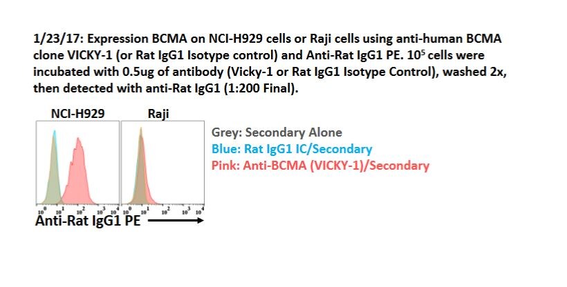
Application: Flow CytometrySample Tested: Raji human Burkitt's lymphoma cell line and H929 cellsSpecies: HumanVerified Customer | Posted 01/27/2017Expression BCMA on NCI-H929 cells or Raji cells using anti-human BCMA clone VICKY-1 (or Rat IgG1 Isotype control) and Anti-Rat IgG1 PE. 105 cells were incubated with 0.5ug of antibody (Vicky-1 or Rat IgG1 IC) and detected with a-Rat IgG1 (1:200 Final).

There are no reviews that match your criteria.
Protocols
Find general support by application which include: protocols, troubleshooting, illustrated assays, videos and webinars.
- 7-Amino Actinomycin D (7-AAD) Cell Viability Flow Cytometry Protocol
- Antigen Retrieval Protocol (PIER)
- Antigen Retrieval for Frozen Sections Protocol
- Appropriate Fixation of IHC/ICC Samples
- Cellular Response to Hypoxia Protocols
- Chromogenic IHC Staining of Formalin-Fixed Paraffin-Embedded (FFPE) Tissue Protocol
- Chromogenic Immunohistochemistry Staining of Frozen Tissue
- ClariTSA™ Fluorophore Kits
- Detection & Visualization of Antibody Binding
- ELISA Sample Preparation & Collection Guide
- ELISA Troubleshooting Guide
- Extracellular Membrane Flow Cytometry Protocol
- Flow Cytometry Protocol for Cell Surface Markers
- Flow Cytometry Protocol for Staining Membrane Associated Proteins
- Flow Cytometry Staining Protocols
- Flow Cytometry Troubleshooting Guide
- Fluorescent IHC Staining of Frozen Tissue Protocol
- Graphic Protocol for Heat-induced Epitope Retrieval
- Graphic Protocol for the Preparation and Fluorescent IHC Staining of Frozen Tissue Sections
- Graphic Protocol for the Preparation and Fluorescent IHC Staining of Paraffin-embedded Tissue Sections
- Graphic Protocol for the Preparation of Gelatin-coated Slides for Histological Tissue Sections
- How to Run an R&D Systems DuoSet ELISA
- How to Run an R&D Systems Quantikine ELISA
- How to Run an R&D Systems Quantikine™ QuicKit™ ELISA
- ICC Cell Smear Protocol for Suspension Cells
- ICC Immunocytochemistry Protocol Videos
- ICC for Adherent Cells
- IHC Sample Preparation (Frozen sections vs Paraffin)
- Immunocytochemistry (ICC) Protocol
- Immunocytochemistry Troubleshooting
- Immunofluorescence of Organoids Embedded in Cultrex Basement Membrane Extract
- Immunofluorescent IHC Staining of Formalin-Fixed Paraffin-Embedded (FFPE) Tissue Protocol
- Immunohistochemistry (IHC) and Immunocytochemistry (ICC) Protocols
- Immunohistochemistry Frozen Troubleshooting
- Immunohistochemistry Paraffin Troubleshooting
- Intracellular Flow Cytometry Protocol Using Alcohol (Methanol)
- Intracellular Flow Cytometry Protocol Using Detergents
- Intracellular Nuclear Staining Flow Cytometry Protocol Using Detergents
- Intracellular Staining Flow Cytometry Protocol Using Alcohol Permeabilization
- Intracellular Staining Flow Cytometry Protocol Using Detergents to Permeabilize Cells
- Preparing Samples for IHC/ICC Experiments
- Preventing Non-Specific Staining (Non-Specific Binding)
- Primary Antibody Selection & Optimization
- Propidium Iodide Cell Viability Flow Cytometry Protocol
- Protocol for Heat-Induced Epitope Retrieval (HIER)
- Protocol for Liperfluo
- Protocol for Making a 4% Formaldehyde Solution in PBS
- Protocol for VisUCyte™ HRP Polymer Detection Reagent
- Protocol for the Characterization of Human Th22 Cells
- Protocol for the Characterization of Human Th9 Cells
- Protocol for the Fluorescent ICC Staining of Cell Smears - Graphic
- Protocol for the Fluorescent ICC Staining of Cultured Cells on Coverslips - Graphic
- Protocol for the Preparation & Fixation of Cells on Coverslips
- Protocol for the Preparation and Chromogenic IHC Staining of Frozen Tissue Sections
- Protocol for the Preparation and Chromogenic IHC Staining of Frozen Tissue Sections - Graphic
- Protocol for the Preparation and Chromogenic IHC Staining of Paraffin-embedded Tissue Sections
- Protocol for the Preparation and Chromogenic IHC Staining of Paraffin-embedded Tissue Sections - Graphic
- Protocol for the Preparation and Fluorescent ICC Staining of Cells on Coverslips
- Protocol for the Preparation and Fluorescent ICC Staining of Non-adherent Cells
- Protocol for the Preparation and Fluorescent ICC Staining of Stem Cells on Coverslips
- Protocol for the Preparation and Fluorescent IHC Staining of Frozen Tissue Sections
- Protocol for the Preparation and Fluorescent IHC Staining of Paraffin-embedded Tissue Sections
- Protocol for the Preparation of Gelatin-coated Slides for Histological Tissue Sections
- Protocol for the Preparation of a Cell Smear for Non-adherent Cell ICC - Graphic
- Protocol: Annexin V and PI Staining by Flow Cytometry
- Protocol: Annexin V and PI Staining for Apoptosis by Flow Cytometry
- Quantikine HS ELISA Kit Assay Principle, Alkaline Phosphatase
- Quantikine HS ELISA Kit Principle, Streptavidin-HRP Polymer
- R&D Systems Quality Control Western Blot Protocol
- Sandwich ELISA (Colorimetric) – Biotin/Streptavidin Detection Protocol
- Sandwich ELISA (Colorimetric) – Direct Detection Protocol
- TUNEL and Active Caspase-3 Detection by IHC/ICC Protocol
- The Importance of IHC/ICC Controls
- Troubleshooting Guide: ELISA
- Troubleshooting Guide: Fluorokine Flow Cytometry Kits
- Troubleshooting Guide: Immunohistochemistry
- Troubleshooting Guide: Western Blot Figures
- Western Blot Conditions
- Western Blot Protocol
- Western Blot Protocol for Cell Lysates
- Western Blot Troubleshooting
- Western Blot Troubleshooting Guide
- View all Protocols, Troubleshooting, Illustrated assays and Webinars
FAQs for BCMA/TNFRSF17 Antibody (Vicky-1) - BSA Free
Showing
1
-
1 的
1 FAQ
Showing All
-
Q: What would be the appropriate negative isotype control for IHC on paraffin embedded human tissue?
A:
A rat IgG1 isotype control is what you will want to use for NBP1-97637. Rat IgG1 contains a light chain and in rat, 99% of antibodies are kappa light chain, so this is what you will want to use. Here is a link to suitable isotype controls.
Loading...
Associated Pathways
