CD3 Antibody (SP7)
Novus Biologicals | Catalog # NB600-1441
![Immunohistochemistry-Paraffin: CD3 Antibody (SP7) [NB600-1441]](https://resources.rndsystems.com/images/products/CD3-Antibody-SP7-Immunohistochemistry-Paraffin-NB600-1441-img0003.jpg "Immunohistochemistry-Paraffin: CD3 Antibody (SP7) [NB600-1441]")
![Immunohistochemistry-Paraffin: CD3 Antibody (SP7) [NB600-1441]](https://resources.rndsystems.com/images/products/CD3-Antibody-SP7-Immunohistochemistry-Paraffin-NB600-1441-img0001.jpg "Immunohistochemistry-Paraffin: CD3 Antibody (SP7) [NB600-1441]")
Loading...
Key Product Details
Validated by
Knockout/Knockdown, Biological Validation
Species Reactivity
Validated:
Human, Mouse, Porcine, Canine
Cited:
Human, Mouse, Porcine
Applications
Validated:
Immunohistochemistry, Immunohistochemistry-Paraffin, Immunohistochemistry-Frozen, Flow Cytometry, Immunocytochemistry/ Immunofluorescence, Knockdown Validated
Cited:
Immunohistochemistry, Immunohistochemistry-Paraffin, Immunohistochemistry-Frozen, Western Blot, Flow Cytometry, Immunocytochemistry/ Immunofluorescence, IF/IHC
Label
Unconjugated
Antibody Source
Monoclonal Rabbit IgG Clone # SP7
Loading...
Product Specifications
Immunogen
This CD3 antibody was developed against a synthetic peptide: KAKAKPVTRGAGA, corresponding to amino acids 156-168 of Human CD3 epsilon chain.
Epitope
aa 156-168 of epsilon chain of human CD3 protein (intracytoplasmic).
Reactivity Notes
Use in Mouse reported in scientific literature (PMID:35111697). Canine reactivity per customer review. Use in Porcine reported in scientific literature (PMID:33839961)
Localization
Type I membrane protein.
Specificity
The CD3 antigen is present on early thymocytes and mature T cells and is generally regarded as a pan-T cell marker. This antibody will help detect CD3 expression in normal and neoplastic tissues. This antibody reacts with the intracytoplasmic portion of the CD3 antigen expressed by T cells. It stains human T cells in both the cortex and medulla of the thymus and in peripheral lymphoid tissues. This antibody is suitable for staining normal and neoplastic T cells in formalin-fixed, paraffin-embedded tissues.
Clonality
Monoclonal
Host
Rabbit
Isotype
IgG
Theoretical MW
23.1 kDa.
Disclaimer note: The observed molecular weight of the protein may vary from the listed predicted molecular weight due to post translational modifications, post translation cleavages, relative charges, and other experimental factors.
Disclaimer note: The observed molecular weight of the protein may vary from the listed predicted molecular weight due to post translational modifications, post translation cleavages, relative charges, and other experimental factors.
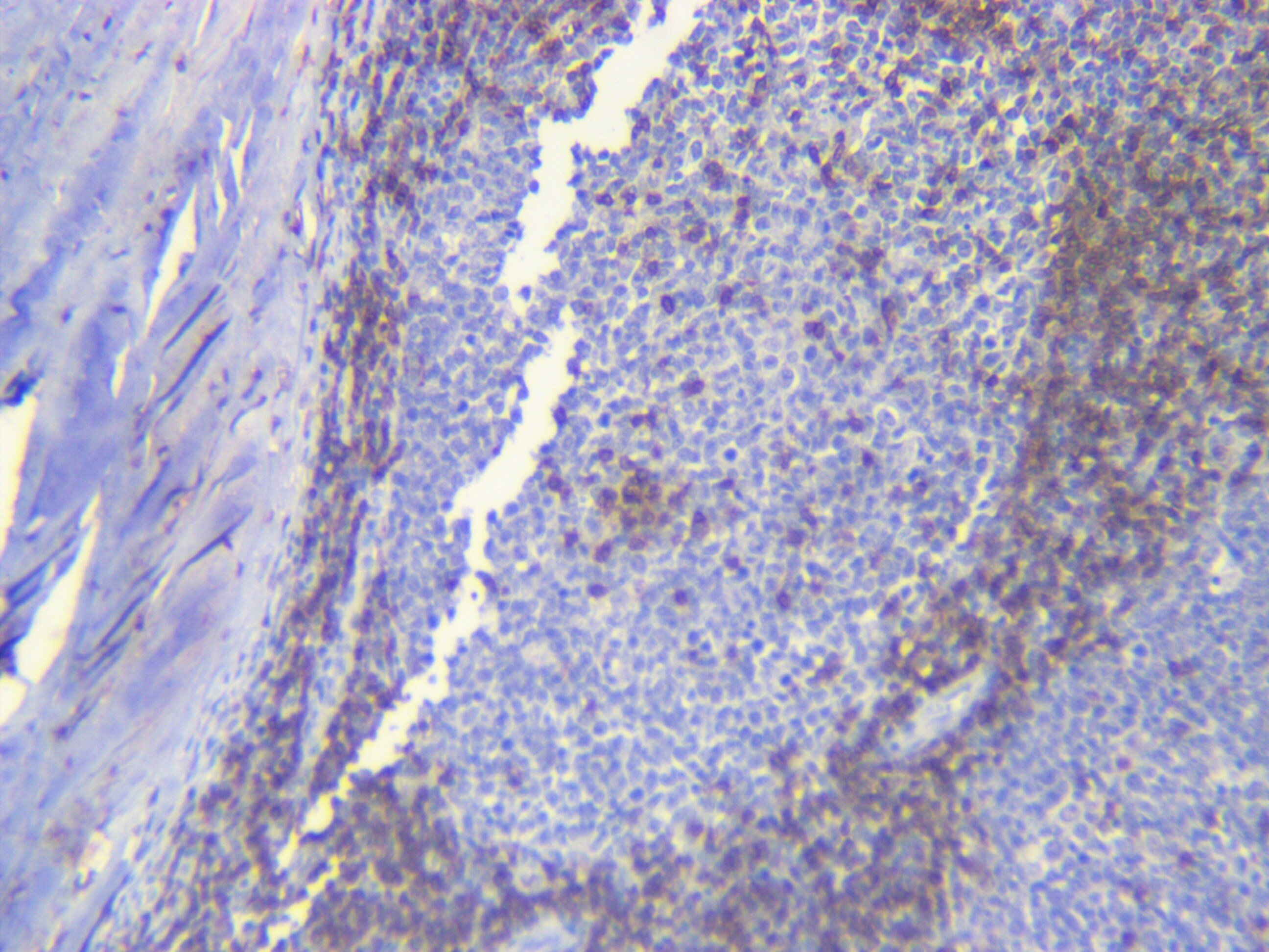
Scientific Data Images for CD3 Antibody (SP7)
Immunohistochemistry-Paraffin: CD3 Antibody (SP7) [NB600-1441]
Immunohistochemistry-Paraffin: CD3 Antibody (SP7) [NB600-1441] - FFPE human tonsil stained with CD3 antibody.![Immunohistochemistry-Frozen: CD3 Antibody (SP7) [NB600-1441]](https://resources.rndsystems.com/images/products/CD3-Antibody-SP7-Immunohistochemistry-Frozen-NB600-1441-img0002.jpg "Immunohistochemistry-Frozen: CD3 Antibody (SP7) [NB600-1441]")
Immunohistochemistry-Frozen: CD3 Antibody (SP7) [NB600-1441]
Immunohistochemistry-Frozen: CD3 Antibody (SP7) [NB600-1441] - T-cells and microglia in mouse spinal cord with acute EAE. Mouse was perfused with 4% paraformaldehyde. Spinal cord was post-fixed overnight, followed by cryoprotection with 30% sucrose for 24h. Spinal cord sections were treated with antigen-retrieval buffer (pH 6.0) at 60C for 10 min, then cooled down to room temperature (this step should not be omitted). Anti-CD3 antibody was diluted at 1:200. Incubation: room temperature overnight. IHC-Fr image submitted by a verified customer review.Applications for CD3 Antibody (SP7)
Application
Recommended Usage
Immunohistochemistry
1:25-1:50
Immunohistochemistry-Frozen
1:10 - 1:500
Immunohistochemistry-Paraffin
1:25-1:50
Application Notes
IHC-P: recommended pretreatment of citrate buffer, pH 6.0. Recommended incubation time of 30-60 min at RT. Use in Immunocytochemistry/immunofluorescence reported in scientific literature (PMID 29037255). Use in FLOW reported in scientific literature (PMID: 31079916).
Reviewed Applications
Read 5 reviews rated 3.6 using NB600-1441 in the following applications:
Flow Cytometry Panel Builder
Bio-Techne Knows Flow Cytometry
Save time and reduce costly mistakes by quickly finding compatible reagents using the Panel Builder Tool.
Advanced Features
- Spectra Viewer - Custom analysis of spectra from multiple fluorochromes
- Spillover Popups - Visualize the spectra of individual fluorochromes
- Antigen Density Selector - Match fluorochrome brightness with antigen density

Formulation, Preparation, and Storage
Purification
Tissue culture supernatant
Formulation
Tissue culture supernatant
Preservative
0.05% Sodium Azide
Concentration
This product is unpurified. The exact concentration of antibody is not quantifiable.
Shipping
The product is shipped with polar packs. Upon receipt, store it immediately at the temperature recommended below.
Stability & Storage
Store at 4C short term. Aliquot and store at -20C long term. Avoid freeze-thaw cycles.
Background: CD3
CD3 proteins are expressed on the surface of thymocytes during thymocyte development, proliferation, and maturation to T-cells (4, 6, 7). During T-cell development CD4-CD8- double negative (DN) cells differentiate to CD4+CD8+ double positive (DP) cells before progressing to single positive (SP) CD4+ helper T-cells or CD8+ cytotoxic T-cells (4, 6, 7). As CD3 plays an important role in thymocyte development, it is understandable that CD3 defects and mutations in CD3 protein chains cause severe combined immunodeficiencies (SCIDs) (8). Additionally, a subset of CD3+ T-cells that co-express CD20 are described in a variety of diseases including rheumatoid arthritis, multiple sclerosis, CD20+ T-cell leukemia/lymphoma, and HIV (9). Clinical trials and animal models have shown that anti-CD3 monoclonal antibodies are a promising treatment modality for inflammatory disorders and autoimmune diseases, such as type I diabetes (10).
References
1. Chetty, R., & Gatter, K. (1994). CD3: structure, function, and role of immunostaining in clinical practice. The Journal of pathology. https://doi.org/10.1002/path.1711730404
2. Mariuzza, R. A., Agnihotri, P., & Orban, J. (2020). The structural basis of T-cell receptor (TCR) activation: An enduring enigma. The Journal of biological chemistry. https://doi.org/10.1074/jbc.REV119.009411
3. Kuhns, M. S., Davis, M. M., & Garcia, K. C. (2006). Deconstructing the form and function of the TCR/CD3 complex. Immunity. https://doi.org/10.1016/j.immuni.2006.01.006
4. Clevers, H., Alarcon, B., Wileman, T., & Terhorst, C. (1988). The T cell receptor/CD3 complex: a dynamic protein ensemble. Annual review of immunology. https://doi.org/10.1146/annurev.iy.06.040188.003213
5. Uniprot: CD3-delta (P04234), CD3-epsilon (P07766), CD3-gamma (P09693), CD3-zeta (P20963)
6. D'Acquisto, F., & Crompton, T. (2011). CD3+CD4-CD8- (double negative) T cells: saviours or villains of the immune response?. Biochemical pharmacology. https://doi.org/10.1016/j.bcp.2011.05.019
7. Dave V. P. (2009). Hierarchical role of CD3 chains in thymocyte development. Immunological reviews. https://doi.org/10.1111/j.1600-065X.2009.00835.x
8. Fischer, A., de Saint Basile, G., & Le Deist, F. (2005). CD3 deficiencies. Current opinion in allergy and clinical immunology. https://doi.org/10.1097/01.all.0000191886.12645.79
9. Chen, Q., Yuan, S., Sun, H., & Peng, L. (2019). CD3+CD20+ T cells and their roles in human diseases. Human immunology. https://doi.org/10.1016/j.humimm.2019.01.001
10. Kuhn, C., & Weiner, H. L. (2016). Therapeutic anti-CD3 monoclonal antibodies: from bench to bedside. Immunotherapy. https://doi.org/10.2217/imt-2016-0049
Alternate Names
CD_antigen: CD3e, CD3 antigen, delta subunit, CD3d antigen, CD3d antigen, delta polypeptide (TiT3 complex), CD3d molecule, delta (CD3-TCR complex), CD3-DELTA, CD3e, CD3e antigen, CD3e antigen, epsilon polypeptide (TiT3 complex), CD3e molecule, epsilon (CD3-TCR complex), CD3-epsilon, CD3G, CD3g antigen, CD3g antigen, gamma polypeptide (TiT3 complex), CD3g molecule, epsilon (CD3-TCR complex), CD3g molecule, gamma (CD3-TCR complex), CD3-GAMMA, FLJ17620, FLJ17664, FLJ18683, FLJ79544, FLJ94613, IMD18, MGC138597, T3DOKT3, delta chain, T3E, T-cell antigen receptor complex, epsilon subunit of T3, T-cell receptor T3 delta chain, T-cell surface antigen T3/Leu-4 epsilon chain, T-cell surface glycoprotein CD3 delta chain, T-cell surface glycoprotein CD3 epsilon chain, TCRE
Gene Symbol
CD3E
Additional CD3 Products
Product Documents for CD3 Antibody (SP7)
Certificate of Analysis
To download a Certificate of Analysis, please enter a lot or batch number in the search box below.
Product Specific Notices for CD3 Antibody (SP7)
This product is for research use only and is not approved for use in humans or in clinical diagnosis. Primary Antibodies are guaranteed for 1 year from date of receipt.
Related Research Areas
Citations for CD3 Antibody (SP7)
 Powered by Bioz
Customer Reviews for CD3 Antibody (SP7) (5)
3.6 out of 5
5 Customer Ratings
Have you used CD3 Antibody (SP7)?
Submit a review and receive an Amazon gift card!
$25/€18/£15/$25CAN/¥2500 Yen for a review with an image
$10/€7/£6/$10CAN/¥1110 Yen for a review without an image
Submit a review
Customer Images
Showing
1
-
5 的
5 reviews
Showing All
Filter By:
-
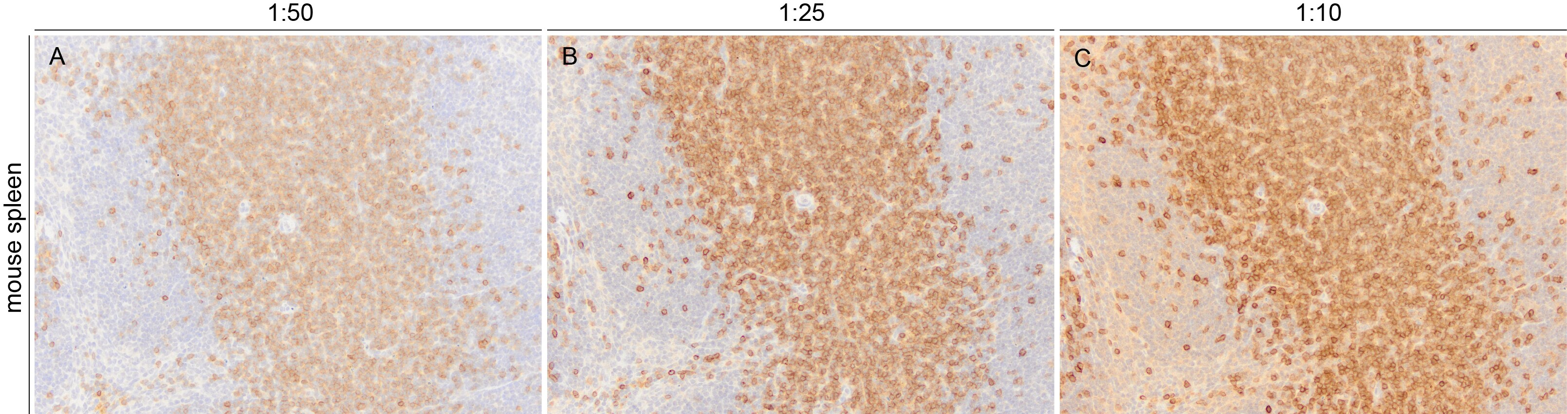
Application: Immunohistochemistry-ParaffinSample Tested: SpleenSpecies: MouseVerified Customer | Posted 03/19/2024CD3 immunoreactivity in sections of mouse FFPE spleen obtained with varying dilutions of NB600-1441. Primary was left on tissue sections for 30m at room temperature.Target Retrieval Solution was used for heat induced epitope retrieval using a vegetable steamer.
 Bio-Techne ResponseThank you for reviewing our product. We are sorry to hear that this product did not perform as expected. We have been in touch with the customer to resolve this issue according to our Product Guarantee and to the customer’s satisfaction.
 -
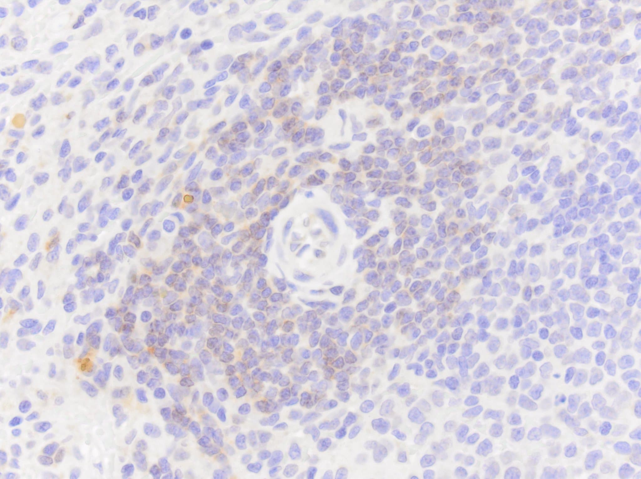
Application: Immunohistochemistry-ParaffinSample Tested: FFPESpecies: DogVerified Customer | Posted 04/06/2023NB600-1441 at 1 to 50 dilution for 1h at room temperature produces weak labeling in dog spleen. Recommend clone CD3-12 instead for detection of dog CD3 in FFPE sections.Tested a variety of concentrations from 1 to 25 to 1 to 800 with an incubation time of 1h at room temperature. NB600-1441 did not produce any signal in any of the 4 sections of dog spleen that were used for testing. The sections were known to contain CD3, as clone CD3-12 produces excellent labeling.
Bio-Techne ResponseThank you for reviewing our product. We are sorry to hear that this product did not perform as expected. We have been in touch with the customer to resolve this issue according to our Product Guarantee and to the customer’s satisfaction.
-
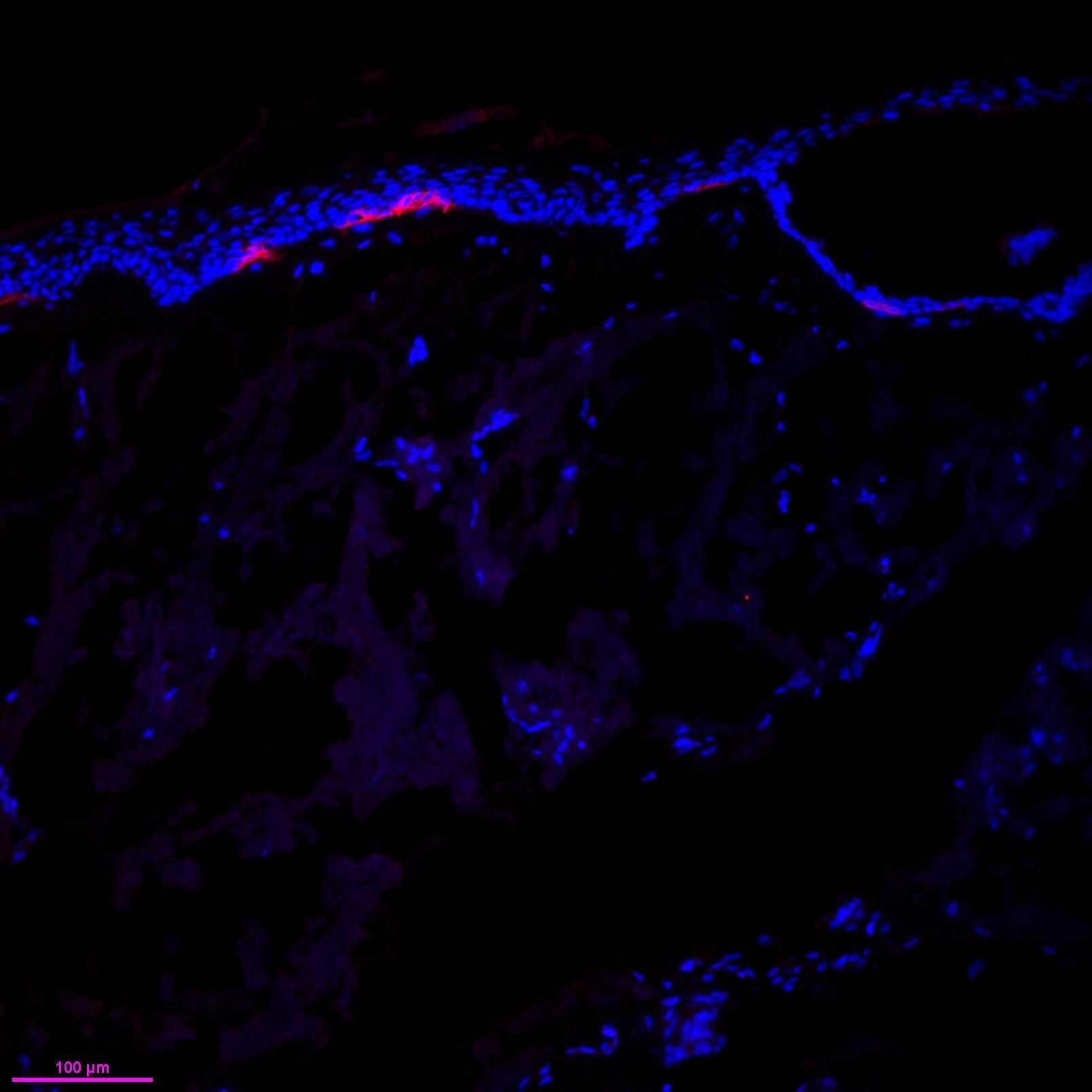
Application: Immunohistochemistry-FrozenSample Tested: Frozen human skin sectionsSpecies: HumanVerified Customer | Posted 08/20/2018CD3 (red, 1:100) stained in frozen human skin section. Counterstained with DAPIHuman skin section fixed with PFA, and permeablized with 0.5% Triton. Sections were blocked with 5% BSA, before anti-CD3 ab was applied at 1:100 for 1hr at 37C. anti-rb-A568 secondary was used, and counterstained with DAPI
-
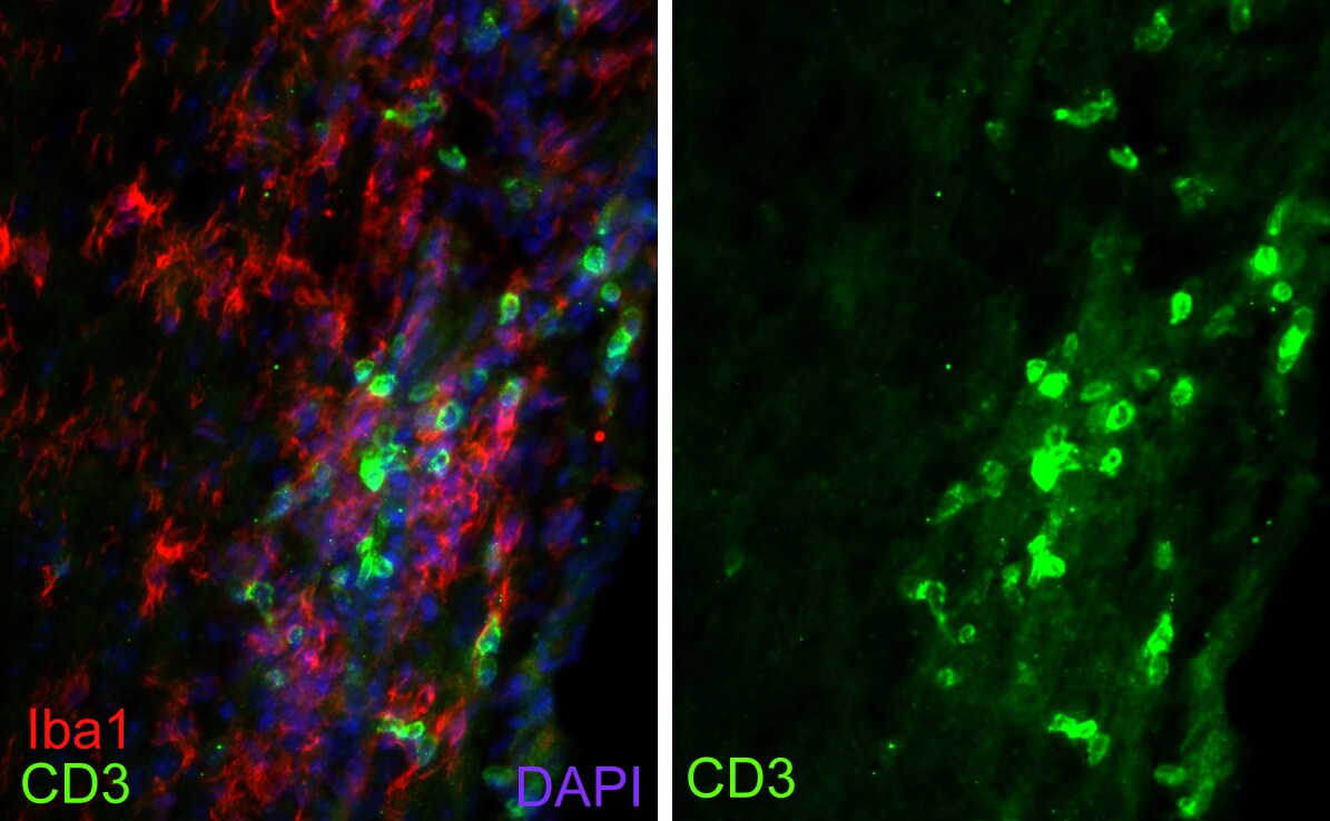
Application: Immunohistochemistry-FrozenSample Tested: mouse spinal cord cryo-sectionsSpecies: MouseVerified Customer | Posted 02/21/2018T-cells and microglia in mouse spinal cord with acute EAEMouse was perfused with 4% paraformaldehyde. Spinal cord was post-fixed overnight, followed by cryoprotection with 30% sucrose for 24h. Spinal cord sections were treated with antigen-retrieval buffer (pH 6.0) at 60C for 10 min, then cooled down to room temperature (this step should not be omitted). Anti-CD3 antibody was diluted at 1:200. Incubation: room temperature overnight.
-
Application: Immunohistochemistry-ParaffinSample Tested: Human Tonsil & Canine lymph paraffin slidesSpecies: Human and CanineVerified Customer | Posted 08/25/2017Human tonsilWorks great in both Human and canine tissues. Sodium citrate pH 6.0 Concentration 1:100
There are no reviews that match your criteria.
Protocols
Find general support by application which include: protocols, troubleshooting, illustrated assays, videos and webinars.
- 7-Amino Actinomycin D (7-AAD) Cell Viability Flow Cytometry Protocol
- Antigen Retrieval Protocol (PIER)
- Antigen Retrieval for Frozen Sections Protocol
- Appropriate Fixation of IHC/ICC Samples
- Cellular Response to Hypoxia Protocols
- Chromogenic IHC Staining of Formalin-Fixed Paraffin-Embedded (FFPE) Tissue Protocol
- Chromogenic Immunohistochemistry Staining of Frozen Tissue
- ClariTSA™ Fluorophore Kits
- Detection & Visualization of Antibody Binding
- Extracellular Membrane Flow Cytometry Protocol
- Flow Cytometry Protocol for Cell Surface Markers
- Flow Cytometry Protocol for Staining Membrane Associated Proteins
- Flow Cytometry Staining Protocols
- Flow Cytometry Troubleshooting Guide
- Fluorescent IHC Staining of Frozen Tissue Protocol
- Graphic Protocol for Heat-induced Epitope Retrieval
- Graphic Protocol for the Preparation and Fluorescent IHC Staining of Frozen Tissue Sections
- Graphic Protocol for the Preparation and Fluorescent IHC Staining of Paraffin-embedded Tissue Sections
- Graphic Protocol for the Preparation of Gelatin-coated Slides for Histological Tissue Sections
- ICC Cell Smear Protocol for Suspension Cells
- ICC Immunocytochemistry Protocol Videos
- ICC for Adherent Cells
- IHC Sample Preparation (Frozen sections vs Paraffin)
- Immunocytochemistry (ICC) Protocol
- Immunocytochemistry Troubleshooting
- Immunofluorescence of Organoids Embedded in Cultrex Basement Membrane Extract
- Immunofluorescent IHC Staining of Formalin-Fixed Paraffin-Embedded (FFPE) Tissue Protocol
- Immunohistochemistry (IHC) and Immunocytochemistry (ICC) Protocols
- Immunohistochemistry Frozen Troubleshooting
- Immunohistochemistry Paraffin Troubleshooting
- Intracellular Flow Cytometry Protocol Using Alcohol (Methanol)
- Intracellular Flow Cytometry Protocol Using Detergents
- Intracellular Nuclear Staining Flow Cytometry Protocol Using Detergents
- Intracellular Staining Flow Cytometry Protocol Using Alcohol Permeabilization
- Intracellular Staining Flow Cytometry Protocol Using Detergents to Permeabilize Cells
- Preparing Samples for IHC/ICC Experiments
- Preventing Non-Specific Staining (Non-Specific Binding)
- Primary Antibody Selection & Optimization
- Propidium Iodide Cell Viability Flow Cytometry Protocol
- Protocol for Heat-Induced Epitope Retrieval (HIER)
- Protocol for Liperfluo
- Protocol for Making a 4% Formaldehyde Solution in PBS
- Protocol for VisUCyte™ HRP Polymer Detection Reagent
- Protocol for the Characterization of Human Th22 Cells
- Protocol for the Characterization of Human Th9 Cells
- Protocol for the Fluorescent ICC Staining of Cell Smears - Graphic
- Protocol for the Fluorescent ICC Staining of Cultured Cells on Coverslips - Graphic
- Protocol for the Preparation & Fixation of Cells on Coverslips
- Protocol for the Preparation and Chromogenic IHC Staining of Frozen Tissue Sections
- Protocol for the Preparation and Chromogenic IHC Staining of Frozen Tissue Sections - Graphic
- Protocol for the Preparation and Chromogenic IHC Staining of Paraffin-embedded Tissue Sections
- Protocol for the Preparation and Chromogenic IHC Staining of Paraffin-embedded Tissue Sections - Graphic
- Protocol for the Preparation and Fluorescent ICC Staining of Cells on Coverslips
- Protocol for the Preparation and Fluorescent ICC Staining of Non-adherent Cells
- Protocol for the Preparation and Fluorescent ICC Staining of Stem Cells on Coverslips
- Protocol for the Preparation and Fluorescent IHC Staining of Frozen Tissue Sections
- Protocol for the Preparation and Fluorescent IHC Staining of Paraffin-embedded Tissue Sections
- Protocol for the Preparation of Gelatin-coated Slides for Histological Tissue Sections
- Protocol for the Preparation of a Cell Smear for Non-adherent Cell ICC - Graphic
- Protocol: Annexin V and PI Staining by Flow Cytometry
- Protocol: Annexin V and PI Staining for Apoptosis by Flow Cytometry
- TUNEL and Active Caspase-3 Detection by IHC/ICC Protocol
- The Importance of IHC/ICC Controls
- Troubleshooting Guide: Fluorokine Flow Cytometry Kits
- Troubleshooting Guide: Immunohistochemistry
- View all Protocols, Troubleshooting, Illustrated assays and Webinars


