CENPF Antibody - BSA Free
Novus Biologicals | Catalog # NB500-101
![Knockdown Validated: CENPF Antibody - BSA Free [NB500-101]](https://resources.rndsystems.com/images/products/CENPF-Antibody-Immunohistochemistry-Paraffin-NB500-101-img0009.jpg "Immunocytochemistry/ Immunofluorescence: CENPF Antibody - BSA Free [NB500-101]")
Key Product Details
Validated by
Species Reactivity
Validated:
Cited:
Applications
Validated:
Cited:
Label
Antibody Source
Format
Product Specifications
Immunogen
Localization
Clonality
Host
Isotype
Scientific Data Images for CENPF Antibody - BSA Free
![Western Blot: CENPF AntibodyBSA Free [NB500-101]](https://resources.rndsystems.com/images/products/CENPF-Antibody-Western-Blot-NB500-101-img0004.jpg "Western Blot: CENPF AntibodyBSA Free [NB500-101]")
![Immunocytochemistry/ Immunofluorescence: CENPF Antibody - BSA Free [NB500-101]](https://resources.rndsystems.com/images/products/CENPF-Antibody-Immunocytochemistry-Immunofluorescence-NB500-101-img0008.jpg "Immunocytochemistry/ Immunofluorescence: CENPF Antibody - BSA Free [NB500-101]")
Immunocytochemistry/ Immunofluorescence: CENPF Antibody - BSA Free [NB500-101]
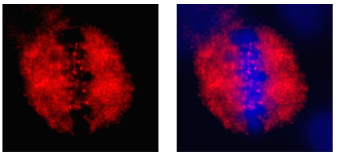
Immunocytochemistry/Immunofluorescence: CENPF Antibody [NB500-101] - Distribution of CENP-F (red), largely associated with the mitotic spindle and a clearly distinguishable frac.![Immunocytochemistry/ Immunofluorescence: CENPF Antibody - BSA Free [NB500-101]](https://resources.rndsystems.com/images/products/CENPF-Antibody-Immunocytochemistry-Immunofluorescence-NB500-101-img0006.jpg "Immunocytochemistry/ Immunofluorescence: CENPF Antibody - BSA Free [NB500-101]")
Immunocytochemistry/ Immunofluorescence: CENPF Antibody - BSA Free [NB500-101]
Immunocytochemistry/Immunofluorescence: CENPF Antibody [NB500-101] - CENPF antibody was tested in HeLa cells with Dylight 488 (green). Nuclei and alpha-tubulin were counterstained with DAPI (blue) and Dylight 550 (red).![Immunocytochemistry/ Immunofluorescence: CENPF Antibody - BSA Free [NB500-101]](https://resources.rndsystems.com/images/products/CENPF-Antibody-Immunocytochemistry-Immunofluorescence-NB500-101-img0007.jpg "Immunocytochemistry/ Immunofluorescence: CENPF Antibody - BSA Free [NB500-101]")
Immunocytochemistry/ Immunofluorescence: CENPF Antibody - BSA Free [NB500-101]
Immunocytochemistry/Immunofluorescence: CENPF Antibody [NB500-101] - Confocal immunofluorescent analysis of HeLa cells using CENPF antibody (NB500-101, 1:5). An Alexa Fluor 488-conjugated Goat to rabbit IgG was used as secondary antibody (green, A). Actin filaments were labeled with Alexa Fluor 568 phalloidin (red, B). DAPI was used to stain the cell nuclei (blue, C).
Western Blot: CENPF Antibody - BSA Free [NB500-101] -
Requirements for CENP-F kinetochore localization.A, representative images of mitotic HeLa cells electroporated with mCherry, mCherry-CENP-F2688-C WT, or mCherry-CENP-F2688-C/C3207A (farnesylation mutant). Scale bar, 5 μm. As for CENP-E, both the WT and the unfarnesylated mutant CENP-F constructs localize at kinetochore. B, mCherry-CENP-F2688-C sample was visualized by EM after glycerol spraying and low-angle platinum shadowing (right panel). The elongated shape of the observed particles is consistent with the secondary structure expected for the mCherry-tag coiled-coil construct (right panel). C and D, SEC elution profiles and SDS-PAGE analysis of binding experiments with 16 μm each of mCherry-CENP-F2688-C WT (C) or C2961S mutant (D) and 4 μm BUB1FL/BUB3 complex. The shift in elution volume of BUB1/BUB3 is observed with both WT and mutant CENP-F, but it is significantly less pronounced for the CENP-F mutant, suggesting that the C2961S mutation reduces the affinity of CENP-F for the BUB1 kinase domain without completely abolishing it. E, SEC elution profile and SDS-PAGE analysis of a binding experiment with 16 μm each of mCherry-CENP-F2688-C and eGFP-CENP-E2070-C. No shift is observed, indicating that the tested constructs do not interact. The elution profile and SDS-PAGE of eGFP-CENP-E2070-C WT in E is the same already shown in Fig. 3, B and D. Similarly, the elution profiles and SDS-PAGE of mCherry-CENP-F2688-C in E is the same as in Fig. 4, D, E, and G. Similarly, the elution profiles and SDS-PAGE of BUB1/BUB3 in C and D are the same. These repetitions were included to facilitate the interpretation of binding experiments by inclusion of elution references. Image collected and cropped by CiteAb from the following open publication (https://pubmed.ncbi.nlm.nih.gov/29748388), licensed under a CC-BY license. Not internally tested by Novus Biologicals.
Immunohistochemistry: CENPF Antibody - BSA Free [NB500-101] -
Kinetochore localization of RZZ and MAD1 are independent of CENP-E and CENP-F.A–H, representative images and quantification of protein kinetochore levels in HeLa cells mock-treated or depleted of Zwilch (A), CENP-E (B, E, and H), CENP-F (C, F, and G), or co-depleted of CENP-E and CENP-F (D). Scale bar, 10 μm. Zwilch depletion does not affect the localization of CENP-E (A). CENP-E depletion does not affect the localization of Zwilch (B), MAD1 (E), and CENP-F (H). Similarly, CENP-F depletion does not interfere with the recruitment of Zwilch (C), MAD1 (F), and CENP-E (G). Co-depletion of CENP-E and CENP-F has no effects on localization of Zwilch (D). The graphs show mean intensity of one (B, C, and E), two (D and F), or three (A, G, and H) experiments; the error bars indicate S.E., and the mean values for nondepleted cells are set to 1. Elements in the left column of A (negative controls of the RNAi experiments) are also shown in Fig. S3C. Elements in G are shown again in Fig. S2E. Image collected and cropped by CiteAb from the following open publication (https://pubmed.ncbi.nlm.nih.gov/29748388), licensed under a CC-BY license. Not internally tested by Novus Biologicals.Applications for CENPF Antibody - BSA Free
Flow Cytometry
Immunocytochemistry/ Immunofluorescence
Immunohistochemistry Whole-Mount
Immunohistochemistry-Frozen
Immunohistochemistry-Paraffin
Immunoprecipitation
Western Blot
Reviewed Applications
Read 2 reviews rated 5 using NB500-101 in the following applications:
Flow Cytometry Panel Builder
Bio-Techne Knows Flow Cytometry
Save time and reduce costly mistakes by quickly finding compatible reagents using the Panel Builder Tool.
Advanced Features
- Spectra Viewer - Custom analysis of spectra from multiple fluorochromes
- Spillover Popups - Visualize the spectra of individual fluorochromes
- Antigen Density Selector - Match fluorochrome brightness with antigen density

Formulation, Preparation, and Storage
Purification
Formulation
Format
Preservative
Concentration
Shipping
Stability & Storage
Background: CENPF
Alternate Names
Gene Symbol
Additional CENPF Products
Product Documents for CENPF Antibody - BSA Free
Certificate of Analysis
To download a Certificate of Analysis, please enter a lot or batch number in the search box below.
Product Specific Notices for CENPF Antibody - BSA Free
This product is for research use only and is not approved for use in humans or in clinical diagnosis. Primary Antibodies are guaranteed for 1 year from date of receipt.
Citations for CENPF Antibody - BSA Free
 Powered by Bioz
Customer Reviews for CENPF Antibody - BSA Free (2)
Have you used CENPF Antibody - BSA Free?
Submit a review and receive an Amazon gift card!
$25/€18/£15/$25CAN/¥2500 Yen for a review with an image
$10/€7/£6/$10CAN/¥1110 Yen for a review without an image
Submit a review
Customer Images
-(005-ml)_NB500-101_8041.bmp)
-
Application: ImmunofluorescenceSample Tested:Species: HumanVerified Customer | Posted 03/24/2015The image shows the distribution of CENP-F (red), largely associated with the mitotic spindle and a clearly distinguishable frac

-
Application: ImmunocytochemistrySample Tested:Species: HumanVerified Customer | Posted 06/05/2014Confocal immunofluorescent analysis of HeLa cells using CENPF antibody (NB500-101, 1:5).
There are no reviews that match your criteria.
Protocols
View specific protocols for CENPF Antibody - BSA Free (NB500-101):
Western Blot Procedure
1) Resolve aliquots (30 mg) of total HeLa cell lysate on a 8.5% lo-crosslinker gel (110:1, acryl:bis) for efficient separation and transfer.
2) Transfer to nitrocellulose membranes in 20 mM Tris-HCL (pH 8.0)/150 mM glycine/20% (vol/vol) methanol. Transfer time for a 1 mm thick gel is 1 hour 15 minutes at 450mA.
3) Block membranes for 1 hour with 5% (vol/vol) nonfat dry milk/TBS-T (20 mM Tris-HCL, pH 7.6/ 137 mM NaCl/0.1% TWEEN 20)
4) Incubate membranes for 1 hour at room temperature (RT) in NB 500-101 diluted 1:3000 in nonfat dry milk/TBS-T.
5) Wash 3X30 minutes at RT with TBS-T.
6) Incubate membranes with alkaline phosphatase conjugated anti-rabbit IgG for 1 hour (RT).
7) Wash 3 x 30 minutes at RT with TBS-T
8) Develop with ECL reagents (Amersham) and autoradiography.
Find general support by application which include: protocols, troubleshooting, illustrated assays, videos and webinars.
- 7-Amino Actinomycin D (7-AAD) Cell Viability Flow Cytometry Protocol
- Antigen Retrieval Protocol (PIER)
- Antigen Retrieval for Frozen Sections Protocol
- Appropriate Fixation of IHC/ICC Samples
- Cellular Response to Hypoxia Protocols
- Chromogenic IHC Staining of Formalin-Fixed Paraffin-Embedded (FFPE) Tissue Protocol
- Chromogenic Immunohistochemistry Staining of Frozen Tissue
- ClariTSA™ Fluorophore Kits
- Detection & Visualization of Antibody Binding
- Extracellular Membrane Flow Cytometry Protocol
- Flow Cytometry Protocol for Cell Surface Markers
- Flow Cytometry Protocol for Staining Membrane Associated Proteins
- Flow Cytometry Staining Protocols
- Flow Cytometry Troubleshooting Guide
- Fluorescent IHC Staining of Frozen Tissue Protocol
- Graphic Protocol for Heat-induced Epitope Retrieval
- Graphic Protocol for the Preparation and Fluorescent IHC Staining of Frozen Tissue Sections
- Graphic Protocol for the Preparation and Fluorescent IHC Staining of Paraffin-embedded Tissue Sections
- Graphic Protocol for the Preparation of Gelatin-coated Slides for Histological Tissue Sections
- ICC Cell Smear Protocol for Suspension Cells
- ICC Immunocytochemistry Protocol Videos
- ICC for Adherent Cells
- IHC Sample Preparation (Frozen sections vs Paraffin)
- Immunocytochemistry (ICC) Protocol
- Immunocytochemistry Troubleshooting
- Immunofluorescence of Organoids Embedded in Cultrex Basement Membrane Extract
- Immunofluorescent IHC Staining of Formalin-Fixed Paraffin-Embedded (FFPE) Tissue Protocol
- Immunohistochemistry (IHC) and Immunocytochemistry (ICC) Protocols
- Immunohistochemistry Frozen Troubleshooting
- Immunohistochemistry Paraffin Troubleshooting
- Immunoprecipitation Protocol
- Intracellular Flow Cytometry Protocol Using Alcohol (Methanol)
- Intracellular Flow Cytometry Protocol Using Detergents
- Intracellular Nuclear Staining Flow Cytometry Protocol Using Detergents
- Intracellular Staining Flow Cytometry Protocol Using Alcohol Permeabilization
- Intracellular Staining Flow Cytometry Protocol Using Detergents to Permeabilize Cells
- Preparing Samples for IHC/ICC Experiments
- Preventing Non-Specific Staining (Non-Specific Binding)
- Primary Antibody Selection & Optimization
- Propidium Iodide Cell Viability Flow Cytometry Protocol
- Protocol for Heat-Induced Epitope Retrieval (HIER)
- Protocol for Liperfluo
- Protocol for Making a 4% Formaldehyde Solution in PBS
- Protocol for VisUCyte™ HRP Polymer Detection Reagent
- Protocol for the Characterization of Human Th22 Cells
- Protocol for the Characterization of Human Th9 Cells
- Protocol for the Fluorescent ICC Staining of Cell Smears - Graphic
- Protocol for the Fluorescent ICC Staining of Cultured Cells on Coverslips - Graphic
- Protocol for the Preparation & Fixation of Cells on Coverslips
- Protocol for the Preparation and Chromogenic IHC Staining of Frozen Tissue Sections
- Protocol for the Preparation and Chromogenic IHC Staining of Frozen Tissue Sections - Graphic
- Protocol for the Preparation and Chromogenic IHC Staining of Paraffin-embedded Tissue Sections
- Protocol for the Preparation and Chromogenic IHC Staining of Paraffin-embedded Tissue Sections - Graphic
- Protocol for the Preparation and Fluorescent ICC Staining of Cells on Coverslips
- Protocol for the Preparation and Fluorescent ICC Staining of Non-adherent Cells
- Protocol for the Preparation and Fluorescent ICC Staining of Stem Cells on Coverslips
- Protocol for the Preparation and Fluorescent IHC Staining of Frozen Tissue Sections
- Protocol for the Preparation and Fluorescent IHC Staining of Paraffin-embedded Tissue Sections
- Protocol for the Preparation of Gelatin-coated Slides for Histological Tissue Sections
- Protocol for the Preparation of a Cell Smear for Non-adherent Cell ICC - Graphic
- Protocol: Annexin V and PI Staining by Flow Cytometry
- Protocol: Annexin V and PI Staining for Apoptosis by Flow Cytometry
- R&D Systems Quality Control Western Blot Protocol
- TUNEL and Active Caspase-3 Detection by IHC/ICC Protocol
- The Importance of IHC/ICC Controls
- Troubleshooting Guide: Fluorokine Flow Cytometry Kits
- Troubleshooting Guide: Immunohistochemistry
- Troubleshooting Guide: Western Blot Figures
- Western Blot Conditions
- Western Blot Protocol
- Western Blot Protocol for Cell Lysates
- Western Blot Troubleshooting
- Western Blot Troubleshooting Guide
- View all Protocols, Troubleshooting, Illustrated assays and Webinars
FAQs for CENPF Antibody - BSA Free
-
Q: Could you please check whether you can provide which region/sequence this antibody is raised against in the C-terminus of CENPF?
A: Unfortunately, I am not able to provide sequence information further than what we have published. I apologize for the inconvenience.
-
Q: What residues of human CENP-F was NB500-101 generated against?
A: The exact immunogen sequence is proprietary and cannot be released.
-
Q: Could you please check whether you can provide which region/sequence this antibody is raised against in the C-terminus of CENPF?
A: Unfortunately, I am not able to provide sequence information further than what we have published. I apologize for the inconvenience.
-
Q: What residues of human CENP-F was NB500-101 generated against?
A: The exact immunogen sequence is proprietary and cannot be released.