A disintegrin and metalloproteinase with thrombospondin motifs 15 (ADAMTS15) is a secreted multi-domain protease that is primarily expressed in fetal liver and kidney. ADAMTS1, 4, 5, 8, and 15 form a subfamily of ADAMTS proteases that possess aggrecanase activity. These proteins are synthesized as zymogens which have a pro-domain that is removed by furin-like protein convertases. ADAMTS15 functions as a suppressor of tumor growth and invasion. It is downregulated in colon cancer, and its expression in breast cancer correlates with poor prognosis. Within amino acids (aa) 18-682 (which includes the propeptide, peptidase, disintegrin, first TSP-1 and most of the Cys-rich domain), human ADAMTS15 shares 95% aa sequence identity with mouse and rat ADAMTS15.
Human ADAMTS15 Antibody (561819)
R&D Systems | Catalog # MAB5149

Key Product Details
Species Reactivity
Validated:
Human
Cited:
Human
Applications
Validated:
Immunohistochemistry, Western Blot, Flow Cytometry, CyTOF-ready
Cited:
Flow Cytometry
Label
Unconjugated
Antibody Source
Monoclonal Mouse IgG2B Clone # 561819
Loading...
Product Specifications
Immunogen
Chinese hamster ovary cell line CHO-derived recombinant human ADAMTS15
Gly18-Cys682
Accession # Q8TE58
Gly18-Cys682
Accession # Q8TE58
Specificity
Detects human ADAMTS15 in direct ELISAs and Western blots.
Clonality
Monoclonal
Host
Mouse
Isotype
IgG2B
Scientific Data Images for Human ADAMTS15 Antibody (561819)
Detection of Human ADAMTS15 by Western Blot.
Western blot shows lysates of MCF-7 human breast cancer cell line. PVDF Membrane was probed with 2 µg/mL of Human ADAMTS15 Monoclonal Antibody (Catalog # MAB5149) followed by HRP-conjugated Anti-Mouse IgG Secondary Antibody (Catalog # HAF007). A specific band was detected for mature ADAMTS15 at approximately 75 kDa (as indicated). This experiment was conducted under reducing conditions and using Immunoblot Buffer Group 1.
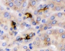
ADAMTS15 in Human Kidney.
ADAMTS15 was detected in immersion fixed paraffin-embedded sections of human kidney using Human ADAMTS15 Monoclonal Antibody (Catalog # MAB5149) at 15 µg/mL overnight at 4 °C. Before incubation with the primary antibody, tissue was subjected to heat-induced epitope retrieval using Antigen Retrieval Reagent-Basic (Catalog # CTS013). Tissue was stained using the Anti-Mouse HRP-DAB Cell & Tissue Staining Kit (brown; Catalog # CTS002) and counterstained with hematoxylin (blue). Specific staining was localized to cytoplasm. View our protocol for Chromogenic IHC Staining of Paraffin-embedded Tissue Sections.
Detection of ADAMTS15 in MCF‑7 Human Cell Line by Flow Cytometry.
MCF-7 human breast cancer cell line was stained with Human ADAMTS15 Monoclonal Antibody (Catalog # MAB5149, filled histogram) or isotype control antibody (Catalog # MAB0041, open histogram), followed by Phycoerythrin-conjugated Anti-Mouse IgG F(ab')2Secondary Antibody (Catalog # F0102B).Applications for Human ADAMTS15 Antibody (561819)
Application
Recommended Usage
CyTOF-ready
Ready to be labeled using established conjugation methods. No BSA or other carrier proteins that could interfere with conjugation.
Flow Cytometry
2.5 µg/106 cells
Sample: MCF‑7 human breast cancer cell line
Sample: MCF‑7 human breast cancer cell line
Immunohistochemistry
8-25 µg/mL
Sample: Immersion fixed paraffin-embedded sections of human kidney
Sample: Immersion fixed paraffin-embedded sections of human kidney
Western Blot
2 µg/mL
Sample: MCF‑7 human breast cancer cell line
Sample: MCF‑7 human breast cancer cell line
Reviewed Applications
Read 3 reviews rated 4.3 using MAB5149 in the following applications:
Flow Cytometry Panel Builder
Bio-Techne Knows Flow Cytometry
Save time and reduce costly mistakes by quickly finding compatible reagents using the Panel Builder Tool.
Advanced Features
- Spectra Viewer - Custom analysis of spectra from multiple fluorochromes
- Spillover Popups - Visualize the spectra of individual fluorochromes
- Antigen Density Selector - Match fluorochrome brightness with antigen density

Formulation, Preparation, and Storage
Purification
Protein A or G purified from hybridoma culture supernatant
Reconstitution
Sterile PBS to a final concentration of 0.5 mg/mL. For liquid material, refer to CoA for concentration.
Loading...
Formulation
Lyophilized from a 0.2 μm filtered solution in PBS with Trehalose. *Small pack size (SP) is supplied either lyophilized or as a 0.2 µm filtered solution in PBS.
Shipping
Lyophilized product is shipped at ambient temperature. Liquid small pack size (-SP) is shipped with polar packs. Upon receipt, store immediately at the temperature recommended below.
Stability & Storage
Use a manual defrost freezer and avoid repeated freeze-thaw cycles.
- 12 months from date of receipt, -20 to -70 °C as supplied.
- 1 month, 2 to 8 °C under sterile conditions after reconstitution.
- 6 months, -20 to -70 °C under sterile conditions after reconstitution.
Calculators
Background: ADAMTS15
Long Name
A Disintegrin-like and Metalloproteinase Domain with Thrombospondin Motifs 15
Alternate Names
A disintegrin and metalloproteinase with thrombospondin motifs 15, a disintegrin-like and metalloprotease (reprolysin type) with thrombospondintype 1 motif, 15, a disintegrin-like and metalloprotease (reprolysin type) with thrombospondintype 1 motif, 15, preproprotein, ADAM metallopeptidase with thrombospondin type 1 motif, 15, ADAM-TS 15, ADAM-TS15, ADAMTS-15, EC 3.4.24, EC 3.4.24.-, EC 3.4.24.14, EC 3.4.24.82, metalloprotease disintegrin 15 with thrombospondin domains, MGC126403
Gene Symbol
ADAMTS15
UniProt
Additional ADAMTS15 Products
Product Documents for Human ADAMTS15 Antibody (561819)
Certificate of Analysis
To download a Certificate of Analysis, please enter a lot or batch number in the search box below.
Note: Certificate of Analysis not available for kit components.
Product Specific Notices for Human ADAMTS15 Antibody (561819)
For research use only
Related Research Areas
Citations for Human ADAMTS15 Antibody (561819)
 Powered by Bioz
Customer Reviews for Human ADAMTS15 Antibody (561819) (3)
4.3 out of 5
3 Customer Ratings
Have you used Human ADAMTS15 Antibody (561819)?
Submit a review and receive an Amazon gift card!
$25/€18/£15/$25CAN/¥2500 Yen for a review with an image
$10/€7/£6/$10CAN/¥1110 Yen for a review without an image
Submit a review
Customer Images


Showing
1
-
3 的
3 reviews
Showing All
Filter By:
-
Application: ImmunohistochemistrySample Tested: Kidney tissueSpecies: HumanVerified Customer | Posted 07/19/2022

-
Application: ELISASample Tested: Serum and PlasmaSpecies: HumanVerified Customer | Posted 09/23/2019I used this antibody for developing a sandwich ELISA in combination with pAb (cat. AF5149) and protein (cat. 5149). This combination works well for a sandwich ELISA detection human ADAMTS 15 in serum.
-
Application: ELISASample Tested: Serum and PlasmaSpecies: HumanVerified Customer | Posted 09/23/2019I used this protein as a standard for developing an ELISA in combination with pAb (cat. AF5149) and mAb (cat. MAB 5149). This combination works well for a sandwich ELISA detection human ADAMTS 14 in serum.
There are no reviews that match your criteria.
Protocols
Find general support by application which include: protocols, troubleshooting, illustrated assays, videos and webinars.
- 7-Amino Actinomycin D (7-AAD) Cell Viability Flow Cytometry Protocol
- Antigen Retrieval Protocol (PIER)
- Antigen Retrieval for Frozen Sections Protocol
- Appropriate Fixation of IHC/ICC Samples
- Cellular Response to Hypoxia Protocols
- Chromogenic IHC Staining of Formalin-Fixed Paraffin-Embedded (FFPE) Tissue Protocol
- Chromogenic Immunohistochemistry Staining of Frozen Tissue
- ClariTSA™ Fluorophore Kits
- Detection & Visualization of Antibody Binding
- Extracellular Membrane Flow Cytometry Protocol
- Flow Cytometry Protocol for Cell Surface Markers
- Flow Cytometry Protocol for Staining Membrane Associated Proteins
- Flow Cytometry Staining Protocols
- Flow Cytometry Troubleshooting Guide
- Fluorescent IHC Staining of Frozen Tissue Protocol
- Graphic Protocol for Heat-induced Epitope Retrieval
- Graphic Protocol for the Preparation and Fluorescent IHC Staining of Frozen Tissue Sections
- Graphic Protocol for the Preparation and Fluorescent IHC Staining of Paraffin-embedded Tissue Sections
- Graphic Protocol for the Preparation of Gelatin-coated Slides for Histological Tissue Sections
- IHC Sample Preparation (Frozen sections vs Paraffin)
- Immunofluorescent IHC Staining of Formalin-Fixed Paraffin-Embedded (FFPE) Tissue Protocol
- Immunohistochemistry (IHC) and Immunocytochemistry (ICC) Protocols
- Immunohistochemistry Frozen Troubleshooting
- Immunohistochemistry Paraffin Troubleshooting
- Intracellular Flow Cytometry Protocol Using Alcohol (Methanol)
- Intracellular Flow Cytometry Protocol Using Detergents
- Intracellular Nuclear Staining Flow Cytometry Protocol Using Detergents
- Intracellular Staining Flow Cytometry Protocol Using Alcohol Permeabilization
- Intracellular Staining Flow Cytometry Protocol Using Detergents to Permeabilize Cells
- Preparing Samples for IHC/ICC Experiments
- Preventing Non-Specific Staining (Non-Specific Binding)
- Primary Antibody Selection & Optimization
- Propidium Iodide Cell Viability Flow Cytometry Protocol
- Protocol for Heat-Induced Epitope Retrieval (HIER)
- Protocol for Liperfluo
- Protocol for Making a 4% Formaldehyde Solution in PBS
- Protocol for VisUCyte™ HRP Polymer Detection Reagent
- Protocol for the Characterization of Human Th22 Cells
- Protocol for the Characterization of Human Th9 Cells
- Protocol for the Preparation & Fixation of Cells on Coverslips
- Protocol for the Preparation and Chromogenic IHC Staining of Frozen Tissue Sections
- Protocol for the Preparation and Chromogenic IHC Staining of Frozen Tissue Sections - Graphic
- Protocol for the Preparation and Chromogenic IHC Staining of Paraffin-embedded Tissue Sections
- Protocol for the Preparation and Chromogenic IHC Staining of Paraffin-embedded Tissue Sections - Graphic
- Protocol for the Preparation and Fluorescent IHC Staining of Frozen Tissue Sections
- Protocol for the Preparation and Fluorescent IHC Staining of Paraffin-embedded Tissue Sections
- Protocol for the Preparation of Gelatin-coated Slides for Histological Tissue Sections
- Protocol: Annexin V and PI Staining by Flow Cytometry
- Protocol: Annexin V and PI Staining for Apoptosis by Flow Cytometry
- R&D Systems Quality Control Western Blot Protocol
- TUNEL and Active Caspase-3 Detection by IHC/ICC Protocol
- The Importance of IHC/ICC Controls
- Troubleshooting Guide: Fluorokine Flow Cytometry Kits
- Troubleshooting Guide: Immunohistochemistry
- Troubleshooting Guide: Western Blot Figures
- Western Blot Conditions
- Western Blot Protocol
- Western Blot Protocol for Cell Lysates
- Western Blot Troubleshooting
- Western Blot Troubleshooting Guide
- View all Protocols, Troubleshooting, Illustrated assays and Webinars
Loading...