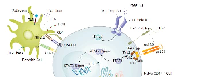
Scientific images may be resized, cropped, or adjusted for brightness or contrast for presentation purposes. These updates are limited to presentation and do not affect the underlying scientific interpretation of the data. Where available, additional source data may be provided upon request.
Key Product Details
Validated by
Species Reactivity
Validated:
Cited:
Applications
Validated:
Cited:
Label
Antibody Source
Product Specifications
Immunogen
Pro29-Met212
Accession # P05231
Specificity
Clonality
Host
Isotype
Endotoxin Level
Scientific Data Images for Human IL-6 Antibody
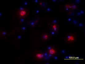
IL‑6 in Human PBMCs.
IL-6 was detected in immersion fixed human peripheral blood mononuclear cells (PBMCs) treated with LPS and monensin using Goat Anti-Human IL-6 Antigen Affinity-purified Polyclonal Antibody (Catalog # AF-206-NA) at 10 µg/mL for 3 hours at room temperature. Cells were stained using the NorthernLights™ 557-conjugated Anti-Goat IgG Secondary Antibody (red; Catalog # NL001) and counter-stained with DAPI (blue). View our protocol for Fluorescent ICC Staining of Non-adherent Cells.
Cell Proliferation Induced by IL‑6 and Neutralization by Human IL‑6 Antibody.
Recombinant Human IL-6 (Catalog # 206-IL) stimulates proliferation in the T1165.85.2.1 mouse plasmacytoma cell line in a dose-dependent manner (orange line). Proliferation elicited by Recombinant Human IL-6 (2.5 ng/mL) is neutralized (green line) by increasing concentrations of Goat Anti-Human IL-6 Antigen Affinity-purified Polyclonal Antibody (Catalog # AF-206-NA). The ND50 is typically ≤ 125 ng/mL.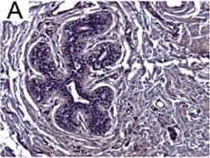
Detection of Human IL-6 by Immunohistochemistry
Breast DCIS cells overexpress proinflammatory markers. Representative images of immunohistochemistry targeting IL-6 protein in normal breast tissue (a), and breast DCIS (b) (N = 61). c and d Hematoxylin staining of serial sections from tissue shown in panel A and B. All images are 20X magnification. Scale bar equals 100 μm. e Evaluation of IL-6 gene expression in DCIS cells via qRT-PCR; the isogenic MCF10.DCIS cells and the non-isogenic SUM102 cell line were analyzed against the non-transformed MCF10A cell line (N = 3). f Secretion of IL-6 protein from DCIS cell lines and non-transformed MCF10A cells as determined by ELISA. *P < 0.05, Student’s t-test; mean ± SD Image collected and cropped by CiteAb from the following publication (https://pubmed.ncbi.nlm.nih.gov/26268945), licensed under a CC-BY license. Not internally tested by R&D Systems.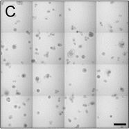
Detection of Human IL-6 by Block/Neutralize
IL-6 neutralizing antibody (nAb) inhibits growth of MCF10.DCIS structures. Representative contiguous 16-tiled DIC images of MCF10.DCIS cells grown in MAME cultures for 8 days. MAME culture of MCF10.DCIS cells treated with an isotype-matched antibody against IgG (control) (8 days) (a), or 1 μg /ml IL-6 nAb (b) (8 days). MCF10.DCIS cells treated with IL-6 nAb followed by a 48 h treatment-free recovery period prior to imaging on day 10 (c). N = 3, Scale bars, 200 μm. d Diameter of MCF10.DCIS structures in the presence of control antibody, IL-6 nAb, or 48 h recovery from IL-6 nAb. N = 20-40 measurements /tiled DIC image (N = 3). ****P ≤ 0.0001, **P ≤ 0.01. e Evaluation of gene expression of invasive tumor cell markers in MAME cultures treated with IL-6 nAb (N = 3). Fold difference as compared to control cultures. Dashed line indicates 2-fold threshold. Student’s t-test; mean ± SD Image collected and cropped by CiteAb from the following publication (https://pubmed.ncbi.nlm.nih.gov/26268945), licensed under a CC-BY license. Not internally tested by R&D Systems.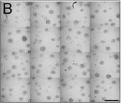
Detection of Human IL-6 by Block/Neutralize
IL-6 nAb inhibits CAF-stimulated MCF10.DCIS structure growth and results in altered morphology. Contiguous 16-tile DIC image of CAFs: WS12Ti (A, B) or CAF40TKi (c, d) co-cultured with MCF10.DCIS cells for 8 days in the presence of isotype matched anti-IgG (a, c) or 1 μg /ml IL-6 nAb (b, d). e Perimeter measurements from DCIS structures in MAME culture in the presence of anti-IgG or IL-6 nAb (N = 100-140 measurements from 6 contiguous tiled DIC images (N = 3). Fluorescent tiled images of CAF40TKi fibroblasts (unlabeled) cultured with MCF10.DCIS-RFP cells in the presence of anti-IgG (f) or 1 μg /ml IL-6 nAb (g). h Quantification of total volume of DCIS-RFP structures in co-culture with CAF40TKi fibroblasts (ratio-paired t-test, N = 3). ***P ≤ 0.001, **P ≤ 0.01; Student’s t-test; mean ± SD Image collected and cropped by CiteAb from the following publication (https://pubmed.ncbi.nlm.nih.gov/26268945), licensed under a CC-BY license. Not internally tested by R&D Systems.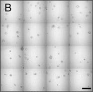
Detection of Human IL-6 by Block/Neutralize
IL-6 neutralizing antibody (nAb) inhibits growth of MCF10.DCIS structures. Representative contiguous 16-tiled DIC images of MCF10.DCIS cells grown in MAME cultures for 8 days. MAME culture of MCF10.DCIS cells treated with an isotype-matched antibody against IgG (control) (8 days) (a), or 1 μg /ml IL-6 nAb (b) (8 days). MCF10.DCIS cells treated with IL-6 nAb followed by a 48 h treatment-free recovery period prior to imaging on day 10 (c). N = 3, Scale bars, 200 μm. d Diameter of MCF10.DCIS structures in the presence of control antibody, IL-6 nAb, or 48 h recovery from IL-6 nAb. N = 20-40 measurements /tiled DIC image (N = 3). ****P ≤ 0.0001, **P ≤ 0.01. e Evaluation of gene expression of invasive tumor cell markers in MAME cultures treated with IL-6 nAb (N = 3). Fold difference as compared to control cultures. Dashed line indicates 2-fold threshold. Student’s t-test; mean ± SD Image collected and cropped by CiteAb from the following publication (https://pubmed.ncbi.nlm.nih.gov/26268945), licensed under a CC-BY license. Not internally tested by R&D Systems.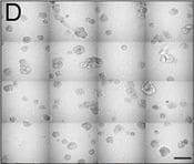
Detection of Human IL-6 by Block/Neutralize
IL-6 nAb inhibits CAF-stimulated MCF10.DCIS structure growth and results in altered morphology. Contiguous 16-tile DIC image of CAFs: WS12Ti (A, B) or CAF40TKi (c, d) co-cultured with MCF10.DCIS cells for 8 days in the presence of isotype matched anti-IgG (a, c) or 1 μg /ml IL-6 nAb (b, d). e Perimeter measurements from DCIS structures in MAME culture in the presence of anti-IgG or IL-6 nAb (N = 100-140 measurements from 6 contiguous tiled DIC images (N = 3). Fluorescent tiled images of CAF40TKi fibroblasts (unlabeled) cultured with MCF10.DCIS-RFP cells in the presence of anti-IgG (f) or 1 μg /ml IL-6 nAb (g). h Quantification of total volume of DCIS-RFP structures in co-culture with CAF40TKi fibroblasts (ratio-paired t-test, N = 3). ***P ≤ 0.001, **P ≤ 0.01; Student’s t-test; mean ± SD Image collected and cropped by CiteAb from the following publication (https://pubmed.ncbi.nlm.nih.gov/26268945), licensed under a CC-BY license. Not internally tested by R&D Systems.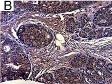
Detection of Human IL-6 by Immunohistochemistry
Breast DCIS cells overexpress proinflammatory markers. Representative images of immunohistochemistry targeting IL-6 protein in normal breast tissue (a), and breast DCIS (b) (N = 61). c and d Hematoxylin staining of serial sections from tissue shown in panel A and B. All images are 20X magnification. Scale bar equals 100 μm. e Evaluation of IL-6 gene expression in DCIS cells via qRT-PCR; the isogenic MCF10.DCIS cells and the non-isogenic SUM102 cell line were analyzed against the non-transformed MCF10A cell line (N = 3). f Secretion of IL-6 protein from DCIS cell lines and non-transformed MCF10A cells as determined by ELISA. *P < 0.05, Student’s t-test; mean ± SD Image collected and cropped by CiteAb from the following publication (https://pubmed.ncbi.nlm.nih.gov/26268945), licensed under a CC-BY license. Not internally tested by R&D Systems.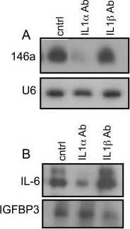
Detection of Human IL-6 by Western Blot
IL-1 alpha upregulates miR-146a in senescent cells.(A) Northern analysis for miR-146a levels in damage-induced senescent HCA2 cells treated with neutralizing antibodies against IL1-alpha and IL1-beta. HCA2 cells (PD 35) were used and induced to senesce by treatment with bleomycin. Cells were harvested for RNA 11 days later. Details of the procedure are described in ‘Experimental Procedures. (B) Western analysis for IL-6 in damage-induced senescent HCA2 cells treated with neutralizing antibodies to IL-1 alpha and IL-1 beta. CM were harvested 11 days after bleomycin treatment. \ Image collected and cropped by CiteAb from the following open publication (https://pubmed.ncbi.nlm.nih.gov/20148189), licensed under a CC-BY license. Not internally tested by R&D Systems.
Human IL-6 ELISA Standard Curve
Recombinant Human IL‑6 (Catalog # 206-IL) was serially diluted and captured by Mouse Anti-Human/Primate IL‑6 Monoclonal Antibody (Catalog # MAB206) coated on a Clear Polystyrene Microplate (Catalog # DY990). Goat Anti-Human IL‑6 Antigen Affinity-purified Polyclonal Antibody (Catalog # AF-206-NA) was biotinylated and incubated with the protein captured on the plate. Detection of the standard curve was achieved by incubating Streptavidin-HRP (Catalog # DY998)Applications for Human IL-6 Antibody
Immunocytochemistry
Sample: Immersion fixed human peripheral blood mononuclear cells treated with LPS and monensin
Neutralization
Reviewed Applications
Read 6 reviews rated 4.5 using AF-206-NA in the following applications:
Formulation, Preparation, and Storage
Purification
Reconstitution
Reconstitute at 0.2 mg/mL in sterile PBS. For liquid material, refer to CoA for concentration.
Formulation
Shipping
Stability & Storage
- 12 months from date of receipt, -20 to -70 °C as supplied.
- 1 month, 2 to 8 °C under sterile conditions after reconstitution.
- 6 months, -20 to -70 °C under sterile conditions after reconstitution.
Calculators
Background: IL-6
Long Name
Alternate Names
Entrez Gene IDs
Gene Symbol
UniProt
Additional IL-6 Products
Product Documents for Human IL-6 Antibody
Certificate of Analysis
To download a Certificate of Analysis, please enter a lot or batch number in the search box below.
Note: Certificate of Analysis not available for kit components.
Product Specific Notices for Human IL-6 Antibody
For research use only
Related Research Areas
Citations for Human IL-6 Antibody
 Powered by Bioz
Customer Reviews for Human IL-6 Antibody (6)
Have you used Human IL-6 Antibody?
Submit a review and receive an Amazon gift card!
$25/€18/£15/$25CAN/¥2500 for a review with an image
$10/€7/£6/$10CAN/¥1110 for a review without an image
Submit a review
Customer Images
-
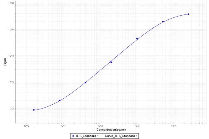
Application: MSD AssaySample Tested: Vitreous humorSpecies: Cynomolgus MonkeyVerified Customer | Posted 11/01/2018After labeling with Sulfo-Tag, used as a detection reagent in MSD assay (Meso Scale Diagnostics LLC). Paired with biotinylated IL-6 antibody (MAB206) as a capture reagent. A standard curve with recombinant human IL-6 from (Cat# 206-IL/CF) is shown (1.6-25,000 pg/ml).

-
Application: Cell depletionSample Tested: Pancreatic cancer cellsSpecies: HumanVerified Customer | Posted 05/11/2018
-
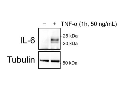
Application: Western BlotSample Tested: Whole cell lysatesSpecies: HumanVerified Customer | Posted 12/22/2016Endothelial cells treated with TNF-a (50 ng/mL) for 1 hour.
-
Application: Western BlotSample Tested: See PMID 21784845Species: HumanVerified Customer | Posted 01/06/2015
-
Application: ImmunoprecipitationSample Tested: HEK293 cells transfected with hIL6Species: HumanVerified Customer | Posted 12/19/2014
-
Application: Western BlotSample Tested: hIL6 transfected cellsSpecies: HumanVerified Customer | Posted 12/19/2014
There are no reviews that match your criteria.
Protocols
Find general support by application which include: protocols, troubleshooting, illustrated assays, videos and webinars.
- Appropriate Fixation of IHC/ICC Samples
- Cellular Response to Hypoxia Protocols
- ClariTSA™ Fluorophore Kits
- Detection & Visualization of Antibody Binding
- ICC Cell Smear Protocol for Suspension Cells
- ICC Immunocytochemistry Protocol Videos
- ICC for Adherent Cells
- Immunocytochemistry (ICC) Protocol
- Immunocytochemistry Troubleshooting
- Immunofluorescence of Organoids Embedded in Cultrex Basement Membrane Extract
- Immunohistochemistry (IHC) and Immunocytochemistry (ICC) Protocols
- Preparing Samples for IHC/ICC Experiments
- Preventing Non-Specific Staining (Non-Specific Binding)
- Primary Antibody Selection & Optimization
- Protocol for VisUCyte™ HRP Polymer Detection Reagent
- Protocol for the Fluorescent ICC Staining of Cell Smears - Graphic
- Protocol for the Fluorescent ICC Staining of Cultured Cells on Coverslips - Graphic
- Protocol for the Preparation and Fluorescent ICC Staining of Cells on Coverslips
- Protocol for the Preparation and Fluorescent ICC Staining of Non-adherent Cells
- Protocol for the Preparation and Fluorescent ICC Staining of Stem Cells on Coverslips
- Protocol for the Preparation of a Cell Smear for Non-adherent Cell ICC - Graphic
- TUNEL and Active Caspase-3 Detection by IHC/ICC Protocol
- The Importance of IHC/ICC Controls
- View all Protocols, Troubleshooting, Illustrated assays and Webinars
FAQs for Human IL-6 Antibody
-
Q: What band size does AF-206-NA detect in Western blot?
A: AF-206-NA detects a 20-22 kDa band in lysate in Western blot.
Associated Pathways