TNF-alpha Antibody
R&D Systems | Catalog # AF-410-NA

Key Product Details
Validated by
Biological Validation
Species Reactivity
Validated:
Human, Mouse
Cited:
Human, Mouse, Rat, Porcine, Transgenic Mouse
Applications
Validated:
Immunohistochemistry, Western Blot, ELISA Capture (Matched Antibody Pair), Neutralization, Intracellular Staining by Flow Cytometry, Immunocytochemistry, CyTOF-ready
Cited:
Immunohistochemistry, Immunohistochemistry-Paraffin, Immunohistochemistry-Frozen, Western Blot, Neutralization, Flow Cytometry, Immunocytochemistry, Immunoprecipitation, Assay Development, Bioassay, Cell Culture, ELISA Capture, ELISA Development, ELISA Development (Capture), In vivo assay, Functional Assay, Luminex Development
Label
Unconjugated
Antibody Source
Polyclonal Goat IgG
Loading...
Product Specifications
Immunogen
E. coli-derived recombinant mouse TNF-alpha
Leu80-Leu235
Accession # P06804
Leu80-Leu235
Accession # P06804
Specificity
Detects mouse TNF-alpha in ELISAs. Detects human and mouse TNF-alpha in Western blots. In sandwich immunoassays, approximately 50% cross-reactivity with recombinant rat TNF‑ alpha is observed.
Clonality
Polyclonal
Host
Goat
Isotype
IgG
Endotoxin Level
<0.10 EU per 1 μg of the antibody by the LAL method.
Scientific Data Images for TNF-alpha Antibody
Detection of Human TNF‑ alpha by Western Blot.
Western blot shows lysates of CHO Chinese hamster ovary cell line either mock transfected or transfected with human TNF- alpha. PVDF membrane was probed with 1 µg/mL of Goat Anti-Human/Mouse TNF-a Antigen Affinity-purified Polyclonal Antibody (Catalog # AF-410-NA) followed by HRP-conjugated Anti-Goat IgG Secondary Antibody (HAF017). Specific bands were detected for TNF-a at approximately 17 and 26 kDa (as indicated). This experiment was conducted under reducing conditions and using Immunoblot Buffer Group 1.
Detection of Mouse TNF‑ alpha by Western Blot.
Western blot shows lysates of RAW 264.7 mouse monocyte/macrophage cell line untreated (-) or treated (+) with 10 µg/mL LPS for 4 hours. PVDF membrane was probed with 2 µg/mL of Goat Anti-Human/Mouse TNF-a Antigen Affinity-purified Polyclonal Antibody (Catalog # AF-410-NA) followed by HRP-conjugated Anti-Goat IgG Secondary Antibody (HAF017). Specific bands were detected for TNF-a at approximately 16 and 25 kDa (as indicated). This experiment was conducted under reducing conditions and using Immunoblot Buffer Group 1.
TNF‑ alpha in RAW 264.7 Mouse Cell Line.
TNF-a was detected in immersion fixed RAW 264.7 mouse monocyte/ macrophage cell line treated with LPS using Human/Mouse TNF-a Antigen Affinity-purified Polyclonal Antibody (Catalog # AF-410-NA) at 10 µg/mL for 3 hours at room temperature. Cells were stained using the NorthernLights™ 557-conjugated Anti-Goat IgG Secondary Antibody (red; NL001) and counterstained with DAPI (blue). View our protocol for Fluorescent ICC Staining of Cells on Coverslips.
TNF‑ alpha in Mouse T Cells.
TNF‑ alpha was detected in immersion fixed activated mouse T Cells using 15 µg/mL Human/Mouse TNF‑ alpha Antigen Affinity-purified Polyclonal Antibody (Catalog # AF‑410‑NA) for 3 hours at room temperature. Cells were stained (red) and counterstained (green). View our protocol for Fluorescent ICC Staining of Non-adherent Cells.
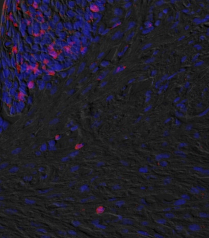
TNF-alpha in Mouse Spleen Tissue.
TNF‑ alpha was detected in immersion fixed paraffin-embedded sections of mouse spleen tissue using Goat Anti-Human/Mouse TNF‑ alpha Antigen Affinity-purified Polyclonal Antibody (Catalog # AF-410-NA) at 3 µg/mL for 1 hour at room temperature followed by incubation with the Anti-Goat IgG VisUCyte™ HRP Polymer Antibody (VC004). Tissue was stained using DAB (brown) and counterstained with hematoxylin (blue). Specific staining was localized to lymphocytes. Staining was performed using our IHC Staining with VisUCyte HRP Polymer Detection Reagents.
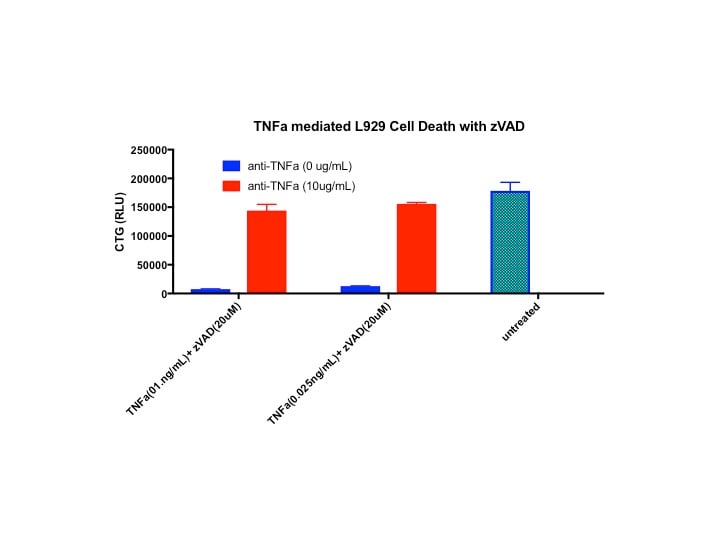
Cytotoxicity Induced by TNF‑ alpha and Neutralization by Mouse TNF‑ alpha Antibody.
Recombinant Mouse TNF-a (410-MT) induces cytotoxicity in the the L-929 mouse fibroblast cell line in a dose-dependent manner (orange line). Cytotoxicity elicited by Recombinant Mouse TNF-a (0.1 ng/mL) is neutralized (green line) by increasing concentrations of Human/Mouse TNF-a Antigen Affinity-purified Polyclonal Antibody (Catalog # AF-410-NA). The ND50 is typically 1.5-10 ng/mL in the presence of the metabolic inhibitor actinomycin D.
Detection of TNF‑ alpha in Human Spleen.
TNF‑ alpha was detected in immersion fixed paraffin-embedded sections of Human Spleen using Goat Anti-Human/Mouse TNF‑ alpha Antigen Affinity-purified Polyclonal Antibody (Catalog # AF-410-NA) at 5 µg/mL for 1 hour at room temperature followed by incubation with the Anti-Goat IgG VisUCyte™ HRP Polymer Antibody (Catalog # VC004). Before incubation with the primary antibody, tissue was subjected to heat-induced epitope retrieval using VisUCyte Antigen Retrieval Reagent-Basic (Catalog # VCTS021). Tissue was stained using DAB (brown) and counterstained with hematoxylin (blue). Specific staining was localized to cytoplasm in lymphocytes. View our protocol for IHC Staining with VisUCyte HRP Polymer Detection Reagents.
Detection of Mouse TNF-alpha by Western Blot
WNT-5A induces a proinflammatory transformation in mouse microglia. (A, B) iNOS, COX-2 and TNF alpha were detected by immunoblotting in lysates from mouse primary microglia after WNT-5A stimulation (ctrl, 300 ng/ml, 6 hours). (B’) shows TNF alpha levels in the supernatant of primary microglia under ctrl conditions and upon WNT-5A stimulation (ctrl, 300 ng/ml, 24 hours; n = 4). At least three experiments are summarized in the bar graphs. Data are normalized to ctrl. Error bars give s.e.m. (C) Microglial proliferation was assessed by an MTT assay monitoring mitochondrial activity, which is proportional to cell number [see Additional file 1: Figure S5]. Stimulation with WNT-5A (300 ng/ml, 24 hours) increased MTT, which was blocked by PTX (100 ng/ml, overnight) or the MEK1/2 (10 μM) inhibitor, SL327. (D). Experiments were done in triplicate and data from three independent experiments are shown. *, P < 0.01; ***, P < 0.001: Error bars show s.e.m.. (E) Cell tracker (red)-stained primary microglia were seeded on top of a collagen matrix in 35 mm glass bottom dishes. One day after ctrl or 300 ng/ml WNT-5A stimulation, invasion was observed by confocal microscopy and Z-stacking using a Zeiss LSM710 microscope and subsequent analysis with the Bitplane Imaris software. The size of the collagen cube shown is 1,000 (Z) x 1,400 (Y) x 1,400 (X) μm. Three invasion experiments in the absence and presence of the MEK1/2 inhibitor SL327 were quantified. Data are presented in a bar graph (F). *, P < 0.05; error bars show s.e.m.. (G) cDNA of ctrl stimulated (−) and WNT-5A stimulated (+) primary microglia was analyzed by QPCR for expression of inflammatory genes. At least three independent experiments are summarized. Gene expression is normalized to the housekeeping gene GAPDH and expressed as arbitrary units (2-delta ct). *, P < 0.05; **, P < 0.01. COX-2, cyclooxygenase 2; iNOS, inducible nitric oxide synthase; MTT, 3-(4,5-dimethylthiazol-2-yl)-2,5-diphenyltetrazolium bromide; n, number; s.e.m., standa
Detection of TNF‑ alpha in RAW 264.7 cells treated with 1µg/mL LPS for 24 hrs by Flow Cytometry
RAW 264.7 cells treated with 1µg/mL LPS for 24 hrs were stained with Goat Anti-Human/Mouse TNF‑ alpha Antigen Affinity-purified Polyclonal Antibody (Catalog # AF-410-NA, filled histogram) or isotype control antibody (Catalog # AB-108-C, open histogram) followed by Allophycocyanin-conjugated Anti-Goat IgG Secondary Antibody (Catalog # F0108). To facilitate intracellular staining, cells were fixed with Flow Cytometry Fixation Buffer (Catalog # FC004) and permeabilized with saponin. View our protocol for Staining Intracellular Molecules.
Mouse TNF-alpha ELISA Standard Curve
Recombinant Mouse TNF‑ alpha aa 80-235 (Catalog # 410-MT) was serially diluted and captured by Goat Anti-Human/Mouse TNF‑ alpha Antigen Affinity-purified Polyclonal Antibody (Catalog # AF-410-NA) coated on a Clear Polystyrene Microplate (Catalog # DY990). Goat Anti-Human/Mouse TNF‑ alpha Antigen Affinity-purified Polyclonal Antibody (Catalog # AF-410-NA) was biotinylated and incubated with the protein captured on the plate. Detection of the standard curve was achieved by incubating Streptavidin-HRP (Catalog # DY998)
Mouse TNF-alpha ELISA Standard Curve
Recombinant Mouse TNF‑ alpha aa 80-235 (Catalog # 410-MT) was serially diluted and captured by Goat Anti-Human/Mouse TNF‑ alpha Antigen Affinity-purified Polyclonal Antibody (Catalog # AF-410-NA) coated on a Clear Polystyrene Microplate (Catalog # DY990). Goat Anti-Human/Mouse TNF‑ alpha Antigen Affinity-purified Polyclonal Antibody (Catalog # AF-410-NA) was biotinylated and incubated with the protein captured on the plate. Detection of the standard curve was achieved by incubating Streptavidin-HRP (Catalog # DY998)Applications for TNF-alpha Antibody
Application
Recommended Usage
CyTOF-ready
Ready to be labeled using established conjugation methods. No BSA or other carrier proteins that could interfere with conjugation.
Immunocytochemistry
5-15 µg/mL
Sample: Immersion fixed RAW 264.7 mouse monocyte/macrophage cell line with or without LPS and monensin treatment, immersion fixed RAW 263 mouse macrophage cell line treated wtih LPS and monensin, and immersion fixed activated mouse T Cells
Sample: Immersion fixed RAW 264.7 mouse monocyte/macrophage cell line with or without LPS and monensin treatment, immersion fixed RAW 263 mouse macrophage cell line treated wtih LPS and monensin, and immersion fixed activated mouse T Cells
Immunohistochemistry
3-15 µg/mL
Sample: Immersion fixed paraffin-embedded sections of Mouse Spleen and Human Spleen.
Sample: Immersion fixed paraffin-embedded sections of Mouse Spleen and Human Spleen.
Intracellular Staining by Flow Cytometry
0.25 µg/106 cells
Sample: RAW 264.7 mouse monocyte/macrophage cell line treated with LPS, fixed with paraformaldehyde, and permeabilized with saponin
Sample: RAW 264.7 mouse monocyte/macrophage cell line treated with LPS, fixed with paraformaldehyde, and permeabilized with saponin
Western Blot
1-2 µg/mL
Sample: CHO Chinese hamster ovary cell line transfected with human TNF- alpha and RAW 264.7 mouse monocyte/macrophage cell line treated with LPS
Sample: CHO Chinese hamster ovary cell line transfected with human TNF- alpha and RAW 264.7 mouse monocyte/macrophage cell line treated with LPS
Neutralization
Measured by its ability to neutralize TNF‑ alpha -induced cytotoxicity in the L‑929 mouse fibroblast cell line. Matthews, N. and M.L. Neale (1987) in Lymphokines and Interferons, A Practical Approach. Clemens, M.J. et al. (eds): IRL Press. 221. The Neutralization Dose (ND50) is typically 1.5-10 ng/mL in the presence of 0.1 ng/mL Recombinant Mouse TNF‑ alpha and actinomycin D.
Mouse TNF-alpha Sandwich Immunoassay
Please Note: Optimal dilutions of this antibody should be experimentally determined.
Reviewed Applications
Read 5 reviews rated 4.4 using AF-410-NA in the following applications:
Flow Cytometry Panel Builder
Bio-Techne Knows Flow Cytometry
Save time and reduce costly mistakes by quickly finding compatible reagents using the Panel Builder Tool.
Advanced Features
- Spectra Viewer - Custom analysis of spectra from multiple fluorochromes
- Spillover Popups - Visualize the spectra of individual fluorochromes
- Antigen Density Selector - Match fluorochrome brightness with antigen density

Formulation, Preparation, and Storage
Purification
Antigen Affinity-purified
Reconstitution
Reconstitute at 0.2 mg/mL in sterile PBS. For liquid material, refer to CoA for concentration.
Loading...
Formulation
Lyophilized from a 0.2 μm filtered solution in PBS with Trehalose. See Certificate of Analysis for details.
*Small pack size (-SP) is supplied either lyophilized or as a 0.2 µm filtered solution in PBS.
*Small pack size (-SP) is supplied either lyophilized or as a 0.2 µm filtered solution in PBS.
Shipping
Lyophilized product is shipped at ambient temperature. Liquid small pack size (-SP) is shipped with polar packs. Upon receipt, store immediately at the temperature recommended below.
Stability & Storage
Use a manual defrost freezer and avoid repeated freeze-thaw cycles.
- 12 months from date of receipt, -20 to -70 °C as supplied.
- 1 month, 2 to 8 °C under sterile conditions after reconstitution.
- 6 months, -20 to -70 °C under sterile conditions after reconstitution.
Calculators
Background: TNF-alpha
Long Name
Tumor Necrosis Factor alpha
Alternate Names
Cachetin, DIF, TNF, TNF-A, TNFA, TNFalpha, TNFG1F, TNFSF1A, TNFSF2
Entrez Gene IDs
Gene Symbol
TNF
UniProt
Additional TNF-alpha Products
Product Documents for TNF-alpha Antibody
Certificate of Analysis
To download a Certificate of Analysis, please enter a lot or batch number in the search box below.
Note: Certificate of Analysis not available for kit components.
Product Specific Notices for TNF-alpha Antibody
For research use only
Related Research Areas
Citations for TNF-alpha Antibody
 Powered by Bioz
Customer Reviews for TNF-alpha Antibody (5)
4.4 out of 5
5 Customer Ratings
Have you used TNF-alpha Antibody?
Submit a review and receive an Amazon gift card!
$25/€18/£15/$25CAN/¥2500 Yen for a review with an image
$10/€7/£6/$10CAN/¥1110 Yen for a review without an image
Submit a review
Customer Images
Showing
1
-
5 的
5 reviews
Showing All
Filter By:
-
Application: ImmunohistochemistrySample Tested: mouse oral tissueSpecies: MouseVerified Customer | Posted 11/23/2020

-
Application: Block/NeutralizeSample Tested: L929Species: MouseVerified Customer | Posted 02/27/2018
-
Application: ELISASample Tested: Recombinant proteinSpecies: MouseVerified Customer | Posted 12/01/2017
-
Application: ELISASample Tested: Liver tissueSpecies: MouseVerified Customer | Posted 10/30/2017
-
Application: Block/NeutralizeSample Tested: RAW 264.7 mouse monocyte/macrophage cell lineSpecies: MouseVerified Customer | Posted 05/10/2017
There are no reviews that match your criteria.
Protocols
Find general support by application which include: protocols, troubleshooting, illustrated assays, videos and webinars.
- 7-Amino Actinomycin D (7-AAD) Cell Viability Flow Cytometry Protocol
- Antigen Retrieval Protocol (PIER)
- Antigen Retrieval for Frozen Sections Protocol
- Appropriate Fixation of IHC/ICC Samples
- Cellular Response to Hypoxia Protocols
- Chromogenic IHC Staining of Formalin-Fixed Paraffin-Embedded (FFPE) Tissue Protocol
- Chromogenic Immunohistochemistry Staining of Frozen Tissue
- ClariTSA™ Fluorophore Kits
- Detection & Visualization of Antibody Binding
- Extracellular Membrane Flow Cytometry Protocol
- Flow Cytometry Protocol for Cell Surface Markers
- Flow Cytometry Protocol for Staining Membrane Associated Proteins
- Flow Cytometry Staining Protocols
- Flow Cytometry Troubleshooting Guide
- Fluorescent IHC Staining of Frozen Tissue Protocol
- Graphic Protocol for Heat-induced Epitope Retrieval
- Graphic Protocol for the Preparation and Fluorescent IHC Staining of Frozen Tissue Sections
- Graphic Protocol for the Preparation and Fluorescent IHC Staining of Paraffin-embedded Tissue Sections
- Graphic Protocol for the Preparation of Gelatin-coated Slides for Histological Tissue Sections
- ICC Cell Smear Protocol for Suspension Cells
- ICC Immunocytochemistry Protocol Videos
- ICC for Adherent Cells
- IHC Sample Preparation (Frozen sections vs Paraffin)
- Immunocytochemistry (ICC) Protocol
- Immunocytochemistry Troubleshooting
- Immunofluorescence of Organoids Embedded in Cultrex Basement Membrane Extract
- Immunofluorescent IHC Staining of Formalin-Fixed Paraffin-Embedded (FFPE) Tissue Protocol
- Immunohistochemistry (IHC) and Immunocytochemistry (ICC) Protocols
- Immunohistochemistry Frozen Troubleshooting
- Immunohistochemistry Paraffin Troubleshooting
- Intracellular Flow Cytometry Protocol Using Alcohol (Methanol)
- Intracellular Flow Cytometry Protocol Using Detergents
- Intracellular Nuclear Staining Flow Cytometry Protocol Using Detergents
- Intracellular Staining Flow Cytometry Protocol Using Alcohol Permeabilization
- Intracellular Staining Flow Cytometry Protocol Using Detergents to Permeabilize Cells
- Preparing Samples for IHC/ICC Experiments
- Preventing Non-Specific Staining (Non-Specific Binding)
- Primary Antibody Selection & Optimization
- Propidium Iodide Cell Viability Flow Cytometry Protocol
- Protocol for Heat-Induced Epitope Retrieval (HIER)
- Protocol for Liperfluo
- Protocol for Making a 4% Formaldehyde Solution in PBS
- Protocol for VisUCyte™ HRP Polymer Detection Reagent
- Protocol for the Characterization of Human Th22 Cells
- Protocol for the Characterization of Human Th9 Cells
- Protocol for the Fluorescent ICC Staining of Cell Smears - Graphic
- Protocol for the Fluorescent ICC Staining of Cultured Cells on Coverslips - Graphic
- Protocol for the Preparation & Fixation of Cells on Coverslips
- Protocol for the Preparation and Chromogenic IHC Staining of Frozen Tissue Sections
- Protocol for the Preparation and Chromogenic IHC Staining of Frozen Tissue Sections - Graphic
- Protocol for the Preparation and Chromogenic IHC Staining of Paraffin-embedded Tissue Sections
- Protocol for the Preparation and Chromogenic IHC Staining of Paraffin-embedded Tissue Sections - Graphic
- Protocol for the Preparation and Fluorescent ICC Staining of Cells on Coverslips
- Protocol for the Preparation and Fluorescent ICC Staining of Non-adherent Cells
- Protocol for the Preparation and Fluorescent ICC Staining of Stem Cells on Coverslips
- Protocol for the Preparation and Fluorescent IHC Staining of Frozen Tissue Sections
- Protocol for the Preparation and Fluorescent IHC Staining of Paraffin-embedded Tissue Sections
- Protocol for the Preparation of Gelatin-coated Slides for Histological Tissue Sections
- Protocol for the Preparation of a Cell Smear for Non-adherent Cell ICC - Graphic
- Protocol: Annexin V and PI Staining by Flow Cytometry
- Protocol: Annexin V and PI Staining for Apoptosis by Flow Cytometry
- R&D Systems Quality Control Western Blot Protocol
- TUNEL and Active Caspase-3 Detection by IHC/ICC Protocol
- The Importance of IHC/ICC Controls
- Troubleshooting Guide: Fluorokine Flow Cytometry Kits
- Troubleshooting Guide: Immunohistochemistry
- Troubleshooting Guide: Western Blot Figures
- Western Blot Conditions
- Western Blot Protocol
- Western Blot Protocol for Cell Lysates
- Western Blot Troubleshooting
- Western Blot Troubleshooting Guide
- View all Protocols, Troubleshooting, Illustrated assays and Webinars
Loading...
Associated Pathways


IL-15 Signaling Pathways and their Primary Biological Effects in Different Immune Cell Types
 Innate Lymphoid Cell Differentiation Pathways
 mTOR Signaling Pathway
 NOD-like Receptor Signaling Pathways
 Th1 Differentiation Pathway
 TNF Superfamily Pathway: Human Ligand-Receptor Interactions & their Associated Functions
 Toll-Like Receptor Signaling Pathways
