Ki67/MKI67 Antibody - BSA Free
Novus Biologicals | Catalog # NB110-89719
![Immunocytochemistry/ Immunofluorescence: Ki67/MKI67 Antibody - BSA Free [NB110-89719]](https://resources.rndsystems.com/images/products/Ki67-MKI67-Antibody-Immunocytochemistry-Immunofluorescence-NB110-89719-img0006.jpg "Immunocytochemistry/ Immunofluorescence: Ki67/MKI67 Antibody - BSA Free [NB110-89719]")
Key Product Details
Validated by
Biological Validation
Species Reactivity
Validated:
Human, Mouse, Rat
Cited:
Human, Mouse, Rat
Applications
Validated:
Immunohistochemistry, Immunohistochemistry-Paraffin, Immunohistochemistry-Frozen, Western Blot, Flow Cytometry, Immunocytochemistry/ Immunofluorescence
Cited:
Immunohistochemistry-Paraffin, Immunohistochemistry-Frozen, Western Blot, Flow Cytometry, Immunocytochemistry/ Immunofluorescence, IF/IHC
Label
Unconjugated
Antibody Source
Polyclonal Rabbit IgG
Format
BSA Free
Loading...
Product Specifications
Immunogen
The immunogen for this KI67/MKI67 Antibody was made using a synthetic peptide from the internal region of Mouse KI67/MKI67, between aminoacids 2000-2050 (2021-2036) Uniprot# E9PVX6.
Reactivity Notes
Rat reactivity reported in scientific literature (PMID: 31286466).
Localization
Nuclear
Marker
Proliferation Marker
Clonality
Polyclonal
Host
Rabbit
Isotype
IgG
Theoretical MW
351 kDa.
Disclaimer note: The observed molecular weight of the protein may vary from the listed predicted molecular weight due to post translational modifications, post translation cleavages, relative charges, and other experimental factors.
Disclaimer note: The observed molecular weight of the protein may vary from the listed predicted molecular weight due to post translational modifications, post translation cleavages, relative charges, and other experimental factors.
Scientific Data Images for Ki67/MKI67 Antibody - BSA Free
Immunocytochemistry/ Immunofluorescence: Ki67/MKI67 Antibody - BSA Free [NB110-89719]
Immunocytochemistry/Immunofluorescence: Ki67/MKI67 Antibody [NB110-89719] - Ki67 antibody was tested in SH-SY5Y cells with DyLight 488 (green). Nuclei and alpha-tubulin were counterstained with DAPI (blue) and Dylight 550 (red).![Immunohistochemistry-Paraffin: Ki67/MKI67 Antibody - BSA Free [NB110-89719]](https://resources.rndsystems.com/images/products/Ki67-MKI67-Antibody-Immunohistochemistry-Paraffin-NB110-89719-img0008.jpg "Immunohistochemistry-Paraffin: Ki67/MKI67 Antibody - BSA Free [NB110-89719]")
Immunohistochemistry-Paraffin: Ki67/MKI67 Antibody - BSA Free [NB110-89719]
Immunohistochemistry-Paraffin: Ki67/MKI67 Antibody [NB110-89719] - IHC analysis of a formalin fixed paraffin embedded tissue section of mouse intestine using 1:200 dilution of rabbit anti-KI67 antibody. The staining was developed with HRP labeled anti-rabbit IgG secondary antibody and DAB reagent, and nuclei of cells were counter-stained with hematoxylin. This antibody generated a specific nuclear staining in epithelial cells and the staining was more intense in the cells close to the bases of crypts. Weak to moderate positivity was found at the cytoplasmic level also.![Immunohistochemistry-Paraffin: Ki67/MKI67 Antibody - BSA Free [NB110-89719]](https://resources.rndsystems.com/images/products/Ki67-MKI67-Antibody-Immunohistochemistry-Paraffin-NB110-89719-img0010.jpg "Immunohistochemistry-Paraffin: Ki67/MKI67 Antibody - BSA Free [NB110-89719]")
![Immunohistochemistry: Ki67/MKI67 Antibody - BSA Free [NB110-89719]](https://resources.rndsystems.com/images/products/Ki67-MKI67-Antibody-Immunohistochemistry-NB110-89719-img0009.jpg "Immunohistochemistry: Ki67/MKI67 Antibody - BSA Free [NB110-89719]")
Immunohistochemistry: Ki67/MKI67 Antibody - BSA Free [NB110-89719]
Immunohistochemistry: Ki67/MKI67 Antibody [NB110-89719] - Detection of Ki67 in formalin-fixed paraffin embedded mouse intestine using NB110-89719.![Immunohistochemistry-Paraffin: Ki67/MKI67 Antibody - BSA Free [NB110-89719]](https://resources.rndsystems.com/images/products/Ki67-MKI67-Antibody-Immunohistochemistry-Paraffin-NB110-89719-img0001.jpg "Immunohistochemistry-Paraffin: Ki67/MKI67 Antibody - BSA Free [NB110-89719]")
Immunohistochemistry-Paraffin: Ki67/MKI67 Antibody - BSA Free [NB110-89719]
Immunohistochemistry-Paraffin: Ki67/MKI67 Antibody [NB110-89719] - FFPE section of human breast carcinoma. Antibody: Affinity purified rabbit anti-Ki-67 (NB110-89719) used at a dilution of 1:250.![Immunohistochemistry: Ki67/MKI67 Antibody - BSA Free [NB110-89719]](https://resources.rndsystems.com/images/products/Ki67-MKI67-Antibody-Immunohistochemistry-NB110-89719-img0007.jpg "Immunohistochemistry: Ki67/MKI67 Antibody - BSA Free [NB110-89719]")
Immunohistochemistry: Ki67/MKI67 Antibody - BSA Free [NB110-89719]
Immunohistochemistry: Ki67/MKI67 Antibody [NB110-89719] - Immunohistochemical analysis of mouse spleen.![Immunohistochemistry-Paraffin: Ki67/MKI67 Antibody - BSA Free [NB110-89719]](https://resources.rndsystems.com/images/products/Ki67-MKI67-Antibody-Immunohistochemistry-Paraffin-NB110-89719-img0005.jpg "Immunohistochemistry-Paraffin: Ki67/MKI67 Antibody - BSA Free [NB110-89719]")
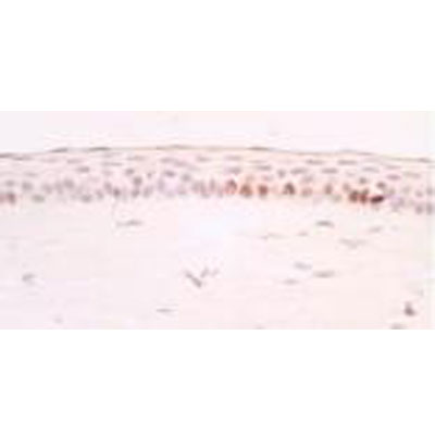
Immunohistochemistry-Paraffin: Ki67/MKI67 Antibody - BSA Free [NB110-89719]
Immunohistochemistry-Paraffin: Ki67/MKI67 Antibody [NB110-89719] - IHC analysis of Ki67 in mouse cornea. Image courtesy of product review by Bo-Yie Chen.
Immunohistochemistry-Paraffin: Ki67/MKI67 Antibody - BSA Free [NB110-89719] -
Effect of saireito on the suppression of cell proliferation induced by 5-fluorouracil (5-FU) in mouse small intestines.5-FU (50 mg/kg) was injected i.p. while saireito (1000 mg/kg) was administered p.o. twice, 30 min before and 8 h after 5-FU injection. The jejunum was excised, sectioned, and Ki67 immunostaining was performed (A). The number of proliferative cells was counted (B). Data are presented as the mean ± SEM of 6 mice. #P < 0.05, versus normal (5-FU-untreated).
Immunocytochemistry/ Immunofluorescence: Ki67/MKI67 Antibody - BSA Free [NB110-89719] -
Immunocytochemistry/ Immunofluorescence: Ki67/MKI67 Antibody - BSA Free [NB110-89719] - The increase in iCM number with SB treatment is not due to increased transgene expression or changes in cell proliferation.Evaluation of gene expression of the transgenes at Day 2 via qPCR is not significantly different between +DMSO (black) control & +SB treatment groups (white, A). Evaluation of overall cell proliferation with Ki67 staining in MEFs with time for +DMSO (black) & +SB treatment groups (grey) (B). A time course evaluation of Troponin T (green) & Ki67 (red) did any dual labeled cells for either the +DMSO (top row) & +SB (bottom row) treated groups (C). Scale bar is 100 µM. Image collected & cropped by CiteAb from the following publication (https://dx.plos.org/10.1371/journal.pone.0089678), licensed under a CC-BY license. Not internally tested by Novus Biologicals.Applications for Ki67/MKI67 Antibody - BSA Free
Application
Recommended Usage
Flow Cytometry
reported in scientific literature (PMID 25733567)
Immunocytochemistry/ Immunofluorescence
1:50-1:200
Immunohistochemistry
1:100-1:500
Immunohistochemistry-Frozen
reported in scientific literature (PMID 23419702)
Immunohistochemistry-Paraffin
1:100-1:500
Western Blot
reported in scientific literature (PMID 23643677)
Reviewed Applications
Read 3 reviews rated 5 using NB110-89719 in the following applications:
Flow Cytometry Panel Builder
Bio-Techne Knows Flow Cytometry
Save time and reduce costly mistakes by quickly finding compatible reagents using the Panel Builder Tool.
Advanced Features
- Spectra Viewer - Custom analysis of spectra from multiple fluorochromes
- Spillover Popups - Visualize the spectra of individual fluorochromes
- Antigen Density Selector - Match fluorochrome brightness with antigen density

Formulation, Preparation, and Storage
Purification
Immunogen affinity purified
Formulation
PBS
Format
BSA Free
Preservative
0.02% Sodium Azide
Concentration
1 mg/ml
Shipping
The product is shipped with polar packs. Upon receipt, store it immediately at the temperature recommended below.
Stability & Storage
Store at 4C. Do not freeze.
Background: Ki67/MKI67
Detection of Ki67 by immunostaining is commonly used as a proliferation marker in solid tumors, as well as certain hematological malignancies (3-5). The Ki67 index, which reports on positive Ki67 stained tumor cell nuclei, has been extensively studied as a prognostic biomarker in cancers such as breast cancer and cervical cancer.
References
1. Gerdes J, Schwab U, Lemke H, Stein H. (1983) Production of a mouse monoclonal antibody reactive with a human nuclear antigen associated with cell proliferation. Int J Cancer. 31:13-20. PMID: 6339421
2. Starborg M, Gell K, Brundell E and Hoog C. (1996) The murine Ki-67 cell proliferation antigen accumulates in the nucleolar and heterochromatic regions of interphase cells and at the periphery of the mitotic chromosomes in a process essential for cell cycle progression. J Cell Sci. 109:143-153. 1996
3. Karamitopoulou E, Perentes E, Tolnay M, Probst A. (1998) Prognostic significance of MIB-1, p53, and bcl-2 immunoreactivity in meningiomas. Hum Pathol. 29(2):140-5. PMID: 9490273
4. Geyer FC, Rodrigues DN, Weigelt B and Reis-Filho JS. (2012) Molecular classification of estrogen receptor-positive/luminal breast cancers. Adv Anat Pathol. 19(1):39-53. PMID: 22156833
5. Ikenberg H, Bergeron C, Schmidt D, Griesser H, Alameda F, Angeloni C, Bogers J, Dachez R, Denton K, Hariri J, Keller T, von Knebel Doeberitz M, Neumann HH, Puig-Tintore LM, Sideri M, Rehm S, Ridder R; PALMS Study Group. (2013) Screening for cervical cancer precursors with p16/Ki-67 dual-stained cytology: results of the PALMS study. J Natl Cancer Inst. 105(20):1550-7. PMID: 24096620
Long Name
Antigen Identified by Monoclonal Antibody Ki67
Alternate Names
Ki-67, KIA, MIB-1, MKI67, PPP1R105, TSG126
Gene Symbol
MKI67
UniProt
Additional Ki67/MKI67 Products
Product Documents for Ki67/MKI67 Antibody - BSA Free
Certificate of Analysis
To download a Certificate of Analysis, please enter a lot or batch number in the search box below.
Product Specific Notices for Ki67/MKI67 Antibody - BSA Free
This product is for research use only and is not approved for use in humans or in clinical diagnosis. Primary Antibodies are guaranteed for 1 year from date of receipt.
Related Research Areas
Citations for Ki67/MKI67 Antibody - BSA Free
 Powered by Bioz
Customer Reviews for Ki67/MKI67 Antibody - BSA Free (3)
5 out of 5
3 Customer Ratings
Have you used Ki67/MKI67 Antibody - BSA Free?
Submit a review and receive an Amazon gift card!
$25/€18/£15/$25CAN/¥2500 Yen for a review with an image
$10/€7/£6/$10CAN/¥1110 Yen for a review without an image
Submit a review
Customer Images
Showing
1
-
3 的
3 reviews
Showing All
Filter By:
-
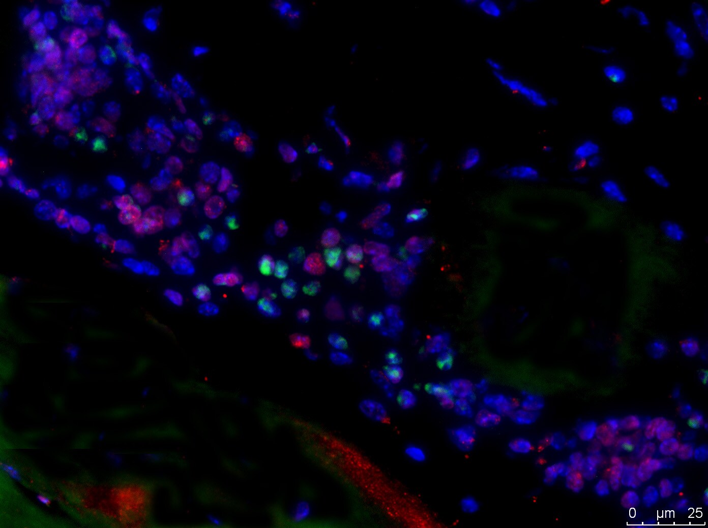
Application: Immunohistochemistry-ParaffinSample Tested: Skeletal muscle tissueSpecies: MouseVerified Customer | Posted 11/30/2016Mouse skeletal muscle tissue stained for Ki67 (green), localized in nuclei. Tissue was co-stained for another muscle marker in red.Heat mediated low pH antigen retrieval was performed on sections prior to use. Permeabilization with 0.1% triton. The antibody was diluted 1/200. An Alexa Fluor anti-rabbit 488 was used to detect Ki67 antibody.

-
Application: Immunohistochemistry-ParaffinSample Tested: Mouse lung tissue that was exposed to cigarette smokeSpecies: MouseVerified Customer | Posted 04/07/2016
-
Application: Immunohistochemistry-ParaffinSample Tested: Mouse corneaSpecies: MouseVerified Customer | Posted 02/06/2012
There are no reviews that match your criteria.
Protocols
View specific protocols for Ki67/MKI67 Antibody - BSA Free (NB110-89719):
Culture cells to appropriate density in 35 mm culture dishes or 6-well plates.
1. Remove culture medium and add 10% formalin to the dish. Fix at room temperature for 30 minutes.
2. Remove the formalin and add ice cold methanol. Incubate for 5-10 minutes.
3. Remove methanol and add washing solution (i.e. PBS). Be sure to not let the specimen dry out. Wash three times for 10 minutes.
4. To block nonspecific antibody binding incubate in 10% normal goat serum from 1 hour to overnight at room temperature.
5. Add primary antibody at appropriate dilution and incubate at room temperature from 2 hours to overnight at room temperature.
6. Remove primary antibody and replace with washing solution. Wash three times for 10 minutes.
7. Add secondary antibody at appropriate dilution. Incubate for 1 hour at room temperature.
8. Remove antibody and replace with wash solution, then wash for 10 minutes. Add Hoechst 33258 to wash solution at 1:25,0000 and incubate for 10 minutes. Wash a third time for 10 minutes.
9. Cells can be viewed directly after washing. The plates can also be stored in PBS containing Azide covered in Parafilm (TM). Cells can also be cover-slipped using Fluoromount, with appropriate sealing.
*The above information is only intended as a guide. The researcher should determine what protocol best meets their needs. Please follow safe laboratory procedures.
Antigen Unmasking
Bring slides to a boil in 10 mM sodium citrate buffer pH 6.0 then maintain at a sub-boiling temperature for 10 minutes. Cool slides on bench top for 30 minutes.
Wash sections in dH2O three times for 5 minutes each.
Wash section in wash buffer for 5 minutes.
Block each section with 100-400 ul blocking solution (1X PBST, 5% goat serum) for 1 hour at room temperature.
Remove blocking solution and add 100-400 ul primary antibody diluted in 1X PBST, 5% goat serum to each section. Incubate overnight at 4C.
Remove antibody solution and wash sections in wash buffer three times for 5 minutes each.
Add 100-400 ul biotinylated secondary antibody, diluted in 1X PBST, 5% goat serum. Incubate 30 minutes at room temperature.
Remove secondary antibody solution and wash sections three times with wash buffer for 5 minutes each.
Add 100-400 ul Striptavidin-HRP reagent to each section and incubate for 30 minutes at room temperature.
Wash sections three times in wash buffer for 5 minutes each.
Add 100-400 ul DAB substrate to each section and monitor staining closely.
As soon as the sections develop, immerse slides in dH2O.
Wash sections in dH2O two times for 5 minutes each.
Mount coverslips.
Find general support by application which include: protocols, troubleshooting, illustrated assays, videos and webinars.
- 7-Amino Actinomycin D (7-AAD) Cell Viability Flow Cytometry Protocol
- Antigen Retrieval Protocol (PIER)
- Antigen Retrieval for Frozen Sections Protocol
- Appropriate Fixation of IHC/ICC Samples
- Cellular Response to Hypoxia Protocols
- Chromogenic IHC Staining of Formalin-Fixed Paraffin-Embedded (FFPE) Tissue Protocol
- Chromogenic Immunohistochemistry Staining of Frozen Tissue
- ClariTSA™ Fluorophore Kits
- Detection & Visualization of Antibody Binding
- Extracellular Membrane Flow Cytometry Protocol
- Flow Cytometry Protocol for Cell Surface Markers
- Flow Cytometry Protocol for Staining Membrane Associated Proteins
- Flow Cytometry Staining Protocols
- Flow Cytometry Troubleshooting Guide
- Fluorescent IHC Staining of Frozen Tissue Protocol
- Graphic Protocol for Heat-induced Epitope Retrieval
- Graphic Protocol for the Preparation and Fluorescent IHC Staining of Frozen Tissue Sections
- Graphic Protocol for the Preparation and Fluorescent IHC Staining of Paraffin-embedded Tissue Sections
- Graphic Protocol for the Preparation of Gelatin-coated Slides for Histological Tissue Sections
- ICC Cell Smear Protocol for Suspension Cells
- ICC Immunocytochemistry Protocol Videos
- ICC for Adherent Cells
- IHC Sample Preparation (Frozen sections vs Paraffin)
- Immunocytochemistry (ICC) Protocol
- Immunocytochemistry Troubleshooting
- Immunofluorescence of Organoids Embedded in Cultrex Basement Membrane Extract
- Immunofluorescent IHC Staining of Formalin-Fixed Paraffin-Embedded (FFPE) Tissue Protocol
- Immunohistochemistry (IHC) and Immunocytochemistry (ICC) Protocols
- Immunohistochemistry Frozen Troubleshooting
- Immunohistochemistry Paraffin Troubleshooting
- Intracellular Flow Cytometry Protocol Using Alcohol (Methanol)
- Intracellular Flow Cytometry Protocol Using Detergents
- Intracellular Nuclear Staining Flow Cytometry Protocol Using Detergents
- Intracellular Staining Flow Cytometry Protocol Using Alcohol Permeabilization
- Intracellular Staining Flow Cytometry Protocol Using Detergents to Permeabilize Cells
- Preparing Samples for IHC/ICC Experiments
- Preventing Non-Specific Staining (Non-Specific Binding)
- Primary Antibody Selection & Optimization
- Propidium Iodide Cell Viability Flow Cytometry Protocol
- Protocol for Heat-Induced Epitope Retrieval (HIER)
- Protocol for Liperfluo
- Protocol for Making a 4% Formaldehyde Solution in PBS
- Protocol for VisUCyte™ HRP Polymer Detection Reagent
- Protocol for the Characterization of Human Th22 Cells
- Protocol for the Characterization of Human Th9 Cells
- Protocol for the Fluorescent ICC Staining of Cell Smears - Graphic
- Protocol for the Fluorescent ICC Staining of Cultured Cells on Coverslips - Graphic
- Protocol for the Preparation & Fixation of Cells on Coverslips
- Protocol for the Preparation and Chromogenic IHC Staining of Frozen Tissue Sections
- Protocol for the Preparation and Chromogenic IHC Staining of Frozen Tissue Sections - Graphic
- Protocol for the Preparation and Chromogenic IHC Staining of Paraffin-embedded Tissue Sections
- Protocol for the Preparation and Chromogenic IHC Staining of Paraffin-embedded Tissue Sections - Graphic
- Protocol for the Preparation and Fluorescent ICC Staining of Cells on Coverslips
- Protocol for the Preparation and Fluorescent ICC Staining of Non-adherent Cells
- Protocol for the Preparation and Fluorescent ICC Staining of Stem Cells on Coverslips
- Protocol for the Preparation and Fluorescent IHC Staining of Frozen Tissue Sections
- Protocol for the Preparation and Fluorescent IHC Staining of Paraffin-embedded Tissue Sections
- Protocol for the Preparation of Gelatin-coated Slides for Histological Tissue Sections
- Protocol for the Preparation of a Cell Smear for Non-adherent Cell ICC - Graphic
- Protocol: Annexin V and PI Staining by Flow Cytometry
- Protocol: Annexin V and PI Staining for Apoptosis by Flow Cytometry
- R&D Systems Quality Control Western Blot Protocol
- TUNEL and Active Caspase-3 Detection by IHC/ICC Protocol
- The Importance of IHC/ICC Controls
- Troubleshooting Guide: Fluorokine Flow Cytometry Kits
- Troubleshooting Guide: Immunohistochemistry
- Troubleshooting Guide: Western Blot Figures
- Western Blot Conditions
- Western Blot Protocol
- Western Blot Protocol for Cell Lysates
- Western Blot Troubleshooting
- Western Blot Troubleshooting Guide
- View all Protocols, Troubleshooting, Illustrated assays and Webinars
FAQs for Ki67/MKI67 Antibody - BSA Free
Showing
1
-
3 的
3 FAQs
Showing All
-
Q: Is there a clone number for this Ki67 antibody?
A: This antibody is polyclonal, and thus it does not have a clone number.
-
Q: Whats the concentration of this Ki67/MKI67 Antibody?
A: The concentration is lot specific available upon request.
-
Q: Whats the concentration of this Ki67/MKI67 Antibody?
A: Optimal concentrations and conditions for each application should be determined by the user.
-
Q: Is there a clone number for this Ki67 antibody?
A: This antibody is polyclonal, and thus it does not have a clone number.
-
Q: Whats the concentration of this Ki67/MKI67 Antibody?
A: The concentration is lot specific available upon request.
-
Q: Whats the concentration of this Ki67/MKI67 Antibody?
A: Optimal concentrations and conditions for each application should be determined by the user.
Loading...