LIN-28A Antibody (14E6-4E6)
Novus Biologicals | Catalog # NBP2-22481
![Western Blot: LIN-28A Antibody (14E6-4E6) [NBP2-22481]](https://resources.rndsystems.com/images/products/LIN-28A-Antibody-14E6-4E6-Western-Blot-NBP2-22481-img0001.jpg "Western Blot: LIN-28A Antibody (14E6-4E6) [NBP2-22481]")
Loading...
Key Product Details
Validated by
Biological Validation
Species Reactivity
Human, Mouse
Applications
Immunohistochemistry, Immunohistochemistry-Paraffin, Western Blot, Flow Cytometry, Immunocytochemistry/ Immunofluorescence, Immunoprecipitation, Chromatin Immunoprecipitation (ChIP)
Label
Unconjugated
Antibody Source
Monoclonal Mouse IgG2A Clone # 14E6-4E6
Loading...
Product Specifications
Immunogen
Full-length human recombinant protein expressed in bacteria
Reactivity Notes
Please note that this antibody is reactive to Mouse and derived from the same host, Mouse. Additional Mouse on Mouse blocking steps may be required for IHC and ICC experiments. Please contact Technical Support for more information.
Marker
Undifferentiated human embryonic stem cell Marker
Clonality
Monoclonal
Host
Mouse
Isotype
IgG2A
Scientific Data Images for LIN-28A Antibody (14E6-4E6)
Western Blot: LIN-28A Antibody (14E6-4E6) [NBP2-22481]
Western Blot: LIN-28A Antibody (14E6-4E6) [NBP2-22481] - Analysis of 75 ug of various whole cell lysates and 10 ul of PageRuler Prestained Protein Ladder onto a 4-20% Tris-HCl polyacrylamide gel.![Immunocytochemistry/ Immunofluorescence: LIN-28A Antibody (14E6-4E6) [NBP2-22481]](https://resources.rndsystems.com/images/products/LIN-28A-Antibody-14E6-4E6-Immunocytochemistry-Immunofluorescence-NBP2-22481-img0005.jpg "Immunocytochemistry/ Immunofluorescence: LIN-28A Antibody (14E6-4E6) [NBP2-22481]")
Immunocytochemistry/ Immunofluorescence: LIN-28A Antibody (14E6-4E6) [NBP2-22481]
Immunocytochemistry/Immunofluorescence: LIN-28A Antibody (14E6-4E6) [NBP2-22481] - Analysis of LIN28 in NCCIT and HeLa cells. Formalin fixed cells were permeabilized with 0.1% Triton X-100 in TBS for 10 minutes at room temperature. Cells were blocked with 1% Blocker BSA for 15 minutes at room temperature. Cells were probed with a LIN28 monoclonal antibody at a dilution of 1:50 for at least 1 hour at room temperature, washed with PBS, and incubated with a DyLight 488-conjugated goat anti-mouse IgG secondary antibody. F-Actin (red) was stained with DyLight-554 Phalloidin and nuclei (blue) were stained with Hoechst 33342 dye.![Immunohistochemistry-Paraffin: LIN-28A Antibody (14E6-4E6) [NBP2-22481]](https://resources.rndsystems.com/images/products/LIN-28A-Antibody-14E6-4E6-Immunohistochemistry-Paraffin-NBP2-22481-img0015.jpg "Immunohistochemistry-Paraffin: LIN-28A Antibody (14E6-4E6) [NBP2-22481]")
Immunohistochemistry-Paraffin: LIN-28A Antibody (14E6-4E6) [NBP2-22481]
Immunohistochemistry-Paraffin: LIN-28A Antibody (14E6-4E6) [NBP2-22481] - Analysis showing staining in the cytoplasm of human seminoma (right) compared with a negative control without primary antibody (left).![Flow Cytometry: LIN-28A Antibody (14E6-4E6) [NBP2-22481]](https://resources.rndsystems.com/images/products/LIN-28A-Antibody-14E6-4E6-Flow-Cytometry-NBP2-22481-img0013.jpg "Flow Cytometry: LIN-28A Antibody (14E6-4E6) [NBP2-22481]")
Flow Cytometry: LIN-28A Antibody (14E6-4E6) [NBP2-22481]
Flow Cytometry: LIN-28A Antibody (14E6-4E6) [NBP2-22481] - Analysis of Lin28 (blue histogram) on H9 embryonic stem cells. To generate single cells suspensions, colonies were treated with TrypLE cell dissociation enzyme for 5 minutes at 37C. Cells were incubated with a Lin28 monoclonal antibody or mouse IgG (green histogram) at a dilution of 1:100 for 1 hour on ice, washed with PBS + 5% fetal calf serum (FACS buffer), and incubated with a fluorescein-conjugated secondary antibody at a dilution of 1:200 for 30 minutes on ice. Cells were washed with cold FACS buffer, resuspended in 500ul of FACS buffer containing 10ul of 4% paraformaldehyde.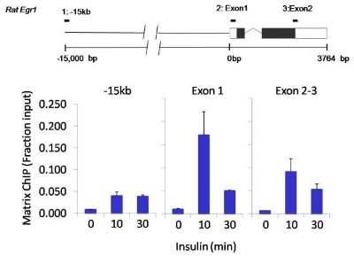
Chromatin Immunoprecipitation: LIN-28A Antibody (14E6-4E6) [NBP2-22481] - Analysis performed using cross-linked chromatin from rat hepatoma cells treated with insulin. IP performed using a multiplex microplate Matrix ChIP assay of LIN28 monoclonal antibody. Chromatin aliquots from cells were used per ChIP pull-down. Quantitative PCR data done in quadruplicate using 1ul of DNA in 2ul SYBR real-time PCR reactions containing primers to amplify -15kb upstream of Egr1 or exon-1 or exon-2-3 of Egr1. Quantitation of immunoprecipitated chromatin is presented as signal relative to the total amount of input chromatin. Results represent the mean +/- SEM. A schematic representation of the rat Egr-1 locus is shown; oxes represent exons (black boxes = translated, white boxes = untranslated), the zigzag line represents an intron, and the straight line represents upstream sequence. Regions amplified by Egr-1 primers are represented by black bars. Data courtesy of the Innovators Program.
![Immunocytochemistry/ Immunofluorescence: LIN-28A Antibody (14E6-4E6) [NBP2-22481]](https://resources.rndsystems.com/images/products/LIN-28A-Antibody-14E6-4E6-Immunocytochemistry-Immunofluorescence-NBP2-22481-img0003.jpg "Immunocytochemistry/ Immunofluorescence: LIN-28A Antibody (14E6-4E6) [NBP2-22481]")
Immunocytochemistry/ Immunofluorescence: LIN-28A Antibody (14E6-4E6) [NBP2-22481]
Immunocytochemistry/Immunofluorescence: LIN-28A Antibody (14E6-4E6) [NBP2-22481] - Analysis of Lin28 (green) in H9 embryonic stem cells grown for a few days on Matrigel-coated chamber slides. Cells fixed in 4% paraformaldehyde were permeabilized with 0.1% Triton X-100 for 15 minutes at room temperature. Cells were probed with a Lin28 monoclonal antibody at a dilution of 1:200 overnight at 4C, washed with PBST, and incubated with a fluorescein-conjugated secondary antibody at a dilution of 1:100 for 1 hour at room temperature. Nuclei (blue) were stained with DAPI and cells were analyzed by fluorescence microscopy at 20X magnification.![Immunocytochemistry/ Immunofluorescence: LIN-28A Antibody (14E6-4E6) [NBP2-22481]](https://resources.rndsystems.com/images/products/LIN-28A-Antibody-14E6-4E6-Immunocytochemistry-Immunofluorescence-NBP2-22481-img0004.jpg "Immunocytochemistry/ Immunofluorescence: LIN-28A Antibody (14E6-4E6) [NBP2-22481]")
Immunocytochemistry/ Immunofluorescence: LIN-28A Antibody (14E6-4E6) [NBP2-22481]
Immunocytochemistry/Immunofluorescence: LIN-28A Antibody (14E6-4E6) [NBP2-22481] - Analysis of Lin28 (green) in HEL 11.4 induced IPS cells grown for a few days on Matrigel-coated chamber slides. Cells fixed in 4% paraformaldehyde were permeabilized with 0.1% Triton X-100 for 15 minutes at room temperature. Cells were probed with a Lin28 monoclonal antibody at a dilution of 1:200 overnight at 4C, washed with PBST, and incubated with a fluorescein-conjugated secondary antibody at a dilution of 1:100 for 1 hour at room temperature. Nuclei (blue) were stained with DAPI and cells were analyzed by fluorescence microscopy at 20X magnification.![Immunohistochemistry-Paraffin: LIN-28A Antibody (14E6-4E6) [NBP2-22481]](https://resources.rndsystems.com/images/products/LIN-28A-Antibody-14E6-4E6-Immunohistochemistry-Paraffin-NBP2-22481-img0014.jpg "Immunohistochemistry-Paraffin: LIN-28A Antibody (14E6-4E6) [NBP2-22481]")
Immunohistochemistry-Paraffin: LIN-28A Antibody (14E6-4E6) [NBP2-22481]
Immunohistochemistry-Paraffin: LIN-28A Antibody (14E6-4E6) [NBP2-22481] - Analysis showing staining in the nucleus and cytoplasm of mouse testis tissue (right) compared with a negative control without primary antibody (left).![Flow Cytometry: LIN-28A Antibody (14E6-4E6) [NBP2-22481]](https://resources.rndsystems.com/images/products/LIN-28A-Antibody-14E6-4E6-Flow-Cytometry-NBP2-22481-img0012.jpg "Flow Cytometry: LIN-28A Antibody (14E6-4E6) [NBP2-22481]")
Flow Cytometry: LIN-28A Antibody (14E6-4E6) [NBP2-22481]
Flow Cytometry: LIN-28A Antibody (14E6-4E6) [NBP2-22481] - Analysis of Lin28 (blue histogram) on HEL 11.4 induced IPS cells. To generate single cells suspensions, colonies were treated with TrypLE cell dissociation enzyme for 5 minutes at 37C. Cells were incubated with a Lin28 monoclonal antibody or mouse IgG (green histogram) at a dilution of 1:100 for 1 hour on ice, washed with PBS + 5% fetal calf serum (FACS buffer), and incubated with a fluorescein-conjugated secondary antibody at a dilution of 1:200 for 30 minutes on ice. Cells were washed with cold FACS buffer, resuspended in 500ul of FACS buffer containing 10ul of 4% paraformaldehyde.![Immunoprecipitation: LIN-28A Antibody (14E6-4E6) [NBP2-22481]](https://resources.rndsystems.com/images/products/LIN-28A-Antibody-14E6-4E6-Immunoprecipitation-NBP2-22481-img0002.jpg "Immunoprecipitation: LIN-28A Antibody (14E6-4E6) [NBP2-22481]")
Immunoprecipitation: LIN-28A Antibody (14E6-4E6) [NBP2-22481]
Immunoprecipitation: LIN-28A Antibody (14E6-4E6) [NBP2-22481] - Analysis of LIN28 was performed. Antigen-antibody complexes were formed by incubating 700ug of lysate with 5 ug of an LIN28 monoclonal antibody overnight on a rocking platform at 4C. The immune complexes were captured on 50 ul Protein A/G Agarose was loaded as a positive control for detection. Samples were resolved on a 4-20% Tris-HCl polyacrylamide gel, transferred to a PVDF membrane, and blocked with 5% BSA/TBS-0.1%Tween for at least 1 hour. The membrane was probed with a LIN28 monoclonal antibody at a dilution of 1:1000 overnight rotating at 4C, washed in TBST, and probed with Clean-blot IP Detection Reagent at a dilution of 1:1000 for at least 1 hour.Applications for LIN-28A Antibody (14E6-4E6)
Application
Recommended Usage
Chromatin Immunoprecipitation (ChIP)
1-3 ul
Flow Cytometry
1:100
Immunocytochemistry/ Immunofluorescence
1:50 - 1:200
Immunohistochemistry
1:20 - 1:200
Immunohistochemistry-Paraffin
1:20 - 1:200
Immunoprecipitation
5 ug
Western Blot
1:1000
Flow Cytometry Panel Builder
Bio-Techne Knows Flow Cytometry
Save time and reduce costly mistakes by quickly finding compatible reagents using the Panel Builder Tool.
Advanced Features
- Spectra Viewer - Custom analysis of spectra from multiple fluorochromes
- Spillover Popups - Visualize the spectra of individual fluorochromes
- Antigen Density Selector - Match fluorochrome brightness with antigen density

Formulation, Preparation, and Storage
Purification
Protein A purified
Formulation
PBS with 1 mg/ml BSA and 30% glycerol
Preservative
0.05% Sodium Azide
Concentration
1 mg/ml
Shipping
The product is shipped with polar packs. Upon receipt, store it immediately at the temperature recommended below.
Stability & Storage
Store at -20C. Avoid freeze-thaw cycles.
Background: LIN-28A
Long Name
RNA-binding Protein LIN-28
Alternate Names
CSDD1, LIN28, LIN28A, Tex17, ZCCHC1
Entrez Gene IDs
79727 (Human)
Gene Symbol
LIN28A
Additional LIN-28A Products
Product Documents for LIN-28A Antibody (14E6-4E6)
Certificate of Analysis
To download a Certificate of Analysis, please enter a lot or batch number in the search box below.
Product Specific Notices for LIN-28A Antibody (14E6-4E6)
This product is for research use only and is not approved for use in humans or in clinical diagnosis. Primary Antibodies are guaranteed for 1 year from date of receipt.
Related Research Areas
Customer Reviews for LIN-28A Antibody (14E6-4E6)
There are currently no reviews for this product. Be the first to review LIN-28A Antibody (14E6-4E6) and earn rewards!
Have you used LIN-28A Antibody (14E6-4E6)?
Submit a review and receive an Amazon gift card!
$25/€18/£15/$25CAN/¥2500 Yen for a review with an image
$10/€7/£6/$10CAN/¥1110 Yen for a review without an image
Submit a review
Protocols
Find general support by application which include: protocols, troubleshooting, illustrated assays, videos and webinars.
- 7-Amino Actinomycin D (7-AAD) Cell Viability Flow Cytometry Protocol
- Antigen Retrieval Protocol (PIER)
- Antigen Retrieval for Frozen Sections Protocol
- Appropriate Fixation of IHC/ICC Samples
- Cellular Response to Hypoxia Protocols
- ChIP Protocol Video
- Chromatin Immunoprecipitation (ChIP) Protocol
- Chromatin Immunoprecipitation Protocol
- Chromogenic IHC Staining of Formalin-Fixed Paraffin-Embedded (FFPE) Tissue Protocol
- Chromogenic Immunohistochemistry Staining of Frozen Tissue
- ClariTSA™ Fluorophore Kits
- Detection & Visualization of Antibody Binding
- Extracellular Membrane Flow Cytometry Protocol
- Flow Cytometry Protocol for Cell Surface Markers
- Flow Cytometry Protocol for Staining Membrane Associated Proteins
- Flow Cytometry Staining Protocols
- Flow Cytometry Troubleshooting Guide
- Fluorescent IHC Staining of Frozen Tissue Protocol
- Graphic Protocol for Heat-induced Epitope Retrieval
- Graphic Protocol for the Preparation and Fluorescent IHC Staining of Frozen Tissue Sections
- Graphic Protocol for the Preparation and Fluorescent IHC Staining of Paraffin-embedded Tissue Sections
- Graphic Protocol for the Preparation of Gelatin-coated Slides for Histological Tissue Sections
- ICC Cell Smear Protocol for Suspension Cells
- ICC Immunocytochemistry Protocol Videos
- ICC for Adherent Cells
- IHC Sample Preparation (Frozen sections vs Paraffin)
- Immunocytochemistry (ICC) Protocol
- Immunocytochemistry Troubleshooting
- Immunofluorescence of Organoids Embedded in Cultrex Basement Membrane Extract
- Immunofluorescent IHC Staining of Formalin-Fixed Paraffin-Embedded (FFPE) Tissue Protocol
- Immunohistochemistry (IHC) and Immunocytochemistry (ICC) Protocols
- Immunohistochemistry Frozen Troubleshooting
- Immunohistochemistry Paraffin Troubleshooting
- Immunoprecipitation Protocol
- Intracellular Flow Cytometry Protocol Using Alcohol (Methanol)
- Intracellular Flow Cytometry Protocol Using Detergents
- Intracellular Nuclear Staining Flow Cytometry Protocol Using Detergents
- Intracellular Staining Flow Cytometry Protocol Using Alcohol Permeabilization
- Intracellular Staining Flow Cytometry Protocol Using Detergents to Permeabilize Cells
- Preparing Samples for IHC/ICC Experiments
- Preventing Non-Specific Staining (Non-Specific Binding)
- Primary Antibody Selection & Optimization
- Propidium Iodide Cell Viability Flow Cytometry Protocol
- Protocol for Heat-Induced Epitope Retrieval (HIER)
- Protocol for Liperfluo
- Protocol for Making a 4% Formaldehyde Solution in PBS
- Protocol for VisUCyte™ HRP Polymer Detection Reagent
- Protocol for the Characterization of Human Th22 Cells
- Protocol for the Characterization of Human Th9 Cells
- Protocol for the Fluorescent ICC Staining of Cell Smears - Graphic
- Protocol for the Fluorescent ICC Staining of Cultured Cells on Coverslips - Graphic
- Protocol for the Preparation & Fixation of Cells on Coverslips
- Protocol for the Preparation and Chromogenic IHC Staining of Frozen Tissue Sections
- Protocol for the Preparation and Chromogenic IHC Staining of Frozen Tissue Sections - Graphic
- Protocol for the Preparation and Chromogenic IHC Staining of Paraffin-embedded Tissue Sections
- Protocol for the Preparation and Chromogenic IHC Staining of Paraffin-embedded Tissue Sections - Graphic
- Protocol for the Preparation and Fluorescent ICC Staining of Cells on Coverslips
- Protocol for the Preparation and Fluorescent ICC Staining of Non-adherent Cells
- Protocol for the Preparation and Fluorescent ICC Staining of Stem Cells on Coverslips
- Protocol for the Preparation and Fluorescent IHC Staining of Frozen Tissue Sections
- Protocol for the Preparation and Fluorescent IHC Staining of Paraffin-embedded Tissue Sections
- Protocol for the Preparation of Gelatin-coated Slides for Histological Tissue Sections
- Protocol for the Preparation of a Cell Smear for Non-adherent Cell ICC - Graphic
- Protocol: Annexin V and PI Staining by Flow Cytometry
- Protocol: Annexin V and PI Staining for Apoptosis by Flow Cytometry
- R&D Systems Quality Control Western Blot Protocol
- TUNEL and Active Caspase-3 Detection by IHC/ICC Protocol
- The Importance of IHC/ICC Controls
- Troubleshooting Guide: Fluorokine Flow Cytometry Kits
- Troubleshooting Guide: Immunohistochemistry
- Troubleshooting Guide: Western Blot Figures
- Western Blot Conditions
- Western Blot Protocol
- Western Blot Protocol for Cell Lysates
- Western Blot Troubleshooting
- Western Blot Troubleshooting Guide
- View all Protocols, Troubleshooting, Illustrated assays and Webinars
Loading...
Associated Pathways
