B7-1 and B7-2, together with their receptors CD28 and CTLA-4, constitute one of the dominant costimulatory pathways that regulate T- and B-cell responses. Although both CTLA-4 and CD28 can bind to the same ligands, CTLA-4 binds to B7-1 and B7-2 with a 20‑100 fold higher affinity than CD28 and is involved in the
down‑regulation of the immune response. B7-1 is expressed on activated B cells, activated T cells, and macrophages. B7-2 is constitutively expressed on interdigitating dendritic cells, Langerhans cells, peripheral blood dendritic cells, memory B cells, and germinal center B cells. Additionally, B7-2 is expressed at low levels on monocytes and can be up-regulated through interferon gamma. B7-1 and B7-2 are both members of the immunoglobulin superfamily. Mouse B7-1 is a 306 amino acid (aa) protein containing a putative 37 aa signal peptide, a 190 aa extracellular domain, a 22 aa transmembrane domain, and a 38 aa cytoplasmic domain. Mouse B7-1 and B7-2 share 28% amino acid identity. Mouse and human B7-1 share 44% amino acid identity. However, it has been observed that both human and mouse
B7‑1 and B7‑2 can bind to either human or mouse CD28 and CTLA-4, suggesting that there are conserved amino acids which form the B7-1/B7-2/CD28/CTLA-4 critical binding sites.

Key Product Details
Validated by
Biological Validation
Species Reactivity
Validated:
Mouse
Cited:
Human, Mouse
Applications
Validated:
Western Blot, ELISA Capture (Matched Antibody Pair), Neutralization, Flow Cytometry, Immunocytochemistry, CyTOF-ready
Cited:
Immunohistochemistry, Western Blot, Flow Cytometry, Immunocytochemistry
Label
Unconjugated
Antibody Source
Polyclonal Goat IgG
Loading...
Product Specifications
Immunogen
Mouse myeloma cell line NS0-derived recombinant mouse B7‑1/CD80
Asp37-Lys245
Accession # Q00609
Asp37-Lys245
Accession # Q00609
Specificity
Detects mouse B7‑1/CD80 in ELISAs and Western blots.
Clonality
Polyclonal
Host
Goat
Isotype
IgG
Endotoxin Level
<0.10 EU per 1 μg of the antibody by the LAL method.
Scientific Data Images for Mouse B7-1/CD80 Antibody
Detection of Mouse B7‑1/CD80 by Western Blot.
Western blot shows lysates of C2C12 mouse myoblast cell line. PVDF membrane was probed with 1 µg/mL of Goat Anti-Mouse B7-1/CD80 Antigen Affinity-purified Polyclonal Antibody (Catalog # AF740) followed by HRP-conjugated Anti-Goat IgG Secondary Antibody (Catalog # HAF017). A specific band was detected for B7-1/CD80 at approximately 60 kDa (as indicated). This experiment was conducted under reducing conditions and using Immunoblot Buffer Group 1.
Detection of B7‑1/CD80 in Mouse Splenocytes by Flow Cytometry.
Mouse splenocytes either treated with 200 ng/mL LPS (filled histogram) or unstimulated (open histogram) were stained with Goat Anti-Mouse B7-1/CD80 Antigen Affinity-purified Polyclonal Antibody (Catalog # AF740), followed by Phycoerythrin-conjugated Anti-Goat IgG Secondary Antibody (Catalog # F0107). View our protocol for Staining Membrane-associated Proteins.
Detection of B7‑1/CD80 in Mouse Splenocytes by Flow Cytometry
Mouse B6 splenocytes treated with 200 ng/mL LPS for 48 hr were stained with (A) Goat Anti-Mouse B7-1/CD80 Antigen Affinity-purified Polyclonal Antibody (Catalog # AF740) or (B) Goat IgG control antibody (AB-108-C) followed by Phycoerythrin-conjugated Anti-Goat IgG Secondary Antibody (F0107) and Rat Anti-Mouse B220/CD45R Alexa Fluor® 488-conjugated Monoclonal Antibody (FAB1217G). View our protocol for Staining Membrane-associated Proteins.
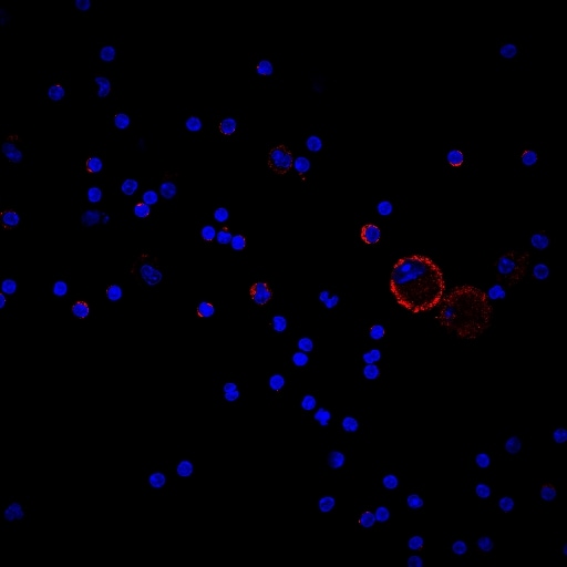
B7‑1/CD80 in Mouse Splenocytes.
B7-1/CD80 was detected in immersion fixed mouse splenocytes using Goat Anti-Mouse B7-1/CD80 Antigen Affinity-purified Polyclonal Antibody (Catalog # AF740) at 15 µg/mL for 3 hours at room temperature. Cells were stained using the NorthernLights™ 557-conjugated Anti-Goat IgG Secondary Antibody (red; Catalog # NL001) and counterstained with DAPI (blue). Specific staining was localized to cytoplasm. View our protocol for Fluorescent ICC Staining of Non-adherent Cells.
IL‑2 secretion Induced by B7‑1/CD80 and Neutralization by Mouse B7‑1/CD80 Antibody.
Recombinant Mouse B7-1/CD80 Fc Chimera (Catalog # 740-B1) co-stimulates IL-2 secretion in the Jurkat human acute T cell leukemia cell line in the presence of PHA in a dose-dependent manner (orange line), as measured by the Human IL-2 Quantikine ELISA Kit (Catalog # D2050). IL-2 secretion elicited by Recombinant Mouse B7-1/CD80 Fc Chimera (0.1 µg/mL) and PHA (10 µg/mL) is neutralized (green line) by increasing concentrations of Goat Anti-Mouse B7-1/CD80 Antigen Affinity-purified Polyclonal Antibody (Catalog # AF740). The ND50 is typically 0.15-0.6 µg/mL.Applications for Mouse B7-1/CD80 Antibody
Application
Recommended Usage
CyTOF-ready
Ready to be labeled using established conjugation methods. No BSA or other carrier proteins that could interfere with conjugation.
Flow Cytometry
0.25 µg/106 cells
Sample: Mouse splenocytes treated with LPS
Sample: Mouse splenocytes treated with LPS
Immunocytochemistry
5-15 µg/mL
Sample: Immersion fixed mouse splenocytes
Sample: Immersion fixed mouse splenocytes
Western Blot
1 µg/mL
Sample: C2C12 mouse myoblast cell line
Sample: C2C12 mouse myoblast cell line
Neutralization
Measured by its ability to neutralize B7‑1/CD80-induced IL‑2 secretion in the Jurkat human acute T cell leukemia cell line. The Neutralization Dose (ND50) is typically 0.15-0.6 µg/mL in the presence of 0.1 µg/mL Recombinant Mouse B7‑1/CD80 Fc Chimera and 10 µg/mL PHA.
Mouse B7-1/CD80 Sandwich Immunoassay
Please Note: Optimal dilutions of this antibody should be experimentally determined.
Reviewed Applications
Read 2 reviews rated 5 using AF740 in the following applications:
Flow Cytometry Panel Builder
Bio-Techne Knows Flow Cytometry
Save time and reduce costly mistakes by quickly finding compatible reagents using the Panel Builder Tool.
Advanced Features
- Spectra Viewer - Custom analysis of spectra from multiple fluorochromes
- Spillover Popups - Visualize the spectra of individual fluorochromes
- Antigen Density Selector - Match fluorochrome brightness with antigen density

Formulation, Preparation, and Storage
Purification
Antigen Affinity-purified
Reconstitution
Reconstitute at 0.2 mg/mL in sterile PBS. For liquid material, refer to CoA for concentration.
Loading...
Formulation
Lyophilized from a 0.2 μm filtered solution in PBS with Trehalose. *Small pack size (SP) is supplied either lyophilized or as a 0.2 µm filtered solution in PBS.
Shipping
Lyophilized product is shipped at ambient temperature. Liquid small pack size (-SP) is shipped with polar packs. Upon receipt, store immediately at the temperature recommended below.
Stability & Storage
Use a manual defrost freezer and avoid repeated freeze-thaw cycles.
- 12 months from date of receipt, -20 to -70 °C as supplied.
- 1 month, 2 to 8 °C under sterile conditions after reconstitution.
- 6 months, -20 to -70 °C under sterile conditions after reconstitution.
Calculators
Background: B7-1/CD80
References
- Azuma, M. et al. (1993) Nature 366:76.
- Freeman, G.J. et al. (1993) Science 262:909.
- Freeman, G. et al. (1991) J. Exp. Med. 174:625.
- Selvakumar, A. et al. (1993) Immunogenetics 38:292.
- Chen, C. et al. (1994) J. Immunol. 152:4929.
- Freeman, G.J. et al. (1993) J. Exp. Med. 178:2185.
Alternate Names
B71, CD80
Gene Symbol
CD80
UniProt
Additional B7-1/CD80 Products
Product Documents for Mouse B7-1/CD80 Antibody
Certificate of Analysis
To download a Certificate of Analysis, please enter a lot or batch number in the search box below.
Note: Certificate of Analysis not available for kit components.
Product Specific Notices for Mouse B7-1/CD80 Antibody
For research use only
Related Research Areas
Citations for Mouse B7-1/CD80 Antibody
 Powered by Bioz
Customer Reviews for Mouse B7-1/CD80 Antibody (2)
5 out of 5
2 Customer Ratings
Have you used Mouse B7-1/CD80 Antibody?
Submit a review and receive an Amazon gift card!
$25/€18/£15/$25CAN/¥2500 Yen for a review with an image
$10/€7/£6/$10CAN/¥1110 Yen for a review without an image
Submit a review
Customer Images
Showing
1
-
2 的
2 reviews
Showing All
Filter By:
-
Application: Immunocytochemistry/ImmunofluorescenceSample Tested: Lung tissueSpecies: MouseVerified Customer | Posted 12/12/2021

-
Application: Flow CytometrySample Tested: Pancreas tissueSpecies: MouseVerified Customer | Posted 12/18/2018
There are no reviews that match your criteria.
Protocols
Find general support by application which include: protocols, troubleshooting, illustrated assays, videos and webinars.
- 7-Amino Actinomycin D (7-AAD) Cell Viability Flow Cytometry Protocol
- Appropriate Fixation of IHC/ICC Samples
- Cellular Response to Hypoxia Protocols
- ClariTSA™ Fluorophore Kits
- Detection & Visualization of Antibody Binding
- Extracellular Membrane Flow Cytometry Protocol
- Flow Cytometry Protocol for Cell Surface Markers
- Flow Cytometry Protocol for Staining Membrane Associated Proteins
- Flow Cytometry Staining Protocols
- Flow Cytometry Troubleshooting Guide
- ICC Cell Smear Protocol for Suspension Cells
- ICC Immunocytochemistry Protocol Videos
- ICC for Adherent Cells
- Immunocytochemistry (ICC) Protocol
- Immunocytochemistry Troubleshooting
- Immunofluorescence of Organoids Embedded in Cultrex Basement Membrane Extract
- Immunohistochemistry (IHC) and Immunocytochemistry (ICC) Protocols
- Intracellular Flow Cytometry Protocol Using Alcohol (Methanol)
- Intracellular Flow Cytometry Protocol Using Detergents
- Intracellular Nuclear Staining Flow Cytometry Protocol Using Detergents
- Intracellular Staining Flow Cytometry Protocol Using Alcohol Permeabilization
- Intracellular Staining Flow Cytometry Protocol Using Detergents to Permeabilize Cells
- Preparing Samples for IHC/ICC Experiments
- Preventing Non-Specific Staining (Non-Specific Binding)
- Primary Antibody Selection & Optimization
- Propidium Iodide Cell Viability Flow Cytometry Protocol
- Protocol for Liperfluo
- Protocol for VisUCyte™ HRP Polymer Detection Reagent
- Protocol for the Characterization of Human Th22 Cells
- Protocol for the Characterization of Human Th9 Cells
- Protocol for the Fluorescent ICC Staining of Cell Smears - Graphic
- Protocol for the Fluorescent ICC Staining of Cultured Cells on Coverslips - Graphic
- Protocol for the Preparation and Fluorescent ICC Staining of Cells on Coverslips
- Protocol for the Preparation and Fluorescent ICC Staining of Non-adherent Cells
- Protocol for the Preparation and Fluorescent ICC Staining of Stem Cells on Coverslips
- Protocol for the Preparation of a Cell Smear for Non-adherent Cell ICC - Graphic
- Protocol: Annexin V and PI Staining by Flow Cytometry
- Protocol: Annexin V and PI Staining for Apoptosis by Flow Cytometry
- R&D Systems Quality Control Western Blot Protocol
- TUNEL and Active Caspase-3 Detection by IHC/ICC Protocol
- The Importance of IHC/ICC Controls
- Troubleshooting Guide: Fluorokine Flow Cytometry Kits
- Troubleshooting Guide: Western Blot Figures
- Western Blot Conditions
- Western Blot Protocol
- Western Blot Protocol for Cell Lysates
- Western Blot Troubleshooting
- Western Blot Troubleshooting Guide
- View all Protocols, Troubleshooting, Illustrated assays and Webinars


