TLR9 Antibody (26C593.2) - BSA Free
Novus Biologicals | Catalog # NBP2-24729
Key Product Details
Validated by
Species Reactivity
Validated:
Cited:
Applications
Validated:
Cited:
Label
Antibody Source
Format
Product Specifications
Immunogen
Reactivity Notes
Clonality
Host
Isotype
Scientific Data Images for TLR9 Antibody (26C593.2) - BSA Free
![Simple Western: TLR9 Antibody (26C593.2)BSA Free [NBP2-24729]](https://resources.rndsystems.com/images/products/TLR9-Antibody-26C593-2-Simple-Western-NBP2-24729-img0029.jpg "Simple Western: TLR9 Antibody (26C593.2)BSA Free [NBP2-24729]")
Simple Western: TLR9 Antibody (26C593.2)BSA Free [NBP2-24729]
Simple Western: TLR9 Antibody (26C593.2) [NBP2-24729] - Lane view shows a specific band for TLR9 in 0.5 mg/ml of Ramos lysate. This experiment was performed under reducing conditions using the 66-440 kDa separation system. Image using the Azide Free format of this antibody.![Flow Cytometry: TLR9 Antibody (26C593.2) - BSA Free [NBP2-24729]](https://resources.rndsystems.com/images/products/TLR9-Antibody-26C593-2-Flow-Cytometry-NBP2-24729-img0025.jpg "Flow Cytometry: TLR9 Antibody (26C593.2) - BSA Free [NBP2-24729]")
Flow Cytometry: TLR9 Antibody (26C593.2) - BSA Free [NBP2-24729]
TLR9-Antibody-26C593-2-Flow-Cytometry-NBP2-24729-img0025.jpg![Western Blot: TLR9 Antibody (26C593.2) - BSA Free [NBP2-24729]](https://resources.rndsystems.com/images/products/TLR9-Antibody-26C593-2-Knockdown-Validated-NBP2-24729-img0031.jpg "Western Blot: TLR9 Antibody (26C593.2) - BSA Free [NBP2-24729]")
![Flow Cytometry: TLR9 Antibody (26C593.2) - BSA Free [NBP2-24729]](https://resources.rndsystems.com/images/products/TLR9-Antibody-26C593-2-Flow-Cytometry-NBP2-24729-img0027.jpg "Flow Cytometry: TLR9 Antibody (26C593.2) - BSA Free [NBP2-24729]")
Flow Cytometry: TLR9 Antibody (26C593.2) - BSA Free [NBP2-24729]
TLR9-Antibody-26C593-2-Flow-Cytometry-NBP2-24729-img0027.jpg![Immunocytochemistry/ Immunofluorescence: TLR9 Antibody (26C593.2) - BSA Free [NBP2-24729]](https://resources.rndsystems.com/images/products/TLR9-Antibody-26C593-2-Immunocytochemistry-Immunofluorescence-NBP2-24729-img0032.jpg "Immunocytochemistry/ Immunofluorescence: TLR9 Antibody (26C593.2) - BSA Free [NBP2-24729]")
Immunocytochemistry/ Immunofluorescence: TLR9 Antibody (26C593.2) - BSA Free [NBP2-24729]
TLR9-Antibody-26C593-2-Immunocytochemistry-Immunofluorescence-NBP2-24729-img0032.jpg![Immunohistochemistry-Paraffin: TLR9 Antibody (26C593.2) - BSA Free [NBP2-24729]](https://resources.rndsystems.com/images/products/TLR9-Antibody-26C593-2-Immunohistochemistry-Paraffin-NBP2-24729-img0023.jpg "Immunohistochemistry-Paraffin: TLR9 Antibody (26C593.2) - BSA Free [NBP2-24729]")
Immunohistochemistry-Paraffin: TLR9 Antibody (26C593.2) - BSA Free [NBP2-24729]
Immunohistochemistry-Paraffin: TLR9 Antibody (26C593.2) [NBP2-24729] - Monkey retina tissue. Image from verified customer review.![Flow Cytometry: TLR9 Antibody (26C593.2) - BSA Free [NBP2-24729]](https://resources.rndsystems.com/images/products/TLR9-Antibody-26C593-2-Flow-Cytometry-NBP2-24729-img0028.jpg "Flow Cytometry: TLR9 Antibody (26C593.2) - BSA Free [NBP2-24729]")
Flow Cytometry: TLR9 Antibody (26C593.2) - BSA Free [NBP2-24729]
TLR9-Antibody-26C593-2-Flow-Cytometry-NBP2-24729-img0028.jpg![Western Blot: TLR9 Antibody (26C593.2)BSA Free [NBP2-24729]](https://resources.rndsystems.com/images/products/TLR9-Antibody-26C593-2-Western-Blot-NBP2-24729-img0033.jpg "Western Blot: TLR9 Antibody (26C593.2)BSA Free [NBP2-24729]")
Western Blot: TLR9 Antibody (26C593.2)BSA Free [NBP2-24729]
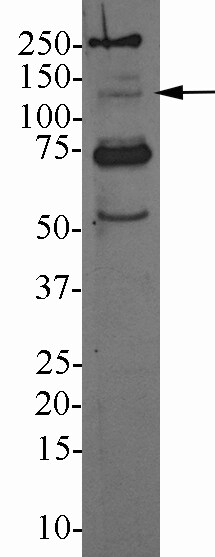
Western Blot: TLR9 Antibody (26C593.2) [NBP2-24729] - Analysis of TLR9 in A) human PBMC, B) human intestine, C) mouse intestine, and D) rat intestine tissue lysates using this antibody at a dilution of 3 ug/ml.![Flow Cytometry: TLR9 Antibody (26C593.2) - BSA Free [NBP2-24729]](https://resources.rndsystems.com/images/products/TLR9-Antibody-26C593-2-Flow-Cytometry-NBP2-24729-img0030.jpg "Flow Cytometry: TLR9 Antibody (26C593.2) - BSA Free [NBP2-24729]")
Flow Cytometry: TLR9 Antibody (26C593.2) - BSA Free [NBP2-24729]
TLR9-Antibody-26C593-2-Flow-Cytometry-NBP2-24729-img0030.jpg in THP-1 Acute Monocytic Leukemia Human Cell Line by Flow Cytometry.")
Detection of TLR9 (26C593.2) in THP-1 Acute Monocytic Leukemia Human Cell Line by Flow Cytometry.
THP-1 human acute monocytic leukemia cell line was stained with Mouse anti-TLR9 (26C593.2) Protein-G purified Monoclonal Antibody conjugated to Janelia Fluor® 549 (Catalog # NBP2-24729JF549, blue histogram) or matched control antibody (orange histogram). - BSA Free [NBP2-24729] -")
Immunocytochemistry/ Immunofluorescence: TLR9 Antibody (26C593.2) - BSA Free [NBP2-24729] -
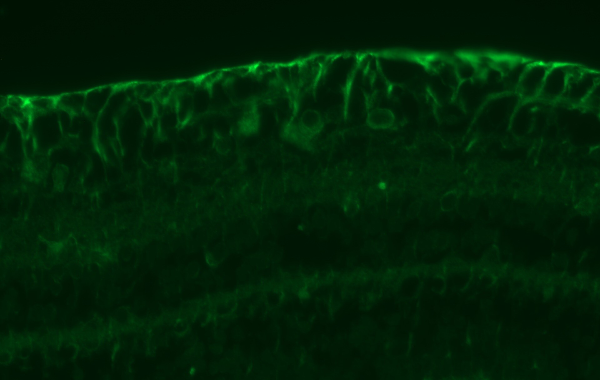
Alveolar epithelial cells (AECs), fibroblasts and fibrocytes all express Toll-like receptor-9. AECs, fibroblasts, fibrocytes and whole lungs were isolated from Balb/c, mice and total RNA prepared and analyzed for TLR-9 expression. The data were normalized to the expression level of TLR-9 in dendritic cells, which was set to 1 (n = 3/group). No TLR-9 message was amplified in cells from TLR-9-/- mice. (B, C) Immunohistochemistry was performed on paraffin wax-embedded whole-lung tissue from normal Balb/c mice. (B) Control staining with secondary antibody only; (C) diffuse TLR-9 staining, with notable staining in the bronchial epithelium, at the alveolar junctions and the interstitium. Fibroblasts (E) and AECs (G) were isolated from Balb/c mice and fixed. Immunofluorescence staining for TLR-9 shows that both cell types expressed TLR-9. The control slides with secondary antibody alone (D and F) did not show any auto-fluorescence or non-specific staining. Image collected and cropped by CiteAb from the following open publication (https://pubmed.ncbi.nlm.nih.gov/21810214), licensed under a CC-BY license. Not internally tested by Novus Biologicals. - BSA Free [NBP2-24729] -")
Immunocytochemistry/ Immunofluorescence: TLR9 Antibody (26C593.2) - BSA Free [NBP2-24729] -
Alveolar epithelial cells (AECs), fibroblasts and fibrocytes all express Toll-like receptor-9. AECs, fibroblasts, fibrocytes and whole lungs were isolated from Balb/c, mice and total RNA prepared and analyzed for TLR-9 expression. The data were normalized to the expression level of TLR-9 in dendritic cells, which was set to 1 (n = 3/group). No TLR-9 message was amplified in cells from TLR-9-/- mice. (B, C) Immunohistochemistry was performed on paraffin wax-embedded whole-lung tissue from normal Balb/c mice. (B) Control staining with secondary antibody only; (C) diffuse TLR-9 staining, with notable staining in the bronchial epithelium, at the alveolar junctions and the interstitium. Fibroblasts (E) and AECs (G) were isolated from Balb/c mice and fixed. Immunofluorescence staining for TLR-9 shows that both cell types expressed TLR-9. The control slides with secondary antibody alone (D and F) did not show any auto-fluorescence or non-specific staining. Image collected and cropped by CiteAb from the following open publication (https://pubmed.ncbi.nlm.nih.gov/21810214), licensed under a CC-BY license. Not internally tested by Novus Biologicals.Applications for TLR9 Antibody (26C593.2) - BSA Free
Block/Neutralize
Dot Blot
ELISA
Flow (Intracellular)
Functional
Immunocytochemistry/ Immunofluorescence
Immunohistochemistry
Immunohistochemistry-Paraffin
Immunoprecipitation
In vitro assay
Knockdown Validated
Simple Western
Western Blot
See Simple Western Antibody Database for Simple Western validation: Tested in Mouse Brain, Ramos lysate 0.5 mg/mL, separated by Size, antibody dilution of 1:50, 30 ug/mL, apparent MW was 90 kDa. Separated by Size-Wes, Sally Sue/Peggy Sue.
Reviewed Applications
Read 4 reviews rated 3.8 using NBP2-24729 in the following applications:
Flow Cytometry Panel Builder
Bio-Techne Knows Flow Cytometry
Save time and reduce costly mistakes by quickly finding compatible reagents using the Panel Builder Tool.
Advanced Features
- Spectra Viewer - Custom analysis of spectra from multiple fluorochromes
- Spillover Popups - Visualize the spectra of individual fluorochromes
- Antigen Density Selector - Match fluorochrome brightness with antigen density

Formulation, Preparation, and Storage
Purification
Formulation
Format
Preservative
Concentration
Shipping
Stability & Storage
Background: TLR9
Additional TLR9 Products
Product Documents for TLR9 Antibody (26C593.2) - BSA Free
Certificate of Analysis
To download a Certificate of Analysis, please enter a lot or batch number in the search box below.
Product Specific Notices for TLR9 Antibody (26C593.2) - BSA Free
This product is for research use only and is not approved for use in humans or in clinical diagnosis. Primary Antibodies are guaranteed for 1 year from date of receipt.
Citations for TLR9 Antibody (26C593.2) - BSA Free
 Powered by Bioz
Customer Reviews for TLR9 Antibody (26C593.2) - BSA Free (4)
Have you used TLR9 Antibody (26C593.2) - BSA Free?
Submit a review and receive an Amazon gift card!
$25/€18/£15/$25CAN/¥2500 Yen for a review with an image
$10/€7/£6/$10CAN/¥1110 Yen for a review without an image
Submit a review
Customer Images
-
Application: Immunohistochemistry-ParaffinSample Tested: paraffin embedded macaque retina tissueSpecies: macaca mulattaVerified Customer | Posted 04/12/2018this antibody recognized TLR9 in paraffin embedded macaque retina tissueHeat mediated antigen retrieval (pH 6 citrate buffer) 1:25 dilution of primary antibody ON at 4C 1:400 dilution of goat anti-mouse Alexa Fluor 488

-
Application: Western BlotSample Tested: human lymphocytes, whole cell lysateSpecies: HumanVerified Customer | Posted 10/20/2017TLR9 in Lympocyte whole cell lysate3ug/mL in Milk-TBS/T 30ug of protein, 4-12% Bis-Tris gel 10 minute exposure, several bands. 250, 120, 75, 55kDA
-
Application: Western BlotSample Tested: Whole cell lysateSpecies: HumanVerified Customer | Posted 01/14/2015
-
Application: Western BlotSample Tested: Primary blood-derived B cellsSpecies: HumanVerified Customer | Posted 11/25/2014Expression of surface TLR9 on human B cells
There are no reviews that match your criteria.
Protocols
Find general support by application which include: protocols, troubleshooting, illustrated assays, videos and webinars.
- 7-Amino Actinomycin D (7-AAD) Cell Viability Flow Cytometry Protocol
- Antigen Retrieval Protocol (PIER)
- Antigen Retrieval for Frozen Sections Protocol
- Appropriate Fixation of IHC/ICC Samples
- Cellular Response to Hypoxia Protocols
- Chromogenic IHC Staining of Formalin-Fixed Paraffin-Embedded (FFPE) Tissue Protocol
- Chromogenic Immunohistochemistry Staining of Frozen Tissue
- ClariTSA™ Fluorophore Kits
- Detection & Visualization of Antibody Binding
- ELISA Sample Preparation & Collection Guide
- ELISA Troubleshooting Guide
- Extracellular Membrane Flow Cytometry Protocol
- Flow Cytometry Protocol for Cell Surface Markers
- Flow Cytometry Protocol for Staining Membrane Associated Proteins
- Flow Cytometry Staining Protocols
- Flow Cytometry Troubleshooting Guide
- Fluorescent IHC Staining of Frozen Tissue Protocol
- Graphic Protocol for Heat-induced Epitope Retrieval
- Graphic Protocol for the Preparation and Fluorescent IHC Staining of Frozen Tissue Sections
- Graphic Protocol for the Preparation and Fluorescent IHC Staining of Paraffin-embedded Tissue Sections
- Graphic Protocol for the Preparation of Gelatin-coated Slides for Histological Tissue Sections
- How to Run an R&D Systems DuoSet ELISA
- How to Run an R&D Systems Quantikine ELISA
- How to Run an R&D Systems Quantikine™ QuicKit™ ELISA
- ICC Cell Smear Protocol for Suspension Cells
- ICC Immunocytochemistry Protocol Videos
- ICC for Adherent Cells
- IHC Sample Preparation (Frozen sections vs Paraffin)
- Immunocytochemistry (ICC) Protocol
- Immunocytochemistry Troubleshooting
- Immunofluorescence of Organoids Embedded in Cultrex Basement Membrane Extract
- Immunofluorescent IHC Staining of Formalin-Fixed Paraffin-Embedded (FFPE) Tissue Protocol
- Immunohistochemistry (IHC) and Immunocytochemistry (ICC) Protocols
- Immunohistochemistry Frozen Troubleshooting
- Immunohistochemistry Paraffin Troubleshooting
- Immunoprecipitation Protocol
- Intracellular Flow Cytometry Protocol Using Alcohol (Methanol)
- Intracellular Flow Cytometry Protocol Using Detergents
- Intracellular Nuclear Staining Flow Cytometry Protocol Using Detergents
- Intracellular Staining Flow Cytometry Protocol Using Alcohol Permeabilization
- Intracellular Staining Flow Cytometry Protocol Using Detergents to Permeabilize Cells
- Preparing Samples for IHC/ICC Experiments
- Preventing Non-Specific Staining (Non-Specific Binding)
- Primary Antibody Selection & Optimization
- Propidium Iodide Cell Viability Flow Cytometry Protocol
- Protocol for Heat-Induced Epitope Retrieval (HIER)
- Protocol for Liperfluo
- Protocol for Making a 4% Formaldehyde Solution in PBS
- Protocol for VisUCyte™ HRP Polymer Detection Reagent
- Protocol for the Characterization of Human Th22 Cells
- Protocol for the Characterization of Human Th9 Cells
- Protocol for the Fluorescent ICC Staining of Cell Smears - Graphic
- Protocol for the Fluorescent ICC Staining of Cultured Cells on Coverslips - Graphic
- Protocol for the Preparation & Fixation of Cells on Coverslips
- Protocol for the Preparation and Chromogenic IHC Staining of Frozen Tissue Sections
- Protocol for the Preparation and Chromogenic IHC Staining of Frozen Tissue Sections - Graphic
- Protocol for the Preparation and Chromogenic IHC Staining of Paraffin-embedded Tissue Sections
- Protocol for the Preparation and Chromogenic IHC Staining of Paraffin-embedded Tissue Sections - Graphic
- Protocol for the Preparation and Fluorescent ICC Staining of Cells on Coverslips
- Protocol for the Preparation and Fluorescent ICC Staining of Non-adherent Cells
- Protocol for the Preparation and Fluorescent ICC Staining of Stem Cells on Coverslips
- Protocol for the Preparation and Fluorescent IHC Staining of Frozen Tissue Sections
- Protocol for the Preparation and Fluorescent IHC Staining of Paraffin-embedded Tissue Sections
- Protocol for the Preparation of Gelatin-coated Slides for Histological Tissue Sections
- Protocol for the Preparation of a Cell Smear for Non-adherent Cell ICC - Graphic
- Protocol: Annexin V and PI Staining by Flow Cytometry
- Protocol: Annexin V and PI Staining for Apoptosis by Flow Cytometry
- Quantikine HS ELISA Kit Assay Principle, Alkaline Phosphatase
- Quantikine HS ELISA Kit Principle, Streptavidin-HRP Polymer
- R&D Systems Quality Control Western Blot Protocol
- Sandwich ELISA (Colorimetric) – Biotin/Streptavidin Detection Protocol
- Sandwich ELISA (Colorimetric) – Direct Detection Protocol
- TUNEL and Active Caspase-3 Detection by IHC/ICC Protocol
- The Importance of IHC/ICC Controls
- Troubleshooting Guide: ELISA
- Troubleshooting Guide: Fluorokine Flow Cytometry Kits
- Troubleshooting Guide: Immunohistochemistry
- Troubleshooting Guide: Western Blot Figures
- Western Blot Conditions
- Western Blot Protocol
- Western Blot Protocol for Cell Lysates
- Western Blot Troubleshooting
- Western Blot Troubleshooting Guide
- View all Protocols, Troubleshooting, Illustrated assays and Webinars
FAQs for TLR9 Antibody (26C593.2) - BSA Free
-
Q: I am a bit unsure about your antibody NBP2-24729 specific against TLR9. I used it for probing mouse B cell lysate on Proteisimple WES. On their website the recommend your antibody and claim that it detects a specific signal at 90 kDa. But on your website simple western detects a band at 200 kDa?
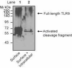
A: This came during the race to 1000 and we had many people generating and reviewing data. In retrospect, this was one that probably should not have made it out. As I reviewed the run data, it looks as though the MW ladder did not assign correctly. When this was corrected, the band at 200 actually shifts higher, up to 280 kDa. This band now overlaps with the 280 kDa fluorescent standard which is prone to have non-specific interactions with some antibodies. Those two things make this non-supportive in Simple Western. We ran Raji lysate on the same run. This antibody detects a very broad band with a peak at ~150-165 kDa, closer to expected MW. However, the baseline for this samples was also very high. If we were presented with this data set today, it would not be considered supportive. The other information on the Protein Simple web site likely came from a customer.
Associated Pathways


