TRPA1 Antibody - BSA Free
Novus Biologicals | Catalog # NB110-40763
![Western Blot: TRPA1 AntibodyBSA Free [NB110-40763]](https://resources.rndsystems.com/images/products/TRPA1-Antibody-Western-Blot-NB110-40763-img0014.jpg "Western Blot: TRPA1 AntibodyBSA Free [NB110-40763]")
Key Product Details
Validated by
Species Reactivity
Validated:
Cited:
Applications
Validated:
Cited:
Label
Antibody Source
Format
Product Specifications
Immunogen
Reactivity Notes
Localization
Specificity
Clonality
Host
Isotype
Theoretical MW
Disclaimer note: The observed molecular weight of the protein may vary from the listed predicted molecular weight due to post translational modifications, post translation cleavages, relative charges, and other experimental factors.
Scientific Data Images for TRPA1 Antibody - BSA Free
Western Blot: TRPA1 AntibodyBSA Free [NB110-40763]
TRPA1-Antibody-Western-Blot-NB110-40763-img0014.jpg![Immunohistochemistry: TRPA1 Antibody - BSA Free [NB110-40763]](https://resources.rndsystems.com/images/products/TRPA1-Antibody-Immunohistochemistry-NB110-40763-img0008.jpg "Immunohistochemistry: TRPA1 Antibody - BSA Free [NB110-40763]")
Immunohistochemistry: TRPA1 Antibody - BSA Free [NB110-40763]
Immunohistochemistry: TRPA1 Antibody [NB110-40763] - Staining TRPA1 in mouse intestine.![Immunocytochemistry/ Immunofluorescence: TRPA1 Antibody - BSA Free [NB110-40763]](https://resources.rndsystems.com/images/products/TRPA1-Antibody-Immunocytochemistry-Immunofluorescence-NB110-40763-img0011.jpg "Immunocytochemistry/ Immunofluorescence: TRPA1 Antibody - BSA Free [NB110-40763]")
Immunocytochemistry/ Immunofluorescence: TRPA1 Antibody - BSA Free [NB110-40763]
TRPA1-Antibody-Immunocytochemistry-Immunofluorescence-NB110-40763-img0011.jpg![Flow Cytometry: TRPA1 Antibody - BSA Free [NB110-40763]](https://resources.rndsystems.com/images/products/TRPA1-Antibody-Flow-Cytometry-NB110-40763-img0010.jpg "Flow Cytometry: TRPA1 Antibody - BSA Free [NB110-40763]")
Flow Cytometry: TRPA1 Antibody - BSA Free [NB110-40763]
Flow Cytometry: TRPA1 Antibody [NB110-40763] - An intracellular stain was performed on A549 cells with NB110-40763C (blue) and a matched isotype control (orange). Cells were fixed with 4% PFA and then permeabilized with 0.1% saponin. Cells were incubated in an antibody dilution of 2.5 ug/mL for 30 minutes at room temperature. Both antibodies were conjugated to DyLight 650.![Flow Cytometry: TRPA1 Antibody - BSA Free [NB110-40763]](https://resources.rndsystems.com/images/products/TRPA1-Antibody-Flow-Cytometry-NB110-40763-img0009.jpg "Flow Cytometry: TRPA1 Antibody - BSA Free [NB110-40763]")
Flow Cytometry: TRPA1 Antibody - BSA Free [NB110-40763]
Flow Cytometry: TRPA1 Antibody [NB110-40763] - An intracellular stain was performed on A549 cells with NB110-40763G (blue) and a matched isotype control (orange). Cells were fixed with 4% PFA and then permeabilized with 0.1% saponin. Cells were incubated in an antibody dilution of 5 ug/mL for 30 minutes at room temperature. Both antibodies were conjugated to DyLight 488![Western Blot: TRPA1 AntibodyBSA Free [NB110-40763]](https://resources.rndsystems.com/images/products/TRPA1-Antibody-Western-Blot-NB110-40763-img0013.jpg "Western Blot: TRPA1 AntibodyBSA Free [NB110-40763]")
![Immunocytochemistry/ Immunofluorescence: TRPA1 Antibody - BSA Free [NB110-40763]](https://resources.rndsystems.com/images/products/TRPA1-Antibody-Immunocytochemistry-Immunofluorescence-NB110-40763-img0006.jpg "Immunocytochemistry/ Immunofluorescence: TRPA1 Antibody - BSA Free [NB110-40763]")
Immunocytochemistry/ Immunofluorescence: TRPA1 Antibody - BSA Free [NB110-40763]
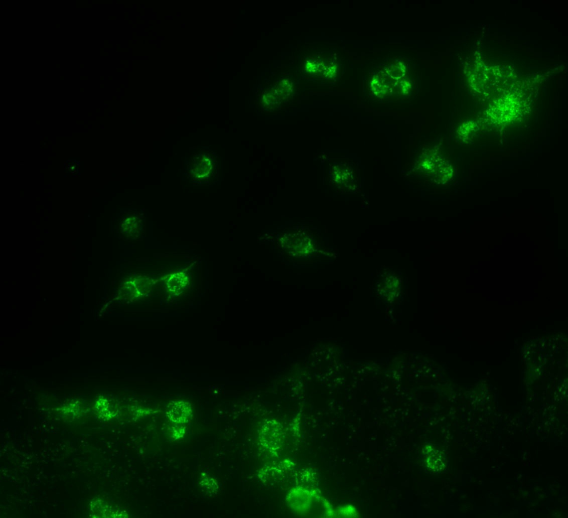
Immunocytochemistry/Immunofluorescence: TRPA1 Antibody [NB110-40763] - Analysis of TRPA1 in HEK 293 cells (Flp In Trex system) cells.
Western Blot: TRPA1 Antibody - BSA Free [NB110-40763] -
Western Blot: TRPA1 Antibody - BSA Free [NB110-40763] - Effects of Lycium barbarum polysaccharides (LBP) & capsaicin (CAP) on colonic (A) Cyclooxygenase-2 (COX-2), (B) Transient receptor potential cation channel V1 (TRPV1), & (C) Transient receptor potential ankyrin 1 (TRPA1) protein expression. (D) Representatives of Western blot for COX-2, TRPV1, TRPA1, & beta -actin. N: control group, U: ulcerative colitis induced group, L: LBP treated group, C: CAP treated group, M: mixed LBP & CAP treated group. Data are presented as mean ± SEM & analyzed by one-way ANOVA & Fisher’s least significant difference test (n = 8). # p < 0.05 compared to the N group. * p < 0.05 compared to the U group. Image collected & cropped by CiteAb from the following publication (https://pubmed.ncbi.nlm.nih.gov/35269566), licensed under a CC-BY license. Not internally tested by Novus Biologicals.
Immunocytochemistry/ Immunofluorescence: TRPA1 Antibody - BSA Free [NB110-40763] -
Immunocytochemistry/ Immunofluorescence: TRPA1 Antibody - BSA Free [NB110-40763] - Expression & localization of TRPA1 channels in wild-type (WT) & APP/PS1 Tg mice. (a) Brains were harvested from WT & APP/PS1 Tg mice at 8 months old. Western blot analysis of protein levels of TRPA1 & alpha -tubulin. Data are mean ± SEM from 6 mice in each group. *, P < 0.05 vs. WT mice. (b, c) Immunohistochemistry of specimens of cortex & hippocampus from 8-month-old WT & APP/PS1 Tg mice with the antibodies anti-TRPA1, anti-vWF (endothelial cell marker), anti-NeuN (neuron marker), anti-GFAP (astrocyte marker) & anti-IBA-1 (microglia marker), then FITC- or Texas red-conjugated secondary antibody. Bar = 50 μm. vWF-positive cells denoted endothelial cells, NeuN-positive cells denoted neurons, & GFAP-positive cells denoted astrocytes, as indicated by arrowheads, stars or arrows, respectively Image collected & cropped by CiteAb from the following publication (http://jneuroinflammation.biomedcentral.com/articles/10.1186/s12974-016…), licensed under a CC-BY license. Not internally tested by Novus Biologicals.
Immunocytochemistry/ Immunofluorescence: TRPA1 Antibody - BSA Free [NB110-40763] -
Immunocytochemistry/ Immunofluorescence: TRPA1 Antibody - BSA Free [NB110-40763] - Evaluation of anti-TRPA1 antibody specificity by western blotting & immunofluorescence. (a) Lysates derived from HEK293T cells transiently transfected with the TRPA1 & GFP expression construct (TRPA1 OE) or TRPM2 & GFP negative control (TRPM2 OE) were probed with the antibodies indicated. Additional untransfected & GFP only controls are presented in the first blot (6G8). Uncropped full length blots are displayed, gaps between the blots are present to indicate the use of a different antibody. Mouse mAbs C-5 & 6G8, & NB110-40763 can detect TRPA1 only in the membrane fraction of lysates at the expected molecular weight (127.5 kDa). Several TRPA1-specific bands are observed above this weight. NB110-40763 also detects several other antigens. Ab58844 & ACC- 037 only appear to detect antigens other than TRPA1 in the conditions used. (b) The performance of the same antibodies was evaluated by immunofluorescence (red) in comparison to the expression of the GFP reporter (green), or control TRPM2 & GFP overexpressing cells as for western blotting. Mouse mAb C-5 again shows high specificity with a strong correlation between the expression of the GFP reporter & antibody staining, & no staining in TRPM2 & GFP overexpressing controls. NB110-40763 also demonstrates some sensitivity but has high background that likely does not correspond to Fc binding of the antibody, as the other two rabbit polyclonal antibodies or isotype control did show different staining patterns. Ab58844 & ACC- 037 only appear to detect antigens other than TRPA1 in the conditions used. Image collected & cropped by CiteAb from the following publication (https://pubmed.ncbi.nlm.nih.gov/31811235), licensed under a CC-BY license. Not internally tested by Novus Biologicals.
Western Blot: TRPA1 Antibody - BSA Free [NB110-40763] -
Western Blot: TRPA1 Antibody - BSA Free [NB110-40763] - Expression & localization of TRPA1 channels in wild-type (WT) & APP/PS1 Tg mice. (a) Brains were harvested from WT & APP/PS1 Tg mice at 8 months old. Western blot analysis of protein levels of TRPA1 & alpha -tubulin. Data are mean ± SEM from 6 mice in each group. *, P < 0.05 vs. WT mice. (b, c) Immunohistochemistry of specimens of cortex & hippocampus from 8-month-old WT & APP/PS1 Tg mice with the antibodies anti-TRPA1, anti-vWF (endothelial cell marker), anti-NeuN (neuron marker), anti-GFAP (astrocyte marker) & anti-IBA-1 (microglia marker), then FITC- or Texas red-conjugated secondary antibody. Bar = 50 μm. vWF-positive cells denoted endothelial cells, NeuN-positive cells denoted neurons, & GFAP-positive cells denoted astrocytes, as indicated by arrowheads, stars or arrows, respectively Image collected & cropped by CiteAb from the following publication (http://jneuroinflammation.biomedcentral.com/articles/10.1186/s12974-016…), licensed under a CC-BY license. Not internally tested by Novus Biologicals.Applications for TRPA1 Antibody - BSA Free
ELISA
Flow Cytometry
Immunocytochemistry/ Immunofluorescence
Immunohistochemistry
Immunohistochemistry-Frozen
Immunohistochemistry-Paraffin
Western Blot
Reviewed Applications
Read 2 reviews rated 4.5 using NB110-40763 in the following applications:
Flow Cytometry Panel Builder
Bio-Techne Knows Flow Cytometry
Save time and reduce costly mistakes by quickly finding compatible reagents using the Panel Builder Tool.
Advanced Features
- Spectra Viewer - Custom analysis of spectra from multiple fluorochromes
- Spillover Popups - Visualize the spectra of individual fluorochromes
- Antigen Density Selector - Match fluorochrome brightness with antigen density

Formulation, Preparation, and Storage
Purification
Formulation
Format
Preservative
Concentration
Shipping
Stability & Storage
Background: TRPA1
Long Name
Alternate Names
Entrez Gene IDs
Gene Symbol
UniProt
Additional TRPA1 Products
Product Documents for TRPA1 Antibody - BSA Free
Certificate of Analysis
To download a Certificate of Analysis, please enter a lot or batch number in the search box below.
Product Specific Notices for TRPA1 Antibody - BSA Free
This product is for research use only and is not approved for use in humans or in clinical diagnosis. Primary Antibodies are guaranteed for 1 year from date of receipt.
Citations for TRPA1 Antibody - BSA Free
 Powered by Bioz
Customer Reviews for TRPA1 Antibody - BSA Free (2)
Have you used TRPA1 Antibody - BSA Free?
Submit a review and receive an Amazon gift card!
$25/€18/£15/$25CAN/¥2500 Yen for a review with an image
$10/€7/£6/$10CAN/¥1110 Yen for a review without an image
Submit a review
Customer Images
-
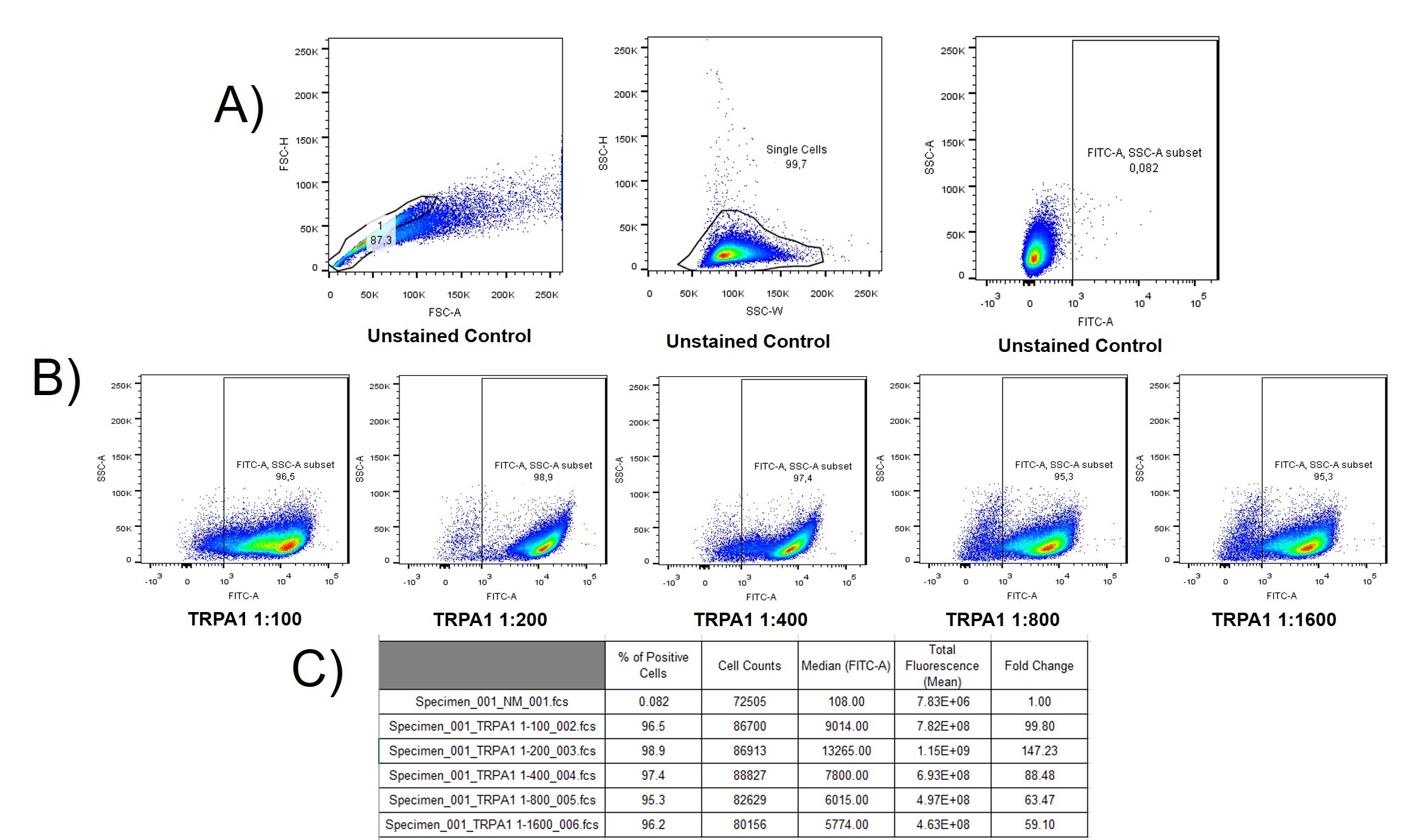
Application: Flow CytometrySample Tested: ZEM2S and ZEM2S (ATCC® CRL-2147™)Species: ZebrafishVerified Customer | Posted 10/28/2019A) Gate strategy is shown and fluorescence (FITC) of unstained cells is demonstrated. B) Fluorescence and percentage of TRPA1 positive cells is shown in a decreasing panel of concentration. C) Table shows several parameters.1. ZEM-2S cells were harvested using Tyrode/EDTA solution; 2. Cells were fixed in 4% paraformaldehyde for 30 min at 4 oC; 3. Cells were washed with PBS and centrifuged at 2000 g for 3 min; 4. Approximately 10^6 cells were added to round-bottom plates and centrifuged; 5. Blocking solution (1% BSA, 22.52 mg/ml glycine in PBS) was added to each well, and cells were kept at 4oC for 30 min; 6. Cells were washed with PBS and incubated with rabbit TRPA1 mouse antibody (NB110-40763) in incubation solution (for each 100 mL 0.25 g of carragenine, 1 g of BSA, and 300 uL of Triton X-100). Cells were incubated overnight. 7. Cells were washed with PBS, centrifuged, and secondary antibody (goat-anti rabbit Alexa 488 - 1:2000) was added at room temperature for 60 min; 8. Cells were washed with PBS and harvested. 9. Acquisition took place in FACS Canto (BD Biosciences)

-
Application: ImmunofluorescenceSample Tested: HEKFLPNTREX Cell lineSpecies: HumanVerified Customer | Posted 08/21/2012
There are no reviews that match your criteria.
Protocols
View specific protocols for TRPA1 Antibody - BSA Free (NB110-40763):
Sample Preparation.
1. Grow cells to 60-85% confluency. Flow cytometry requires between 2 x 10⁵ and 1 x 10⁶ cells for optimal performance.
2. If cells are adherent, harvest gently by washing once with staining buffer and then scraping. Avoid using trypsin as this can disrupt certain epitopes of interest. If enzymatic harvest is required, use Accutase, Collagenase, or TrypLE Express for a less damaging option.
3. Reserve 100 uL for counting, then transfer cell volume into a 50 mL conical tube and centrifuge for 8 minutes at 400 RCF.
a. Count cells using a hemocytometer and a 1:1 trypan blue exclusion stain to determine cell viability before starting the flow protocol. If cells appear blue, do not proceed.
4. Re-suspend cells to a concentration of 1 x 10⁶ cells/mL in staining buffer (NBP2-26247).
5. Aliquot out 100 uL samples in accordance with your experimental samples.
Tip: When cell surface and intracellular staining are required in the same sample, it is advisable that the cell surface staining be performed first since the fixation and permeabilization steps might reduce the availability of surface antigens.
Intracellular Staining.
Tip: When performing intracellular staining, it is important to use appropriate fixation and permeabilization reagents based upon the target and its subcellular location. Generally, our Intracellular Flow Assay Kit (NBP2-29450) is a good place to start as it contains an optimized combination of reagents for intracellular staining as well as an inhibitor of intracellular protein transport (necessary if staining secreted proteins). Certain targets may require more gentle or transient permeabilization protocols such as the commonly employed methanol or saponin-based methods.
Protocol for Cytoplasmic Targets:
1. Fix the cells by adding 100 uL fixation solution (such as 4% PFA) to each sample for 10-15 minutes.
2. Permeabilize cells by adding 100 uL of a permeabilization buffer to every 1 x 10⁶ cells present in the sample. Mix well and incubate at room temperature for 15 minutes.
a. For cytoplasmic targets, use a gentle permeabilization solution such as 1X PBS + 0.5% Saponin or 1X PBS + 0.5% Tween-20.
b. To maintain the permeabilized state throughout your experiment, use staining buffer + 0.1% of the permeabilization reagent (i.e. 0.1% Tween-20 or 0.1% Saponin).
3. Following the 15 minute incubation, add 2 mL of the staining buffer + 0.1% permeabilizer to each sample.
4. Centrifuge for 1 minute at 400 RCF.
5. Discard supernatant and re-suspend in 100 uL of staining buffer + 0.1% permeabilizer.
6. Add appropriate amount of each antibody (eg. 1 test or 1 ug per sample, as experimentally determined).
7. Mix well and incubate at room temperature for 30 minutes- 1 hour. Gently mix samples every 10-15 minutes.
8. Following the primary/conjugate incubation, add 1-2 mL/sample of staining buffer +0.1% permeabilizer and centrifuge for 1 minute at 400 RCF.
9. Wash twice by re-suspending cells in staining buffer (2 mL for tubes or 200 uL for wells) and centrifuging at 400 RCF for 5 minutes. Discard supernatant.
10. Add appropriate amount of secondary antibody (as experimentally determined) to each sample.
11. Incubate at room temperature in dark for 20 minutes.
12. Add 1-2 mL of staining buffer and centrifuge at 400 RCF for 1 minute and discard supernatant.
13. Wash twice by re-suspending cells in staining buffer (2 mL for tubes or 200 uL for wells) and centrifuging at 400 RCF for 5 minutes. Discard supernatant.
14. Resuspend in an appropriate volume of staining buffer (usually 500 uL per sample) and proceed with analysis on your flow cytometer.
Culture cells to appropriate density in 35 mm culture dishes or 6-well plates.
1. Remove culture medium and wash the cells briefly in PBS. Add 10% formalin to the dish and fix at room temperature for 10 minutes.
2. Remove the formalin and wash the cells in PBS.
3. Permeablize the cells with 0.1% Triton X100 or other suitable detergent for 10 min.
4. Remove the permeablization buffer and wash three times for 10 minutes each in PBS. Be sure to not let the specimen dry out.
5. To block nonspecific antibody binding, incubate in 10% normal goat serum from 1 hour to overnight at room temperature.
6. Add primary antibody at appropriate dilution and incubate overnight at 4C.
7. Remove primary antibody and replace with PBS. Wash three times for 10 minutes each.
8. Add secondary antibody at appropriate dilution. Incubate for 1 hour at room temperature.
9. Remove secondary antibody and replace with PBS. Wash three times for 10 minutes each.
10. Counter stain DNA with DAPi if required.
Antigen Unmasking:
Bring slides to a boil in 10 mM sodium citrate buffer (pH 6.0) then maintain at a sub-boiling temperature for 10 minutes. Cool slides on bench-top for 30 minutes (keep slides in the sodium citrate buffer at all times).
Staining:
1. Wash sections in deionized water three times for 5 minutes each.
2. Wash sections in PBS for 5 minutes.
3. Block each section with 100-400 ul blocking solution (1% BSA in PBS) for 1 hour at room temperature.
4. Remove blocking solution and add 100-400 ul diluted primary antibody. Incubate overnight at 4 C.
5. Remove antibody solution and wash sections in wash buffer three times for 5 minutes each.
6. Add 100-400 ul HRP polymer conjugated secondary antibody. Incubate 30 minutes at room temperature.
7. Wash sections three times in wash buffer for 5 minutes each.
8. Add 100-400 ul DAB substrate to each section and monitor staining closely.
9. As soon as the sections develop, immerse slides in deionized water.
10. Counterstain sections in hematoxylin.
11. Wash sections in deionized water two times for 5 minutes each.
12. Dehydrate sections.
13. Mount coverslips.
1. Perform SDS-PAGE on samples to be analyzed, loading 10-25 ug of total protein per lane.
2. Transfer proteins to PVDF membrane according to the instructions provided by the manufacturer of the membrane and transfer apparatus.
3. Stain the membrane with Ponceau S (or similar product) to assess transfer success, and mark molecular weight standards where appropriate.
4. Rinse the blot TBS -0.05% Tween 20 (TBST).
5. Block the membrane in 5% Non-fat milk in TBST (blocking buffer) for at least 1 hour.
6. Wash the membrane in TBST three times for 10 minutes each.
7. Dilute primary antibody in blocking buffer and incubate overnight at 4C with gentle rocking.
8. Wash the membrane in TBST three times for 10 minutes each.
9. Incubate the membrane in diluted HRP conjugated secondary antibody in blocking buffer (as per manufacturer's instructions) for 1 hour at room temperature.
10. Wash the blot in TBST three times for 10 minutes each (this step can be repeated as required to reduce background).
11. Apply the detection reagent of choice in accordance with the manufacturer's instructions.
Find general support by application which include: protocols, troubleshooting, illustrated assays, videos and webinars.
- 7-Amino Actinomycin D (7-AAD) Cell Viability Flow Cytometry Protocol
- Antigen Retrieval Protocol (PIER)
- Antigen Retrieval for Frozen Sections Protocol
- Appropriate Fixation of IHC/ICC Samples
- Cellular Response to Hypoxia Protocols
- Chromogenic IHC Staining of Formalin-Fixed Paraffin-Embedded (FFPE) Tissue Protocol
- Chromogenic Immunohistochemistry Staining of Frozen Tissue
- ClariTSA™ Fluorophore Kits
- Detection & Visualization of Antibody Binding
- ELISA Sample Preparation & Collection Guide
- ELISA Troubleshooting Guide
- Extracellular Membrane Flow Cytometry Protocol
- Flow Cytometry Protocol for Cell Surface Markers
- Flow Cytometry Protocol for Staining Membrane Associated Proteins
- Flow Cytometry Staining Protocols
- Flow Cytometry Troubleshooting Guide
- Fluorescent IHC Staining of Frozen Tissue Protocol
- Graphic Protocol for Heat-induced Epitope Retrieval
- Graphic Protocol for the Preparation and Fluorescent IHC Staining of Frozen Tissue Sections
- Graphic Protocol for the Preparation and Fluorescent IHC Staining of Paraffin-embedded Tissue Sections
- Graphic Protocol for the Preparation of Gelatin-coated Slides for Histological Tissue Sections
- How to Run an R&D Systems DuoSet ELISA
- How to Run an R&D Systems Quantikine ELISA
- How to Run an R&D Systems Quantikine™ QuicKit™ ELISA
- ICC Cell Smear Protocol for Suspension Cells
- ICC Immunocytochemistry Protocol Videos
- ICC for Adherent Cells
- IHC Sample Preparation (Frozen sections vs Paraffin)
- Immunocytochemistry (ICC) Protocol
- Immunocytochemistry Troubleshooting
- Immunofluorescence of Organoids Embedded in Cultrex Basement Membrane Extract
- Immunofluorescent IHC Staining of Formalin-Fixed Paraffin-Embedded (FFPE) Tissue Protocol
- Immunohistochemistry (IHC) and Immunocytochemistry (ICC) Protocols
- Immunohistochemistry Frozen Troubleshooting
- Immunohistochemistry Paraffin Troubleshooting
- Intracellular Flow Cytometry Protocol Using Alcohol (Methanol)
- Intracellular Flow Cytometry Protocol Using Detergents
- Intracellular Nuclear Staining Flow Cytometry Protocol Using Detergents
- Intracellular Staining Flow Cytometry Protocol Using Alcohol Permeabilization
- Intracellular Staining Flow Cytometry Protocol Using Detergents to Permeabilize Cells
- Preparing Samples for IHC/ICC Experiments
- Preventing Non-Specific Staining (Non-Specific Binding)
- Primary Antibody Selection & Optimization
- Propidium Iodide Cell Viability Flow Cytometry Protocol
- Protocol for Heat-Induced Epitope Retrieval (HIER)
- Protocol for Liperfluo
- Protocol for Making a 4% Formaldehyde Solution in PBS
- Protocol for VisUCyte™ HRP Polymer Detection Reagent
- Protocol for the Characterization of Human Th22 Cells
- Protocol for the Characterization of Human Th9 Cells
- Protocol for the Fluorescent ICC Staining of Cell Smears - Graphic
- Protocol for the Fluorescent ICC Staining of Cultured Cells on Coverslips - Graphic
- Protocol for the Preparation & Fixation of Cells on Coverslips
- Protocol for the Preparation and Chromogenic IHC Staining of Frozen Tissue Sections
- Protocol for the Preparation and Chromogenic IHC Staining of Frozen Tissue Sections - Graphic
- Protocol for the Preparation and Chromogenic IHC Staining of Paraffin-embedded Tissue Sections
- Protocol for the Preparation and Chromogenic IHC Staining of Paraffin-embedded Tissue Sections - Graphic
- Protocol for the Preparation and Fluorescent ICC Staining of Cells on Coverslips
- Protocol for the Preparation and Fluorescent ICC Staining of Non-adherent Cells
- Protocol for the Preparation and Fluorescent ICC Staining of Stem Cells on Coverslips
- Protocol for the Preparation and Fluorescent IHC Staining of Frozen Tissue Sections
- Protocol for the Preparation and Fluorescent IHC Staining of Paraffin-embedded Tissue Sections
- Protocol for the Preparation of Gelatin-coated Slides for Histological Tissue Sections
- Protocol for the Preparation of a Cell Smear for Non-adherent Cell ICC - Graphic
- Protocol: Annexin V and PI Staining by Flow Cytometry
- Protocol: Annexin V and PI Staining for Apoptosis by Flow Cytometry
- Quantikine HS ELISA Kit Assay Principle, Alkaline Phosphatase
- Quantikine HS ELISA Kit Principle, Streptavidin-HRP Polymer
- R&D Systems Quality Control Western Blot Protocol
- Sandwich ELISA (Colorimetric) – Biotin/Streptavidin Detection Protocol
- Sandwich ELISA (Colorimetric) – Direct Detection Protocol
- TUNEL and Active Caspase-3 Detection by IHC/ICC Protocol
- The Importance of IHC/ICC Controls
- Troubleshooting Guide: ELISA
- Troubleshooting Guide: Fluorokine Flow Cytometry Kits
- Troubleshooting Guide: Immunohistochemistry
- Troubleshooting Guide: Western Blot Figures
- Western Blot Conditions
- Western Blot Protocol
- Western Blot Protocol for Cell Lysates
- Western Blot Troubleshooting
- Western Blot Troubleshooting Guide
- View all Protocols, Troubleshooting, Illustrated assays and Webinars
FAQs for TRPA1 Antibody - BSA Free
-
Q: Has the specificity of NB110-40763 has been confirmed by mouse dorsal root ganglion (DRG) tissue in immunohistochemistry? Do you know what sample types have been tested?
A: We have a couple of publications with the base product catalog number NB110-40763 (which is just the unconjugated version of NB110-40763G), which have utilized our product with mouse colonic dorsal root ganglion. (PMID 19875705 and PMID 21689654). The image on our website was taken of a mouse intestine sample.
-
Q: Human brain membrane fraction is mentioned to be used as a positive control for this antibody. Do you sell the positive control too?
A: The Western blot data that is shown on our product page for the TRPA1 antibody with catalogue number NB110-40763 was, as you have pointed out, generated from human brain membrane lysate. We do sell a human brain membrane fraction that is suitable for Western blotting, and you can see this here: TRPA1 antibody datasheet. This lysate is supplied as native protein, and we recommend adding 1 x sample buffer with 5% BME (or other reducing agent) prior to use.
-
Q: I read the first publication (Yu S et al. 2009) referenced for NB110-40763. This product reacts with human and mouse according to your datasheet, but in that publication it was used for western blot in guinea pig. Does this mean this antibody is suitable to use for guinea pig samples?
A: Yes, according to this paper this antibody has been successfully used on guinea pig samples. This is the only testing that has been done in guinea pig though as we have done no in-house testing. It should work just fine for you.
-
Q: What are the other two bands in the image on the datasheet? UniProt finds a theoretical MW of 127.5kDa. You find a MW of 110kDa in WB application. There are two more bands at 60kDa and 17kDa. What are these? Unspecific staining or fragments or variants of the protein? I did not find any info on variants when looking into the UniProt data (http://www.uniprot.org/uniprot/O75762).
A: We generally use gradient gels in our lab and for proteins which are multi-pass membrane protein, it is not uncommon to see an approximate 10-15% change in expected vs observed molecular weight in WB assay. The band observed at approximately 110kD is highly specific and there is no doubt that the antibody is detecting the right band. No, we have not characterized the additional low molecular weight bands yet which are considerably mild bands when compared to the expected target band. During a brief literature research this morning, I came across some papers wherein other researchers have also observed additional lower molecular weight bands and some of those references are: J Biol Chem. 2010 May 14; 285(20): 15167-15177; J Biol Chem. 2006 October 27; 281(43): 32879-32890; J Clin Invest. 2007 July 2; 117(7): 1979-1987.
-
Q: Has the specificity of NB110-40763 has been confirmed by mouse dorsal root ganglion (DRG) tissue in immunohistochemistry? Do you know what sample types have been tested?
A: We have a couple of publications with the base product catalog number NB110-40763 (which is just the unconjugated version of NB110-40763G), which have utilized our product with mouse colonic dorsal root ganglion. (PMID 19875705 and PMID 21689654). The image on our website was taken of a mouse intestine sample.
-
Q: Human brain membrane fraction is mentioned to be used as a positive control for this antibody. Do you sell the positive control too?
A: The Western blot data that is shown on our product page for the TRPA1 antibody with catalogue number NB110-40763 was, as you have pointed out, generated from human brain membrane lysate. We do sell a human brain membrane fraction that is suitable for Western blotting, and you can see this here: TRPA1 antibody datasheet. This lysate is supplied as native protein, and we recommend adding 1 x sample buffer with 5% BME (or other reducing agent) prior to use.
-
Q: I read the first publication (Yu S et al. 2009) referenced for NB110-40763. This product reacts with human and mouse according to your datasheet, but in that publication it was used for western blot in guinea pig. Does this mean this antibody is suitable to use for guinea pig samples?
A: Yes, according to this paper this antibody has been successfully used on guinea pig samples. This is the only testing that has been done in guinea pig though as we have done no in-house testing. It should work just fine for you.
-
Q: What are the other two bands in the image on the datasheet? UniProt finds a theoretical MW of 127.5kDa. You find a MW of 110kDa in WB application. There are two more bands at 60kDa and 17kDa. What are these? Unspecific staining or fragments or variants of the protein? I did not find any info on variants when looking into the UniProt data (http://www.uniprot.org/uniprot/O75762).
A: We generally use gradient gels in our lab and for proteins which are multi-pass membrane protein, it is not uncommon to see an approximate 10-15% change in expected vs observed molecular weight in WB assay. The band observed at approximately 110kD is highly specific and there is no doubt that the antibody is detecting the right band. No, we have not characterized the additional low molecular weight bands yet which are considerably mild bands when compared to the expected target band. During a brief literature research this morning, I came across some papers wherein other researchers have also observed additional lower molecular weight bands and some of those references are: J Biol Chem. 2010 May 14; 285(20): 15167-15177; J Biol Chem. 2006 October 27; 281(43): 32879-32890; J Clin Invest. 2007 July 2; 117(7): 1979-1987.