Human/Mouse FoxP3 Antibody
R&D Systems | Catalog # MAB8214

Key Product Details
Validated by
Species Reactivity
Validated:
Cited:
Applications
Validated:
Cited:
Label
Antibody Source
Product Specifications
Immunogen
Met1-Leu71
Accession # Q9BZS1
Specificity
Clonality
Host
Isotype
Scientific Data Images for Human/Mouse FoxP3 Antibody
Detection of FOXP3 in Hodgkin's Lymphoma via Multiplex Immunofluorescence staining on COMET™
FOXP3 was detected in immersion fixed paraffin-embedded sections of Hodgkin's Lymphoma using Rabbit Anti-Human FOXP3 Monoclonal Antibody (Catalog # MAB8214) at 20 µg/mL at 37 ° Celsius for 4 minutes. Before incubation with the primary antibody, tissue underwent an all-in-one dewaxing and antigen retrieval preprocessing using PreTreatment Module (PT Module) and Dewax and HIER Buffer H (pH 9). Tissue was stained using the Alexa Fluor™ Plus 647 Goat anti-Rabbit IgG Secondary Antibody at 1:200 at 37 ° Celsius for 2 minutes. (Yellow; Lunaphore Catalog # DR647RB) and counterstained with DAPI (blue; Lunaphore Catalog # DR100). Specific staining was localized to the nucleus. Protocol available in COMET™ Panel Builder.

Detection of FOXP3 in Mouse Thymus via seqIF™ staining on COMET™
FOXP3 Antibody was detected in immersion fixed paraffin-embedded sections of mouse Thymus using Rabbit Anti-Mouse FOXP3 Monoclonal Antibody (Catalog # MAB8214) at 20ug/mL at 37 ° Celsius for 4 minutes. Before incubation with the primary antibody, tissue underwent an all-in-one dewaxing and antigen retrieval preprocessing using PreTreatment Module (PT Module) and Dewax and HIER Buffer H (pH 9; Epredia Catalog # TA-999-DHBH). Tissue was stained using the Alexa Fluor™ Plus 647 Goat anti-Rabbit IgG Secondary Antibody at 1:200 at 37 ° Celsius for 2 minutes. (Yellow; Lunaphore Catalog # DR647RB) and counterstained with DAPI (blue; Lunaphore Catalog # DR100). Specific staining was localized to the nucleus. Protocol available in COMET™ Panel Builder.

Detection of FoxP3 in Human PBMCs stimulated to induce Tregs by Flow Cytometry.
Human peripheral blood mononuclear cells (PBMCs) either (A) untreated or (B) stimulated to induce Regulatory T Cells (Tregs) with Recombinant Human TGF-beta 1 (Catalog # 240-B) and Recombinant Human IL-2 (Catalog # 202-IL) for 2 days were stained with Rabbit Anti-Human/Mouse FoxP3 Monoclonal Antibody (Catalog # MAB8214) followed by Phycoerythrin-conjugated Anti-Rabbit IgG Secondary Antibody (Catalog # F0110) and Mouse Anti-Human CD4 APC-conjugated Monoclonal Antibody (Catalog # FAB3791A). Quadrant markers were set based on control antibody staining (Catalog # AB-105-C). To facilitate intracellular staining, cells were fixed and permeabilized with FlowX FoxP3 Fixation & Permeabilization Buffer Kit (Catalog # FC012).

Detection of FoxP3 in Mouse Splenocytes by Flow Cytometry.
Mouse splenocytes were stained with Rat Anti-Mouse CD4 APC-conjugated Monoclonal Antibody (Catalog # FAB554A) and either (A) Rabbit Anti-Human/Mouse FoxP3 Monoclonal Antibody (Catalog # MAB8214) or (B) Normal Rabbit IgG Control (Catalog # AB-105-C) followed by Phycoerythrin-conjugated Anti-Rabbit IgG Secondary Antibody (Catalog # F0110). To facilitate intracellular staining, cells were fixed and permeabilized with FlowX FoxP3 Fixation & Permeabilization Buffer Kit (Catalog # FC012).

FoxP3 in HeLa Human Cell Line.
FoxP3 was detected in immersion fixed HeLa human cervical epithelial carcinoma cell line using Rabbit Anti-Human/Mouse FoxP3 Monoclonal Antibody (Catalog # MAB8214) at 8 µg/mL for 3 hours at room temperature. Cells were stained using the NorthernLights™ 557-conjugated Anti-Rabbit IgG Secondary Antibody (red, upper panel; Catalog # NL004) and counterstained with DAPI (blue, lower panel). Specific staining was localized to nuclei. View our protocol for Fluorescent ICC Staining of Cells on Coverslips.

FoxP3 in Human Tonsil.
FoxP3 was detected in immersion fixed paraffin-embedded sections of human tonsil using Rabbit Anti-Human/Mouse FoxP3 Monoclonal Antibody (Catalog # MAB8214) at 15 µg/mL overnight at 4 °C. Tissue was stained using the Anti-Rabbit HRP-DAB Cell & Tissue Staining Kit (brown; Catalog # CTS005) and counterstained with hematoxylin (blue). Specific staining was localized to nuclei. View our protocol for Chromogenic IHC Staining of Paraffin-embedded Tissue Sections.

FoxP3 in Human Ovarian Cancer Tissue.
FoxP3 was detected in immersion fixed paraffin-embedded sections of human ovarian cancer tissue using Rabbit Anti-Human/Mouse FoxP3 Monoclonal Antibody (Catalog # MAB8214) at 5 µg/mL for 1 hour at room temperature followed by incubation with the Anti-Rabbit IgG VisUCyte™ HRP Polymer Antibody (Catalog # VC003). Tissue was stained using DAB (brown) and counterstained with hematoxylin (blue). Specific staining was localized to nuclei. View our protocol for IHC Staining with VisUCyte HRP Polymer Detection Reagents.

FoxP3 in Human Tonsil Using Dual RNAscope®ISH and IHC.
FoxP3 mRNA (red) and protein (green) was detected in formalin-fixed paraffin-embedded tissue sections of human tonsil probed with ACD RNAScope®Probe (Catalog # 418471) followed by immunohistochemistry using R&D Systems Rabbit Anti-Human/Mouse FoxP3 Monoclonal Antibody (Catalog# MAB8214) at 5ug/mL for 1 hour at room temperature followed by incubation with the Anti-Rabbit IgG VisUCyte HRP Polymer Antibody (R&D Systems, Catalog # VC003). Tissue was stained using ACD RNAscope®2.5 HD Duplex Detection Reagents (Catalog # 322500).

Detection of Human FoxP3 by Immunocytochemistry/Immunofluorescence
Multiplex fluorescent immunohistochemistry staining. Image collected and cropped by CiteAb from the following publication (https://pubmed.ncbi.nlm.nih.gov/31192136), licensed under a CC-BY license. Not internally tested by R&D Systems.
Detection of FoxP3 in Mouse Thymus.
FoxP3 was detected in immersion fixed paraffin-embedded sections of mouse thymus using Rabbit Anti-Human/Mouse FoxP3 Monoclonal Antibody (Catalog # MAB8214) at 15 µg/ml for 1 hour at room temperature followed by incubation with the Anti-Rabbit IgG VisUCyte™ HRP Polymer Antibody (Catalog # VC003). Before incubation with the primary antibody, tissue was subjected to heat-induced epitope retrieval using VisUCyte Antigen Retrieval Reagent-Basic (Catalog # VCTS021). Tissue was stained using DAB (brown) and counterstained with hematoxylin (blue). Specific staining was localized to the nucleus. View our protocol for IHC Staining with VisUCyte HRP Polymer Detection Reagents.
Applications for Human/Mouse FoxP3 Antibody
COMET
Dual RNAscope ISH-IHC Compatible
Sample: Immersion fixed paraffin-embedded sections of human tonsil
Flow Cytometry
Sample: Human peripheral blood mononuclear cells (PBMCs) stimulated to induce Regulatory T Cells (Tregs) with Recombinant Human TGF‑ beta 1 (Catalog # 240-B) and Recombinant Human IL‑2 (Catalog # 202-IL) and mouse splenocytes were fixed and permeabilized with FlowX FoxP3 Fixation & Permeabilization Buffer Kit (Catalog # FC012)
Immunocytochemistry
Sample: Immersion fixed HeLa human cervical epithelial carcinoma cell line
Immunohistochemistry
Sample: Immersion fixed paraffin-embedded sections of human tonsil, human ovarian cancer tissue, and mouse thymus tissue
Multiplex Immunofluorescence
Sample: Immersion fixed paraffin-embedded sections of Hodgkin's Lymphoma and Mouse Thymus
Reviewed Applications
Read 2 reviews rated 4 using MAB8214 in the following applications:
Flow Cytometry Panel Builder
Bio-Techne Knows Flow Cytometry
Save time and reduce costly mistakes by quickly finding compatible reagents using the Panel Builder Tool.
Advanced Features
- Spectra Viewer - Custom analysis of spectra from multiple fluorochromes
- Spillover Popups - Visualize the spectra of individual fluorochromes
- Antigen Density Selector - Match fluorochrome brightness with antigen density

Formulation, Preparation, and Storage
Purification
Reconstitution
Reconstitute at 0.5 mg/mL in sterile PBS. For liquid material, refer to CoA for concentration.
Formulation
*Small pack size (-SP) is supplied either lyophilized or as a 0.2 µm filtered solution in PBS.
Shipping
Stability & Storage
- 12 months from date of receipt, -20 to -70 °C as supplied.
- 1 month, 2 to 8 °C under sterile conditions after reconstitution.
- 6 months, -20 to -70 °C under sterile conditions after reconstitution.
Calculators
Background: FoxP3
Long Name
Alternate Names
Gene Symbol
UniProt
Additional FoxP3 Products
Product Documents for Human/Mouse FoxP3 Antibody
Certificate of Analysis
To download a Certificate of Analysis, please enter a lot or batch number in the search box below.
Note: Certificate of Analysis not available for kit components.
Product Specific Notices for Human/Mouse FoxP3 Antibody
For research use only
Related Research Areas
Citations for Human/Mouse FoxP3 Antibody
 Powered by Bioz
Customer Reviews for Human/Mouse FoxP3 Antibody (2)
Have you used Human/Mouse FoxP3 Antibody?
Submit a review and receive an Amazon gift card!
$25/€18/£15/$25CAN/¥2500 Yen for a review with an image
$10/€7/£6/$10CAN/¥1110 Yen for a review without an image
Submit a review
Customer Images
-
Application: Western BlotSample Tested: MCF-7 human breast cancer cell lineSpecies: HumanVerified Customer | Posted 05/25/2017
-
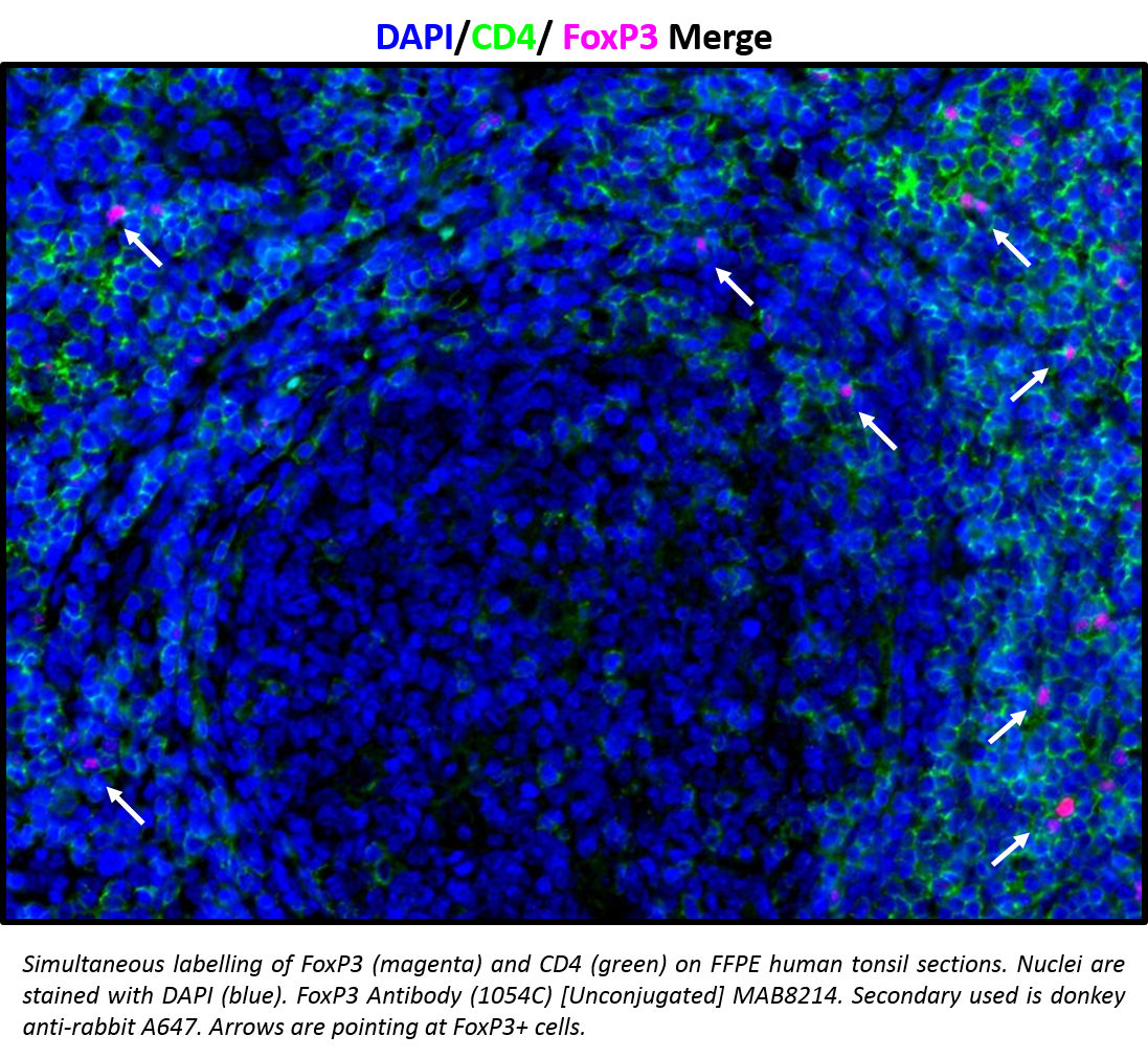
Application: Immunohistochemistry-ParaffinSample Tested: FFPE Tonsil and FFPE human tonsilSpecies: HumanVerified Customer | Posted 03/03/2017Simultaneous labelling of FoxP3 (magenta) and CD4 (green) on FFPE human tonsil sections. Nuclei stained with DAPI (blue). FoxP3 Antibody (1054C) MAB8214. Secondary Ab used is donkey anti-rabbit A647. Arrows pointing at FoxP3+ cells.

There are no reviews that match your criteria.
Protocols
Find general support by application which include: protocols, troubleshooting, illustrated assays, videos and webinars.
- 7-Amino Actinomycin D (7-AAD) Cell Viability Flow Cytometry Protocol
- Antigen Retrieval Protocol (PIER)
- Antigen Retrieval for Frozen Sections Protocol
- Appropriate Fixation of IHC/ICC Samples
- Cellular Response to Hypoxia Protocols
- Chromogenic IHC Staining of Formalin-Fixed Paraffin-Embedded (FFPE) Tissue Protocol
- Chromogenic Immunohistochemistry Staining of Frozen Tissue
- ClariTSA™ Fluorophore Kits
- Detection & Visualization of Antibody Binding
- Extracellular Membrane Flow Cytometry Protocol
- Flow Cytometry Protocol for Cell Surface Markers
- Flow Cytometry Protocol for Staining Membrane Associated Proteins
- Flow Cytometry Staining Protocols
- Flow Cytometry Troubleshooting Guide
- Fluorescent IHC Staining of Frozen Tissue Protocol
- Graphic Protocol for Heat-induced Epitope Retrieval
- Graphic Protocol for the Preparation and Fluorescent IHC Staining of Frozen Tissue Sections
- Graphic Protocol for the Preparation and Fluorescent IHC Staining of Paraffin-embedded Tissue Sections
- Graphic Protocol for the Preparation of Gelatin-coated Slides for Histological Tissue Sections
- ICC Cell Smear Protocol for Suspension Cells
- ICC Immunocytochemistry Protocol Videos
- ICC for Adherent Cells
- IHC Sample Preparation (Frozen sections vs Paraffin)
- ISH-IHC Protocol for Chromogenic Detection on Formalin Fixed Paraffin Embedded (FFPE) Tissue
- Immunocytochemistry (ICC) Protocol
- Immunocytochemistry Troubleshooting
- Immunofluorescence of Organoids Embedded in Cultrex Basement Membrane Extract
- Immunofluorescent IHC Staining of Formalin-Fixed Paraffin-Embedded (FFPE) Tissue Protocol
- Immunohistochemistry (IHC) and Immunocytochemistry (ICC) Protocols
- Immunohistochemistry Frozen Troubleshooting
- Immunohistochemistry Paraffin Troubleshooting
- Intracellular Flow Cytometry Protocol Using Alcohol (Methanol)
- Intracellular Flow Cytometry Protocol Using Detergents
- Intracellular Nuclear Staining Flow Cytometry Protocol Using Detergents
- Intracellular Staining Flow Cytometry Protocol Using Alcohol Permeabilization
- Intracellular Staining Flow Cytometry Protocol Using Detergents to Permeabilize Cells
- Preparing Samples for IHC/ICC Experiments
- Preventing Non-Specific Staining (Non-Specific Binding)
- Primary Antibody Selection & Optimization
- Propidium Iodide Cell Viability Flow Cytometry Protocol
- Protocol for Heat-Induced Epitope Retrieval (HIER)
- Protocol for Liperfluo
- Protocol for Making a 4% Formaldehyde Solution in PBS
- Protocol for VisUCyte™ HRP Polymer Detection Reagent
- Protocol for the Characterization of Human Th22 Cells
- Protocol for the Characterization of Human Th9 Cells
- Protocol for the Fluorescent ICC Staining of Cell Smears - Graphic
- Protocol for the Fluorescent ICC Staining of Cultured Cells on Coverslips - Graphic
- Protocol for the Preparation & Fixation of Cells on Coverslips
- Protocol for the Preparation and Chromogenic IHC Staining of Frozen Tissue Sections
- Protocol for the Preparation and Chromogenic IHC Staining of Frozen Tissue Sections - Graphic
- Protocol for the Preparation and Chromogenic IHC Staining of Paraffin-embedded Tissue Sections
- Protocol for the Preparation and Chromogenic IHC Staining of Paraffin-embedded Tissue Sections - Graphic
- Protocol for the Preparation and Fluorescent ICC Staining of Cells on Coverslips
- Protocol for the Preparation and Fluorescent ICC Staining of Non-adherent Cells
- Protocol for the Preparation and Fluorescent ICC Staining of Stem Cells on Coverslips
- Protocol for the Preparation and Fluorescent IHC Staining of Frozen Tissue Sections
- Protocol for the Preparation and Fluorescent IHC Staining of Paraffin-embedded Tissue Sections
- Protocol for the Preparation of Gelatin-coated Slides for Histological Tissue Sections
- Protocol for the Preparation of a Cell Smear for Non-adherent Cell ICC - Graphic
- Protocol: Annexin V and PI Staining by Flow Cytometry
- Protocol: Annexin V and PI Staining for Apoptosis by Flow Cytometry
- TUNEL and Active Caspase-3 Detection by IHC/ICC Protocol
- The Importance of IHC/ICC Controls
- Troubleshooting Guide: Fluorokine Flow Cytometry Kits
- Troubleshooting Guide: Immunohistochemistry
- View all Protocols, Troubleshooting, Illustrated assays and Webinars
FAQs for Human/Mouse FoxP3 Antibody
-
Q: What detection reagent was used for IHC, to create the Dual RNAscope ISH-IHC image on the datasheet for Catalog # MAB8214?
A: Green Chromogen from ACD (ACD Catalog # 322550) was ued for IHC detection. See ACD website (https://acdbio.com/store/catalog/product/view/id/22/) for protocol and handling recommendations. Extensive washing with buffers containing PBS is discouraged as it may cause decolorization. It is critical that after staining with green chromogen, slides with tissues are allowed to dry at room temperature prior to using a permanent mounting media.
-
Q: What is the immunogen sequence of this FoxP3 antibody?
A: A sequence from Met1 - Leu71.
-
Q: What detection reagent was used for IHC, to create the Dual RNAscope ISH-IHC image on the datasheet for Catalog # MAB8214?
A: Green Chromogen from ACD (ACD Catalog # 322550) was ued for IHC detection. See ACD website (https://acdbio.com/store/catalog/product/view/id/22/) for protocol and handling recommendations. Extensive washing with buffers containing PBS is discouraged as it may cause decolorization. It is critical that after staining with green chromogen, slides with tissues are allowed to dry at room temperature prior to using a permanent mounting media.
-
Q: What is the immunogen sequence of this FoxP3 antibody?
A: A sequence from Met1 - Leu71.

