ABCG2/CD338 Antibody (3G8) - BSA Free
Novus Biologicals | Catalog # NBP2-22124
Key Product Details
Species Reactivity
Validated:
Human, Mouse, Primate
Cited:
Human
Applications
Validated:
Immunohistochemistry, Immunohistochemistry-Paraffin, Western Blot, ELISA, Flow Cytometry, Immunocytochemistry/ Immunofluorescence
Cited:
Immunohistochemistry-Paraffin, Western Blot
Label
Unconjugated
Antibody Source
Monoclonal Mouse IgG1 Clone # 3G8
Format
BSA Free
Loading...
Product Specifications
Immunogen
Purified recombinant fragment of human ABCG2 expressed in E. coli. [Uniprot: Q9UNQ0]
Marker
Stem Cell Marker
Clonality
Monoclonal
Host
Mouse
Isotype
IgG1
Theoretical MW
60-70 kDa.
Disclaimer note: The observed molecular weight of the protein may vary from the listed predicted molecular weight due to post translational modifications, post translation cleavages, relative charges, and other experimental factors.
Disclaimer note: The observed molecular weight of the protein may vary from the listed predicted molecular weight due to post translational modifications, post translation cleavages, relative charges, and other experimental factors.
Scientific Data Images for ABCG2/CD338 Antibody (3G8) - BSA Free
![Western Blot: ABCG2/CD338 Antibody (3G8)BSA Free [NBP2-22124]](https://resources.rndsystems.com/images/products/ABCG2-Antibody-3G8-Western-Blot-NBP2-22124-img0006.jpg "Western Blot: ABCG2/CD338 Antibody (3G8)BSA Free [NBP2-22124]")
Western Blot: ABCG2/CD338 Antibody (3G8)BSA Free [NBP2-22124]
Western Blot: ABCG2/CD338 Antibody (3G8) [NBP2-22124] - Analysis of ABCG2 expression in (1) human small intestine, (2) human placenta and (3) mouse placenta tissue extracts.![Immunocytochemistry/ Immunofluorescence: ABCG2/CD338 Antibody (3G8) - BSA Free [NBP2-22124]](https://resources.rndsystems.com/images/products/ABCG2-Antibody-3G8-Immunocytochemistry-Immunofluorescence-NBP2-22124-img0004.jpg "Immunocytochemistry/ Immunofluorescence: ABCG2/CD338 Antibody (3G8) - BSA Free [NBP2-22124]")
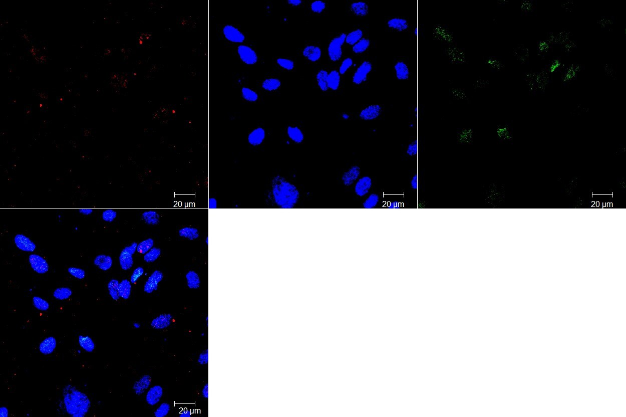
Immunocytochemistry/ Immunofluorescence: ABCG2/CD338 Antibody (3G8) - BSA Free [NBP2-22124]
Immunocytochemistry/Immunofluorescence: ABCG2/CD338 Antibody (3G8) [NBP2-22124] - Analysis of Hela cells using ABCG2 mouse mAb (green). DRAQ5 fluorescent DNA dye (blue). Actin filaments have been labeled with Alexa Fluor-555 phalloidin (red).![Immunohistochemistry-Paraffin: ABCG2/CD338 Antibody (3G8) - BSA Free [NBP2-22124]](https://resources.rndsystems.com/images/products/ABCG2-Antibody-3G8-Immunohistochemistry-Paraffin-NBP2-22124-img0005.jpg "Immunohistochemistry-Paraffin: ABCG2/CD338 Antibody (3G8) - BSA Free [NBP2-22124]")
Immunohistochemistry-Paraffin: ABCG2/CD338 Antibody (3G8) - BSA Free [NBP2-22124]
Immunohistochemistry-Paraffin: ABCG2/CD338 Antibody (3G8) [NBP2-22124] - Analysis of paraffin-embedded bladder cancer tissues (left) and skeletal muscle tissues (right) using ABCG2 mouse mAb with DAB staining.![Flow Cytometry: ABCG2/CD338 Antibody (3G8) - BSA Free [NBP2-22124]](https://resources.rndsystems.com/images/products/ABCG2-Antibody-3G8-Flow-Cytometry-NBP2-22124-img0003.jpg "Flow Cytometry: ABCG2/CD338 Antibody (3G8) - BSA Free [NBP2-22124]")
Flow Cytometry: ABCG2/CD338 Antibody (3G8) - BSA Free [NBP2-22124]
Flow Cytometry: ABCG2/CD338 Antibody (3G8) [NBP2-22124] - Analysis of HepG2 cells using ABCG2 mouse mAb (green) and negative control (purple).![ELISA: ABCG2/CD338 Antibody (3G8) - BSA Free [NBP2-22124]](https://resources.rndsystems.com/images/products/ABCG2-Antibody-3G8-ELISA-NBP2-22124-img0001.jpg "ELISA: ABCG2/CD338 Antibody (3G8) - BSA Free [NBP2-22124]")
ELISA: ABCG2/CD338 Antibody (3G8) - BSA Free [NBP2-22124]
ELISA: ABCG2/CD338 Antibody (3G8) [NBP2-22124] - Red: Control Antigen (100ng), Purple: Antigen (10ng), Green: Antigen (50ng), Blue: Antigen (100ng). [NBP2-22124]")
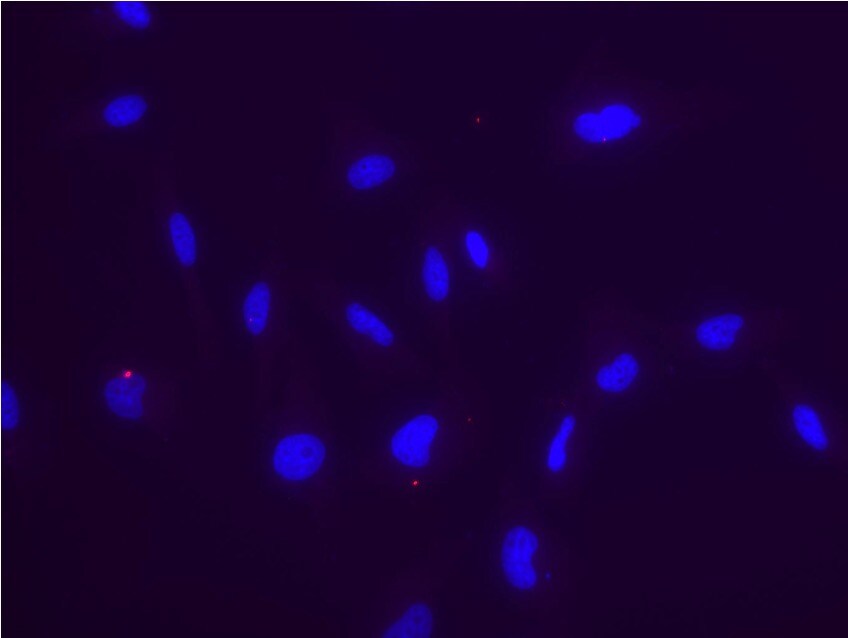
Immunocytochemistry/Immunofluorescence: Mouse Monoclonal ABCG2/CD338 Antibody (3G8) [NBP2-22124]
Immunocytochemistry/Immunofluorescence: Mouse Monoclonal ABCG2/CD338 Antibody (3G8) [NBP2-22124] - Human brain microvascular endothelial cells stained for cilia (red) and ABCG2 (green) and nuclear DAPI (blue). Image from a verified customer review. - BSA Free [NBP2-22124] -")
Western Blot: ABCG2/CD338 Antibody (3G8) - BSA Free [NBP2-22124] -
(A–D) Gene expression of drug transporters in breast cancer patients (A) ABCC1, (B) ABCG2 in local cohort (C) ABCC1, (D) ABCG2 in TCGA cohort and (E–G) Protein expression of drug transporters (E) Representative blots in adjacent normal (N) and tumor (T) tissues, (F) Densitometric analysis of ABCG2 and (G) ABCC1 levels in adjacent normal and tumor tissues. Image collected and cropped by CiteAb from the following open publication (https://pubmed.ncbi.nlm.nih.gov/36309544), licensed under a CC-BY license. Not internally tested by Novus Biologicals.Applications for ABCG2/CD338 Antibody (3G8) - BSA Free
Application
Recommended Usage
ELISA
1:10000
Flow Cytometry
1:200-1:400
Immunocytochemistry/ Immunofluorescence
1:200-1:1000
Immunohistochemistry
1:10-1:500
Immunohistochemistry-Paraffin
1:200-1:1000
Western Blot
1:500-1:2000
Application Notes
In Western blot, a dimer can be seen at 60-70 kDa representing ABCG2.
Reviewed Applications
Read 2 reviews rated 3 using NBP2-22124 in the following applications:
Flow Cytometry Panel Builder
Bio-Techne Knows Flow Cytometry
Save time and reduce costly mistakes by quickly finding compatible reagents using the Panel Builder Tool.
Advanced Features
- Spectra Viewer - Custom analysis of spectra from multiple fluorochromes
- Spillover Popups - Visualize the spectra of individual fluorochromes
- Antigen Density Selector - Match fluorochrome brightness with antigen density

Formulation, Preparation, and Storage
Purification
Ammonium sulfate precipitation
Formulation
PBS
Format
BSA Free
Preservative
0.03% Sodium Azide
Concentration
1.0 mg/ml
Shipping
The product is shipped with polar packs. Upon receipt, store it immediately at the temperature recommended below.
Stability & Storage
Store at 4C short term. Aliquot and store at -20C long term. Avoid freeze-thaw cycles.
Background: ABCG2
Long Name
ATP-binding Cassette Transporter G2
Alternate Names
ABCP, BCRP, CD338, MXR
Entrez Gene IDs
9429 (Human)
Gene Symbol
ABCG2
UniProt
Additional ABCG2 Products
Product Documents for ABCG2/CD338 Antibody (3G8) - BSA Free
Certificate of Analysis
To download a Certificate of Analysis, please enter a lot or batch number in the search box below.
Product Specific Notices for ABCG2/CD338 Antibody (3G8) - BSA Free
This product is for research use only and is not approved for use in humans or in clinical diagnosis. Primary Antibodies are guaranteed for 1 year from date of receipt.
Citations for ABCG2/CD338 Antibody (3G8) - BSA Free
 Powered by Bioz
Customer Reviews for ABCG2/CD338 Antibody (3G8) - BSA Free (2)
3 out of 5
2 Customer Ratings
Have you used ABCG2/CD338 Antibody (3G8) - BSA Free?
Submit a review and receive an Amazon gift card!
$25/€18/£15/$25CAN/¥2500 Yen for a review with an image
$10/€7/£6/$10CAN/¥1110 Yen for a review without an image
Submit a review
Customer Images
Showing
1
-
2 of
2 reviews
Showing All
Filter By:
-
Application: ImmunocytochemistrySample Tested: human brain microvascular endothelial cellsSpecies: HumanVerified Customer | Posted 12/20/2024Human brain microvascular endothelial cells stained for cilia (red) and ABCG2 (green) and nuclear DAPI (blue).

-
Application: ImmunocytochemistrySample Tested: A2058 human melanoma cell lineSpecies: HumanVerified Customer | Posted 11/16/2023A2058 melanoma cell line stained with NBP2-22124 dilution 1:100. No signal
Bio-Techne ResponseThank you for reviewing our product. We are sorry to hear that this product did not perform as expected. We have been in touch with the customer to resolve this issue according to our Product Guarantee and to the customer’s satisfaction.
There are no reviews that match your criteria.
Protocols
View specific protocols for ABCG2/CD338 Antibody (3G8) - BSA Free (NBP2-22124):
ABCG2/CD338 Antibody (3G8):
Flow Cytometry Protocol
Solutions and Reagents:
1X PBS
Blocking buffer: 0.5% BSA in 1X PBS
Filter buffer: 0.1%Triton-X100 and Blocking buffer
Ice cold 4% paraformaldehyde (1% solution -optional for storing samples)
Fluorescently-conjugated secondary antibody (various forms)
Protocol
2.1. Collect 1x10^6 cells/sample.
2.2. Wash cells once with blocking buffer.
2.3. Fix cells with 4% paraformaldehyde and incubate at 4C for 30 min.
2.4 Permeabilize cells: Add 0.5 ml filter buffer(0.1%Triton-X100 and Blocking buffer) and incubate at 0 degree C for 15 min.
2.4. Wash cells once with blocking buffer.
2.5. Add 0.5 ml filter buffer and incubate at 0C for 15 min.
2.6. Wash cells twice with blocking buffer.
2.7. Incubate cells in blocking buffer for 10 min at room temperature.
2.8. Add primary antibody at the appropriate dilution and incubate for 30 min at room temperature.
2.9. Wash twice with blocking buffer and incubate with fluorescently-conjugated secondary antibody for 30 min at room temperature.
2.10. Wash cells twice with blocking buffer.
2.11. Re-suspend cells in 1X PBS and analyze on flow cytometry. Samples can be kept in 1% paraformaldehyde at 4C overnight.
Advice: Keep the cells in the dark on ice or at 4C in a fridge until your scheduled time for analysis. Analyze the cells on the flow cytometer as soon as possible.
Find general support by application which include: protocols, troubleshooting, illustrated assays, videos and webinars.
- 7-Amino Actinomycin D (7-AAD) Cell Viability Flow Cytometry Protocol
- Antigen Retrieval Protocol (PIER)
- Antigen Retrieval for Frozen Sections Protocol
- Appropriate Fixation of IHC/ICC Samples
- Cellular Response to Hypoxia Protocols
- Chromogenic IHC Staining of Formalin-Fixed Paraffin-Embedded (FFPE) Tissue Protocol
- Chromogenic Immunohistochemistry Staining of Frozen Tissue
- ClariTSA™ Fluorophore Kits
- Detection & Visualization of Antibody Binding
- ELISA Sample Preparation & Collection Guide
- ELISA Troubleshooting Guide
- Extracellular Membrane Flow Cytometry Protocol
- Flow Cytometry Protocol for Cell Surface Markers
- Flow Cytometry Protocol for Staining Membrane Associated Proteins
- Flow Cytometry Staining Protocols
- Flow Cytometry Troubleshooting Guide
- Fluorescent IHC Staining of Frozen Tissue Protocol
- Graphic Protocol for Heat-induced Epitope Retrieval
- Graphic Protocol for the Preparation and Fluorescent IHC Staining of Frozen Tissue Sections
- Graphic Protocol for the Preparation and Fluorescent IHC Staining of Paraffin-embedded Tissue Sections
- Graphic Protocol for the Preparation of Gelatin-coated Slides for Histological Tissue Sections
- How to Run an R&D Systems DuoSet ELISA
- How to Run an R&D Systems Quantikine ELISA
- How to Run an R&D Systems Quantikine™ QuicKit™ ELISA
- ICC Cell Smear Protocol for Suspension Cells
- ICC Immunocytochemistry Protocol Videos
- ICC for Adherent Cells
- IHC Sample Preparation (Frozen sections vs Paraffin)
- Immunocytochemistry (ICC) Protocol
- Immunocytochemistry Troubleshooting
- Immunofluorescence of Organoids Embedded in Cultrex Basement Membrane Extract
- Immunofluorescent IHC Staining of Formalin-Fixed Paraffin-Embedded (FFPE) Tissue Protocol
- Immunohistochemistry (IHC) and Immunocytochemistry (ICC) Protocols
- Immunohistochemistry Frozen Troubleshooting
- Immunohistochemistry Paraffin Troubleshooting
- Intracellular Flow Cytometry Protocol Using Alcohol (Methanol)
- Intracellular Flow Cytometry Protocol Using Detergents
- Intracellular Nuclear Staining Flow Cytometry Protocol Using Detergents
- Intracellular Staining Flow Cytometry Protocol Using Alcohol Permeabilization
- Intracellular Staining Flow Cytometry Protocol Using Detergents to Permeabilize Cells
- Preparing Samples for IHC/ICC Experiments
- Preventing Non-Specific Staining (Non-Specific Binding)
- Primary Antibody Selection & Optimization
- Propidium Iodide Cell Viability Flow Cytometry Protocol
- Protocol for Heat-Induced Epitope Retrieval (HIER)
- Protocol for Liperfluo
- Protocol for Making a 4% Formaldehyde Solution in PBS
- Protocol for VisUCyte™ HRP Polymer Detection Reagent
- Protocol for the Characterization of Human Th22 Cells
- Protocol for the Characterization of Human Th9 Cells
- Protocol for the Fluorescent ICC Staining of Cell Smears - Graphic
- Protocol for the Fluorescent ICC Staining of Cultured Cells on Coverslips - Graphic
- Protocol for the Preparation & Fixation of Cells on Coverslips
- Protocol for the Preparation and Chromogenic IHC Staining of Frozen Tissue Sections
- Protocol for the Preparation and Chromogenic IHC Staining of Frozen Tissue Sections - Graphic
- Protocol for the Preparation and Chromogenic IHC Staining of Paraffin-embedded Tissue Sections
- Protocol for the Preparation and Chromogenic IHC Staining of Paraffin-embedded Tissue Sections - Graphic
- Protocol for the Preparation and Fluorescent ICC Staining of Cells on Coverslips
- Protocol for the Preparation and Fluorescent ICC Staining of Non-adherent Cells
- Protocol for the Preparation and Fluorescent ICC Staining of Stem Cells on Coverslips
- Protocol for the Preparation and Fluorescent IHC Staining of Frozen Tissue Sections
- Protocol for the Preparation and Fluorescent IHC Staining of Paraffin-embedded Tissue Sections
- Protocol for the Preparation of Gelatin-coated Slides for Histological Tissue Sections
- Protocol for the Preparation of a Cell Smear for Non-adherent Cell ICC - Graphic
- Protocol: Annexin V and PI Staining by Flow Cytometry
- Protocol: Annexin V and PI Staining for Apoptosis by Flow Cytometry
- Quantikine HS ELISA Kit Assay Principle, Alkaline Phosphatase
- Quantikine HS ELISA Kit Principle, Streptavidin-HRP Polymer
- R&D Systems Quality Control Western Blot Protocol
- Sandwich ELISA (Colorimetric) – Biotin/Streptavidin Detection Protocol
- Sandwich ELISA (Colorimetric) – Direct Detection Protocol
- TUNEL and Active Caspase-3 Detection by IHC/ICC Protocol
- The Importance of IHC/ICC Controls
- Troubleshooting Guide: ELISA
- Troubleshooting Guide: Fluorokine Flow Cytometry Kits
- Troubleshooting Guide: Immunohistochemistry
- Troubleshooting Guide: Western Blot Figures
- Western Blot Conditions
- Western Blot Protocol
- Western Blot Protocol for Cell Lysates
- Western Blot Troubleshooting
- Western Blot Troubleshooting Guide
- View all Protocols, Troubleshooting, Illustrated assays and Webinars


