Acetylcholinesterase/ACHE Antibody
Novus Biologicals | Catalog # NB100-1519
![Western Blot: Acetylcholinesterase/ACHE Antibody [NB100-1519]](https://resources.rndsystems.com/images/products/Acetylcholinesterase-ACHE-Antibody-Western-Blot-NB100-1519-img0006.jpg "Western Blot: Acetylcholinesterase/ACHE Antibody [NB100-1519]")
Loading...
Key Product Details
Species Reactivity
Human, Mouse, Rat
Applications
Immunohistochemistry Free-Floating, Western Blot, Peptide ELISA, Flow Cytometry, Immunocytochemistry/ Immunofluorescence
Label
Unconjugated
Antibody Source
Polyclonal Goat IgG
Loading...
Product Specifications
Immunogen
Peptide with sequence QFDHYSKQDRCSDL corresponding to C-Terminus according to NP_000656.1.
Reactivity Notes
Mouse reactivity reported from a verified customer review.
Marker
Early Neuronal Development Marker
Specificity
This antibody is expected to recognise isoform NP_000656.1 only (the ubiquitously expressed, hydrophillic form).
Clonality
Polyclonal
Host
Goat
Isotype
IgG
Scientific Data Images for Acetylcholinesterase/ACHE Antibody
Western Blot: Acetylcholinesterase/ACHE Antibody [NB100-1519]
Western Blot: Acetylcholinesterase/ACHE Antibody [NB100-1519] - Staining (0.3ug/ml) of Jurkat (A) and (0.5ug/ml) HepG2 (B) cell lysate (35ug protein in RIPA buffer). Detected by chemiluminescence.![Immunocytochemistry/ Immunofluorescence: Acetylcholinesterase/ACHE Antibody [NB100-1519]](https://resources.rndsystems.com/images/products/Acetylcholinesterase-ACHE-Antibody-Immunocytochemistry-Immunofluorescence-NB100-1519-img0005.jpg "Immunocytochemistry/ Immunofluorescence: Acetylcholinesterase/ACHE Antibody [NB100-1519]")
Immunocytochemistry/ Immunofluorescence: Acetylcholinesterase/ACHE Antibody [NB100-1519]
Immunocytochemistry/Immunofluorescence: Acetylcholinesterase/ACHE Antibody [NB100-1519] - Immunofluorescence analysis of paraformaldehyde fixed U2OS cells, permeabilized with 0.15% Triton. Primary incubation 1hr (10ug/ml) followed by Alexa Fluor 488 secondary antibody (2ug/ml), showing nuclear, membrane and cytoplasmic staining. The nuclear stain is DAPI (blue). Negative control: Unimmunized goat IgG (10ug/ml) followed by Alexa Fluor 488 secondary antibody (2ug/ml).![Flow Cytometry: Acetylcholinesterase/ACHE Antibody [NB100-1519]](https://resources.rndsystems.com/images/products/Acetylcholinesterase-ACHE-Antibody-Flow-Cytometry-NB100-1519-img0004.jpg "Flow Cytometry: Acetylcholinesterase/ACHE Antibody [NB100-1519]")
Flow Cytometry: Acetylcholinesterase/ACHE Antibody [NB100-1519]
Flow Cytometry: Acetylcholinesterase/ACHE Antibody [NB100-1519] - Flow cytometric analysis of paraformaldehyde fixed HeLa cells (blue line), permeabilized with 0.5% Triton. Primary incubation 1hr (10ug/ml) followed by Alexa Fluor 488 secondary antibody (1ug/ml). IgG control: Unimmunized goat IgG (black line) followed by Alexa Fluor 488 secondary antibody.Applications for Acetylcholinesterase/ACHE Antibody
Application
Recommended Usage
Flow Cytometry
10 ug/mL
Immunocytochemistry/ Immunofluorescence
10 ug/mL
Peptide ELISA
Detection limit 1:32000
Western Blot
0.3 - 1 ug/mL
Application Notes
This Acetylcholinesterase/ACHE antibody is validated for IHC-FrFl from a verified customer review.
Reviewed Applications
Read 1 review rated 5 using NB100-1519 in the following applications:
Flow Cytometry Panel Builder
Bio-Techne Knows Flow Cytometry
Save time and reduce costly mistakes by quickly finding compatible reagents using the Panel Builder Tool.
Advanced Features
- Spectra Viewer - Custom analysis of spectra from multiple fluorochromes
- Spillover Popups - Visualize the spectra of individual fluorochromes
- Antigen Density Selector - Match fluorochrome brightness with antigen density

Formulation, Preparation, and Storage
Purification
Immunogen affinity purified
Formulation
Tris saline (20 mM Tris pH 7.3, 150 mM NaCl), 0.5% BSA
Preservative
0.02% Sodium Azide
Concentration
0.5 mg/ml
Shipping
The product is shipped with polar packs. Upon receipt, store it immediately at the temperature recommended below.
Stability & Storage
Store at -20C. Avoid freeze-thaw cycles.
Background: Acetylcholinesterase/ACHE
Alternate Names
ACHE, ARACHE, N-ACHE
Gene Symbol
ACHE
UniProt
Additional Acetylcholinesterase/ACHE Products
Product Documents for Acetylcholinesterase/ACHE Antibody
Certificate of Analysis
To download a Certificate of Analysis, please enter a lot or batch number in the search box below.
Product Specific Notices for Acetylcholinesterase/ACHE Antibody
This product is for research use only and is not approved for use in humans or in clinical diagnosis. Primary Antibodies are guaranteed for 1 year from date of receipt.
Citations for Acetylcholinesterase/ACHE Antibody
 Powered by Bioz
Customer Reviews for Acetylcholinesterase/ACHE Antibody (1)
5 out of 5
1 Customer Rating
Have you used Acetylcholinesterase/ACHE Antibody?
Submit a review and receive an Amazon gift card!
$25/€18/£15/$25CAN/¥2500 Yen for a review with an image
$10/€7/£6/$10CAN/¥1110 Yen for a review without an image
Submit a review
Customer Images
Showing
1
-
1 of
1 review
Showing All
Filter By:
-
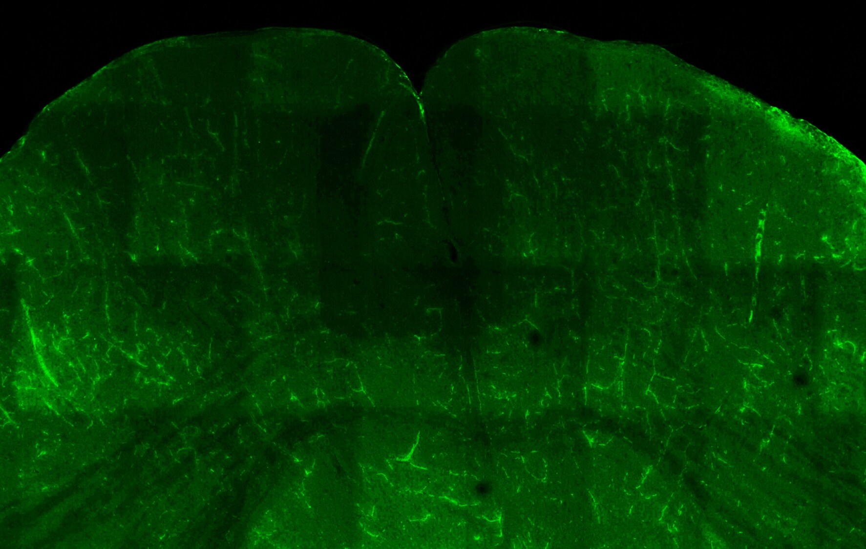
Application: IHC-Floating sectionSample Tested: Mouse brainSpecies: MouseVerified Customer | Posted 11/21/2016Positive ACHE staining (green) in neurons of the mouse brain. Fixed in 4% paraformaldehyde, and sectioned at 50um using a vibratomeFloating sections were incubated overnight at 4C with a 1:200 dilution of the above ACHE antibody, washed and then incubated for 1 hour at room temperature with a 1:500 dilution of donkey anti-rabbit DyLight 594 secondary antibody (Cat#NBP1-75637).

There are no reviews that match your criteria.
Protocols
Find general support by application which include: protocols, troubleshooting, illustrated assays, videos and webinars.
- 7-Amino Actinomycin D (7-AAD) Cell Viability Flow Cytometry Protocol
- Antigen Retrieval Protocol (PIER)
- Antigen Retrieval for Frozen Sections Protocol
- Appropriate Fixation of IHC/ICC Samples
- Cellular Response to Hypoxia Protocols
- Chromogenic IHC Staining of Formalin-Fixed Paraffin-Embedded (FFPE) Tissue Protocol
- Chromogenic Immunohistochemistry Staining of Frozen Tissue
- ClariTSA™ Fluorophore Kits
- Detection & Visualization of Antibody Binding
- ELISA Sample Preparation & Collection Guide
- ELISA Troubleshooting Guide
- Extracellular Membrane Flow Cytometry Protocol
- Flow Cytometry Protocol for Cell Surface Markers
- Flow Cytometry Protocol for Staining Membrane Associated Proteins
- Flow Cytometry Staining Protocols
- Flow Cytometry Troubleshooting Guide
- Fluorescent IHC Staining of Frozen Tissue Protocol
- Graphic Protocol for Heat-induced Epitope Retrieval
- Graphic Protocol for the Preparation and Fluorescent IHC Staining of Frozen Tissue Sections
- Graphic Protocol for the Preparation and Fluorescent IHC Staining of Paraffin-embedded Tissue Sections
- Graphic Protocol for the Preparation of Gelatin-coated Slides for Histological Tissue Sections
- How to Run an R&D Systems DuoSet ELISA
- How to Run an R&D Systems Quantikine ELISA
- How to Run an R&D Systems Quantikine™ QuicKit™ ELISA
- ICC Cell Smear Protocol for Suspension Cells
- ICC Immunocytochemistry Protocol Videos
- ICC for Adherent Cells
- IHC Sample Preparation (Frozen sections vs Paraffin)
- Immunocytochemistry (ICC) Protocol
- Immunocytochemistry Troubleshooting
- Immunofluorescence of Organoids Embedded in Cultrex Basement Membrane Extract
- Immunofluorescent IHC Staining of Formalin-Fixed Paraffin-Embedded (FFPE) Tissue Protocol
- Immunohistochemistry (IHC) and Immunocytochemistry (ICC) Protocols
- Immunohistochemistry Frozen Troubleshooting
- Immunohistochemistry Paraffin Troubleshooting
- Intracellular Flow Cytometry Protocol Using Alcohol (Methanol)
- Intracellular Flow Cytometry Protocol Using Detergents
- Intracellular Nuclear Staining Flow Cytometry Protocol Using Detergents
- Intracellular Staining Flow Cytometry Protocol Using Alcohol Permeabilization
- Intracellular Staining Flow Cytometry Protocol Using Detergents to Permeabilize Cells
- Preparing Samples for IHC/ICC Experiments
- Preventing Non-Specific Staining (Non-Specific Binding)
- Primary Antibody Selection & Optimization
- Propidium Iodide Cell Viability Flow Cytometry Protocol
- Protocol for Heat-Induced Epitope Retrieval (HIER)
- Protocol for Liperfluo
- Protocol for Making a 4% Formaldehyde Solution in PBS
- Protocol for VisUCyte™ HRP Polymer Detection Reagent
- Protocol for the Characterization of Human Th22 Cells
- Protocol for the Characterization of Human Th9 Cells
- Protocol for the Fluorescent ICC Staining of Cell Smears - Graphic
- Protocol for the Fluorescent ICC Staining of Cultured Cells on Coverslips - Graphic
- Protocol for the Preparation & Fixation of Cells on Coverslips
- Protocol for the Preparation and Chromogenic IHC Staining of Frozen Tissue Sections
- Protocol for the Preparation and Chromogenic IHC Staining of Frozen Tissue Sections - Graphic
- Protocol for the Preparation and Chromogenic IHC Staining of Paraffin-embedded Tissue Sections
- Protocol for the Preparation and Chromogenic IHC Staining of Paraffin-embedded Tissue Sections - Graphic
- Protocol for the Preparation and Fluorescent ICC Staining of Cells on Coverslips
- Protocol for the Preparation and Fluorescent ICC Staining of Non-adherent Cells
- Protocol for the Preparation and Fluorescent ICC Staining of Stem Cells on Coverslips
- Protocol for the Preparation and Fluorescent IHC Staining of Frozen Tissue Sections
- Protocol for the Preparation and Fluorescent IHC Staining of Paraffin-embedded Tissue Sections
- Protocol for the Preparation of Gelatin-coated Slides for Histological Tissue Sections
- Protocol for the Preparation of a Cell Smear for Non-adherent Cell ICC - Graphic
- Protocol: Annexin V and PI Staining by Flow Cytometry
- Protocol: Annexin V and PI Staining for Apoptosis by Flow Cytometry
- Quantikine HS ELISA Kit Assay Principle, Alkaline Phosphatase
- Quantikine HS ELISA Kit Principle, Streptavidin-HRP Polymer
- R&D Systems Quality Control Western Blot Protocol
- Sandwich ELISA (Colorimetric) – Biotin/Streptavidin Detection Protocol
- Sandwich ELISA (Colorimetric) – Direct Detection Protocol
- TUNEL and Active Caspase-3 Detection by IHC/ICC Protocol
- The Importance of IHC/ICC Controls
- Troubleshooting Guide: ELISA
- Troubleshooting Guide: Fluorokine Flow Cytometry Kits
- Troubleshooting Guide: Immunohistochemistry
- Troubleshooting Guide: Western Blot Figures
- Western Blot Conditions
- Western Blot Protocol
- Western Blot Protocol for Cell Lysates
- Western Blot Troubleshooting
- Western Blot Troubleshooting Guide
- View all Protocols, Troubleshooting, Illustrated assays and Webinars
Loading...