Best Seller
alpha-Smooth Muscle Actin Antibody (1A4/asm-1) - BSA Free
Novus Biologicals | Catalog # NBP2-33006
Key Product Details
Species Reactivity
Validated:
Human, Mouse, Rat, Porcine, Baboon, Bovine, Canine, Chicken, Equine, Feline, Goat, Guinea Pig, Monkey, Rabbit, Sheep
Cited:
Human, Mouse, Rat, Porcine, Feline
Applications
Validated:
Multiplex Immunofluorescence, Immunohistochemistry, Immunohistochemistry-Paraffin, Immunohistochemistry-Frozen, Western Blot, Flow Cytometry, Flow (Intracellular), Immunocytochemistry/ Immunofluorescence, Simple Western, Immunoprecipitation, COMET
Cited:
Immunohistochemistry-Paraffin, Immunohistochemistry-Frozen, Western Blot, Flow Cytometry, Immunocytochemistry/ Immunofluorescence, Simple Western, IF/IHC
Label
Unconjugated
Antibody Source
Monoclonal Mouse IgG2a Kappa Clone # 1A4/asm-1
Format
BSA Free
Loading...
Product Specifications
Immunogen
Alpha-Smooth Muscle Actin Antibody (1A4/asm-1) is made to the N-Terminal decapeptide of Alpha smooth muscle isoform of actin and conjugated to KLH.
Reactivity Notes
Feline and Equine reactivity reported from a verified customer review.
Localization
Cytoplasmic
Marker
Leiomyosarcoma Marker
Specificity
Actin is a major component of the cytoskeleton and is present in most cell types. This MAb is highly specific to actin from smooth muscles. Its epitope lies in the first four N-terminal amino acids. This MAb does not stain cardiac or skeletal muscle; however, it does stain myofibroblasts and myoepithelial cells. This antibody could be used together with anti-muscle specific actin and myogenin in making a diagnosis of smooth muscle and skeletal muscle tumors. In most cases of rhabdomyosarcoma, this antibody yields negative results whereas anti-muscle specific actin and myogenin are positive. Leiomyosarcomas are positive only with anti-muscle specific actin and anti-smooth muscle actin and are negative with anti-myogenin.
Clonality
Monoclonal
Host
Mouse
Isotype
IgG2a Kappa
Theoretical MW
42 kDa.
Disclaimer note: The observed molecular weight of the protein may vary from the listed predicted molecular weight due to post translational modifications, post translation cleavages, relative charges, and other experimental factors.
Disclaimer note: The observed molecular weight of the protein may vary from the listed predicted molecular weight due to post translational modifications, post translation cleavages, relative charges, and other experimental factors.
Scientific Data Images for alpha-Smooth Muscle Actin Antibody (1A4/asm-1) - BSA Free

Detection of alpha -Smooth Muscle Actin in Human Colon Cancer via Multiplex Immunofluorescence staining on COMET™
alpha -Smooth Muscle Actin was detected in immersion fixed paraffin-embedded sections of human colon cancer using Mouse Anti-Human/Mouse/Rat alpha -Smooth Muscle Actin Monoclonal Antibody (Novus Catalog # NBP2-33006) at 0.25ug/mL at 37 ° Celsius for 2 minutes. Before incubation with the primary antibody, tissue underwent an all-in-one dewaxing and antigen retrieval preprocessing using PreTreatment Module (PT Module) and Dewax and HIER Buffer H (pH 9). Tissue was stained using the Alexa Fluor™ 555 Goat anti-Mouse IgG Secondary Antibody at 1:100 at 37 ° Celsius for 2 minutes. (Yellow; Lunaphore Catalog # DR555MS) and counterstained with DAPI (blue; Lunaphore Catalog # DR100). Specific staining was localized to the cytoplasm and cytoskeleton. Protocol available in COMET™ Panel Builder.
Detection of alpha -Smooth Muscle Actin in Human Colon via Multiplex Immunofluorescence staining on COMET™
alpha -Smooth Muscle Actin was detected in immersion fixed paraffin-embedded sections of human colon using Mouse Anti-Human/Mouse/Rat alpha -Smooth Muscle Actin Monoclonal Antibody (Novus Catalog # NBP2-33006) at 0.25ug/mL at 37 ° Celsius for 2 minutes. Before incubation with the primary antibody, tissue underwent an all-in-one dewaxing and antigen retrieval preprocessing using PreTreatment Module (PT Module) and Dewax and HIER Buffer H (pH 9). Tissue was stained using the Alexa Fluor™ 555 Goat anti-Mouse IgG Secondary Antibody at 1:100 at 37 ° Celsius for 2 minutes. (Yellow; Lunaphore Catalog # DR555MS) and counterstained with DAPI (blue; Lunaphore Catalog # DR100). Specific staining was localized to the cytoplasm and cytoskeleton. Protocol available in COMET™ Panel Builder..png "Detection of alpha -Smooth Muscle Actin in Human Breast Cancer via Multiplex Immunofluorescence staining on COMET™")
Detection of alpha -Smooth Muscle Actin in Human Breast Cancer via Multiplex Immunofluorescence staining on COMET™
alpha -Smooth Muscle Actin was detected in immersion fixed paraffin-embedded sections of human breast cancer using Mouse Anti-Human/Mouse/Rat alpha -Smooth Muscle Actin Monoclonal Antibody (Novus Catalog # NBP2-33006) at 0.25ug/mL at 37 ° Celsius for 2 minutes. Before incubation with the primary antibody, tissue underwent an all-in-one dewaxing and antigen retrieval preprocessing using PreTreatment Module (PT Module) and Dewax and HIER Buffer H (pH 9). Tissue was stained using the Alexa Fluor™ 555 Goat anti-Mouse IgG Secondary Antibody at 1:100 at 37 ° Celsius for 2 minutes. (Yellow; Lunaphore Catalog # DR555MS) and counterstained with DAPI (blue; Lunaphore Catalog # DR100). Specific staining was localized to the cytoplasm and cytoskeleton. Protocol available in COMET™ Panel Builder.
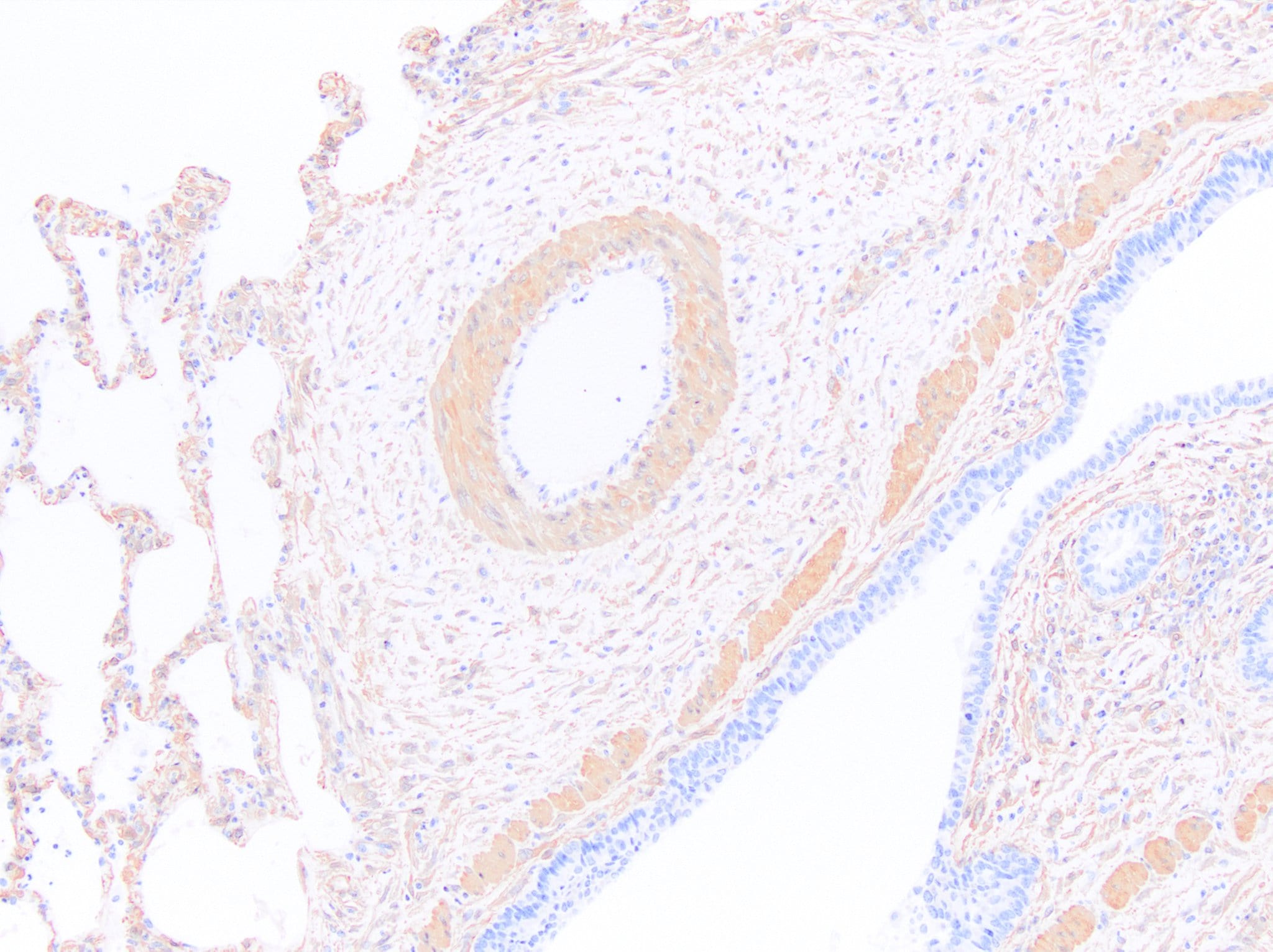
Detection of SMA in Mouse Lung via seqIF™ staining on COMET™
SMA Antibody was detected in immersion fixed paraffin-embedded sections of mouse Lung using Mouse Anti-Human/Mouse SMA, Monoclonal Antibody (Catalog #NBP2-33006) at 0.25ug/mL at 37 ° Celsius for 2 minutes. Before incubation with the primary antibody, tissue underwent an all-in-one dewaxing and antigen retrieval preprocessing using PreTreatment Module (PT Module) and Dewax and HIER Buffer H (pH 9; Epredia Catalog # TA-999-DHBH). Tissue was stained using the Alexa Fluor™ 555 Goat anti-Mouse IgG Secondary Antibody at 1:100 at 37 ° Celsius for 2 minutes. (Yellow; Lunaphore Catalog # DR555MS) and counterstained with DAPI (blue; Lunaphore Catalog # DR100). Specific staining was localized to the cytoplasm. Protocol available in COMET™ Panel Builder.
Western Blot Detection of alpha-Smooth Muscle Actin in Human Heart Tissue and iBJ6 hiPSCs
Western blot shows lysates of human heart tissue and iBJ6 human induced pluripotent stem cell line undifferentiated or cardiac differentiated. PVDF membrane was probed with 0.25 ug/mL of Mouse Anti-alpha-Smooth Muscle Actin Monoclonal Antibody (Catalog # NBP2-33006) followed by HRP-conjugated Anti-Mouse IgG Secondary Antibody (Catalog # HAF018). The observed molecular weight was detected for alpha Smooth Muscle Actin at approximately 40 kDa (as indicated). This experiment was conducted under reducing conditions.
Immunohistological Imaging of alpha-Smooth Muscle Actin in Frozen Feline Tissue
Feline tissue stained with alpha-Smooth Muscle Actin Antibody at dilution of 1:600. Vascular wall was stained. IHC image submitted by a verified customer review.
Simple Western Analysis of alpha-Smooth Muscle Actin in Aorta Lysate
Simple Western lane view shows a specific band for an observed molecular weight at ~45 kDa for alpha-Smooth Muscle Actin in 0.2 mg/ml of h. Aorta lysate. This experiment was performed under reducing conditions using the 12-230 kDa separation system.
Flow Cytometry of HeLa Cells Stained with Alexa Fluor 700 Conjugated alpha-Smooth Muscle Actin Antibody
An intracellular stain was performed on HeLa cells with NBP2-34522AF700 (blue) and a matched isotype control (orange). Cells were fixed with 4% PFA and then permeabilized with 0.1% saponin. Cells were incubated in an antibody dilution of 10 ug/mL for 30 minutes at room temperature. Both antibodies were conjugated to Alexa Fluor 700.
Immunohistological Staining of alpha-Smooth Muscle Actin in Frozen Canine Brain/Spinal Cord
Canine brain/spinal cord section. Alpha-Smooth Muscle Actin Antibody (1A4/asm-1) staining in red. Endothelial cell marker in green. DAPI in blue. Alexa Fluor conjugated antibodies used for secondary antibodies. IHC image submitted by a verified customer review.
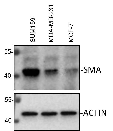
Western Blot Detection of alpha-Smooth Muscle Actin in Multiple Cell Lines
Whole cell lysates from SUM159, MDA-MB-231 and MCF-7 cells were loaded with 50 ug/lane. 10% SDS-PAGE. Alpha-Smooth Muscle Actin antibody (NBP2-33006) was used for primary antibody: 1:1000, 4C, overnight. Image from verified customer review.
Immunohistological Staining of alpha-Smooth Muscle Actin in Paraffin Embedded Cancer Tissues
alpha-Smooth-Muscle-Actin-Antibody-1A4-asm-1-Immunohistochemistry-Paraffin-NBP2-33006-img0017.jpg
Flow Cytometry of HeLa Cells Stained with Allophycocyanin Conjugated alpha-Smooth Muscle Actin Antibody
An intracellular stain was performed on HeLa cells with alpha-Smooth Muscle Actin Antibody (1A4/asm-1) NBP2-34522APC (blue) and a matched isotype control (orange). Cells were fixed with 4% PFA and then permeabilized with 0.1% saponin. Cells were incubated in an antibody dilution of 1 ug/mL for 30 minutes at room temperature. Both antibodies were conjugated to Allophycocyanin.
Immunohistological Detection of alpha-Smooth Muscle Actin in Paraffin Embedded Human Leiomyosarcoma
Staining of human Leiomyosarcoma statined with the Azide and BSA-free version of alpha-Smooth Muscle Actin Antibody (1A4/asm-1).
Flow Cytometry of HeLa Cells Stained with Alexa Fluor 647 Conjugated alpha-Smooth Muscle Actin Antibody
An intracellular stain was performed on HeLa cells with alpha-Smooth Muscle Actin Antibody (1A4/asm-1) NBP2-34522AF647 (blue) and a matched isotype control (orange). Cells were fixed with 4% PFA and then permeabilized with 0.1% saponin. Cells were incubated in an antibody dilution of 2.5 ug/mL for 30 minutes at room temperature. Both antibodies were conjugated to Alexa Fluor 647. Image from the Alexa Fluor 647 version of this antibody.
Detection of alpha-Smooth Muscle Actin in Uterus Lysate by Simple Western
Simple Western lane view shows a specific band for an observed molecular weight for Smooth Muscle Actin in 0.2 mg/ml of h. Uterus lysate. This experiment was performed under reducing conditions using the 12-230 kDa separation system.
Electropherogram Representation of alpha-Smooth Muscle Actin Detection in Aorta and Uterus Lysates by Simple Western
Electropherogram image of the corresponding Simple Western lane. alpha-Smooth Muscle Actin antibody was used at 10 ug/ml dilution of h. Aorta and h. Uterus lysates(s) respectively. - BSA Free [NBP2-33006] -")
Flow Cytometry: alpha-Smooth Muscle Actin Antibody (1A4/asm-1) - BSA Free [NBP2-33006] -
Flow Cytometry: alpha-Smooth Muscle Actin Antibody (1A4/asm-1) - BSA Free [NBP2-33006] - An intracellular stain was performed on HeLa cells with alpha-Smooth Muscle Actin Antibody (1A4/asm-1) NBP2-34522PCP (blue) and a matched isotype control (orange). Cells were fixed with 4% PFA and then permeabilized with 0.1% saponin. Cells were incubated in an antibody dilution of 10 ug/mL for 30 minutes at room temperature. Both antibodies were conjugated to PerCP. - BSA Free [NBP2-33006] -")
Flow Cytometry: alpha-Smooth Muscle Actin Antibody (1A4/asm-1) - BSA Free [NBP2-33006] -
Flow Cytometry: alpha-Smooth Muscle Actin Antibody (1A4/asm-1) - BSA Free [NBP2-33006] - An intracellular stain was performed on HeLa cells with alpha Smooth Muscle Actin (1A4/asm-1) Antibody NBP2-34522JF549 (blue) and a matched isotype control (orange). Cells were fixed with 4% PFA and then permeabilized with 0.1% saponin. Cells were incubated in an antibody dilution of 5 ug/mL for 30 minutes at room temperature. Both antibodies were conjugated to Janelia Fluor 549. - BSA Free [NBP2-33006] -")
Flow Cytometry: alpha-Smooth Muscle Actin Antibody (1A4/asm-1) - BSA Free [NBP2-33006] -
Flow Cytometry: alpha-Smooth Muscle Actin Antibody (1A4/asm-1) - BSA Free [NBP2-33006] - Human tonsil frozen tissue section stained for alpha-Smooth Muscle Actin (red) with the Janelia Fluor 549 conjugate of the Azide and BSA-Free version of alpha-Smooth Muscle Actin Antibody (1A4/asm-1), and counterstained with DAPI (blue). Image from verified customer review. - BSA Free [NBP2-33006] -")
Flow Cytometry: alpha-Smooth Muscle Actin Antibody (1A4/asm-1) - BSA Free [NBP2-33006] -
Flow Cytometry: alpha-Smooth Muscle Actin Antibody (1A4/asm-1) - BSA Free [NBP2-33006] - An intracellular stain was performed on HeLa cells with alpha-Smooth Muscle Actin Antibody (1A4/asm-1) NBP2-34522PE (blue) and a matched isotype control (orange). Cells were fixed with 4% PFA and then permeabilized with 0.1% saponin. Cells were incubated in an antibody dilution of 2.5 ug/mL for 30 minutes at room temperature. Both antibodies were conjugated to phycoerythrin. - BSA Free [NBP2-33006] -")
Flow Cytometry: alpha-Smooth Muscle Actin Antibody (1A4/asm-1) - BSA Free [NBP2-33006] -
Flow Cytometry: alpha-Smooth Muscle Actin Antibody (1A4/asm-1) - BSA Free [NBP2-33006] - alpha-Smooth Muscle Actin expression in colorectal tumor cells. Using Allophycocyanin conjugated version of antibody (NBP2-34522APC). Image from verified customer review. - BSA Free [NBP2-33006] -")
Flow Cytometry: alpha-Smooth Muscle Actin Antibody (1A4/asm-1) - BSA Free [NBP2-33006] -
Flow Cytometry: alpha-Smooth Muscle Actin Antibody (1A4/asm-1) - BSA Free [NBP2-33006] - An intracellular stain was performed on HeLa cells with alpha-Smooth Muscle Actin Antibody (1A4/asm-1) NBP2-34522AF647 (blue) and a matched isotype control (orange). Cells were fixed with 4% PFA and then permeabilized with 0.1% saponin. Cells were incubated in an antibody dilution of 2.5 ug/mL for 30 minutes at room temperature. Both antibodies were conjugated to Alexa Fluor 647. - BSA Free [NBP2-33006]")
Flow Cytometry: alpha-Smooth Muscle Actin Antibody (1A4/asm-1) - BSA Free [NBP2-33006]
Flow Cytometry: alpha-Smooth Muscle Actin Antibody (1A4/asm-1) - BSA Free [NBP2-33006] - Analysis of alpha-Smooth Muscle Actin Antibody (1A4/asm-1) [Alexa Fluor® 647] (Catalog Number: NBP2-34522AF647) using two lung cancer cell lines to acquire the subcutaneous tumors. MMT status of macrophages was then analyzed. Image from a verified customer review. - BSA Free [NBP2-33006]")
Immunohistochemistry-Paraffin: alpha-Smooth Muscle Actin Antibody (1A4/asm-1) - BSA Free [NBP2-33006]
Immunohistochemistry-Paraffin: alpha-Smooth Muscle Actin Antibody (1A4/asm-1) - BSA Free [NBP2-33006] - aSMA stained over night in human FFPE tonsil section ((Catalog # NBP2-33006AF750)). Heat mediated antigen retrieval at pH 9. 1:50 Dilution in 3% BSA, incubation over night at 4 °C. Image from a verified customer review. [NBP2-33006]")
Immunohistochemistry-Paraffin: Mouse Monoclonal alpha-Smooth Muscle Actin Antibody (1A4/asm-1) [NBP2-33006]
Immunohistochemistry-Paraffin: Mouse Monoclonal alpha-Smooth Muscle Actin Antibody (1A4/asm-1) [NBP2-33006] - Images demonstrating SMA immunoreactivity in a variety of human FFPE tissue sections. NBP2-33006 was used at a concentration of 125ng/mL and was left on tissue sections for 30m at room temperature. Formalin fixed paraffin embedded sections were deparaffinized and rehydrated. Sections then underwent heat induced epitope retrieval in a citrate-based solution for 5min in a food steamer. Endogenous peroxidase activity was blocked by incubating slides for 15min in tris buffered saline containing 3% (volume/volume) hydrogen peroxide. Tissue sections were then washed, blocked with normal horse serum for 20 min, and then incubated with the primary antibody (125ng/mL) for 30min at room temperature. Sections were then washed and incubated with a horse anti-mouse HRP polymer for 30m at room temperature. Sections were washed and incubated with DAB chromogen for approximately 2.5min. Sections were then washed, counterstained with hematoxylin, dehydrated, cleared and mounted with a coverslip. Image from a verified customer review. [NBP2-33006]")
Immunohistochemistry-Paraffin: Mouse Monoclonal alpha-Smooth Muscle Actin Antibody (1A4/asm-1) [NBP2-33006]
Immunohistochemistry-Paraffin: Mouse Monoclonal alpha-Smooth Muscle Actin Antibody (1A4/asm-1) [NBP2-33006] - Images demonstrating SMA immunoreactivity in a variety of canine FFPE tissue sections. NBP2-33006 was used at a concentration of 125ng/mL and was left on tissue sections for 30m at room temperature. Formalin fixed paraffin embedded sections were deparaffinized and rehydrated. Sections then underwent heat induced epitope retrieval in a citrate-based solution for 5min in a food steamer. Endogenous peroxidase activity was blocked by incubating slides for 15min in tris buffered saline containing 3% (volume/volume) hydrogen peroxide. Tissue sections were then washed, blocked with normal horse serum for 20 min, and then incubated with the primary antibody (125ng/mL) for 30min at room temperature. Sections were then washed and incubated with a horse anti-mouse HRP polymer for 30m at room temperature. Sections were washed and incubated with DAB chromogen for approximately 2.5min. Sections were then washed, counterstained with hematoxylin, dehydrated, cleared and mounted with a coverslip. Image from a verified customer review. [NBP2-33006]")
Immunohistochemistry-Paraffin: Mouse Monoclonal alpha-Smooth Muscle Actin Antibody (1A4/asm-1) [NBP2-33006]
Immunohistochemistry-Paraffin: Mouse Monoclonal alpha-Smooth Muscle Actin Antibody (1A4/asm-1) [NBP2-33006] - Images demonstrating SMA immunoreactivity in a variety of feline FFPE tissue sections. NBP2-33006 was used at a concentration of 125ng/mL and was left on tissue sections for 30m at room temperature. Formalin fixed paraffin embedded sections were deparaffinized and rehydrated. Sections then underwent heat induced epitope retrieval in a citrate-based solution for 5min in a food steamer. Endogenous peroxidase activity was blocked by incubating slides for 15min in tris buffered saline containing 3% (volume/volume) hydrogen peroxide. Tissue sections were then washed, blocked with normal horse serum for 20 min, and then incubated with the primary antibody (125ng/mL) for 30min at room temperature. Sections were then washed and incubated with a horse anti-mouse HRP polymer for 30m at room temperature. Sections were washed and incubated with DAB chromogen for approximately 2.5min. Sections were then washed, counterstained with hematoxylin, dehydrated, cleared and mounted with a coverslip. Image from a verified customer review. [NBP2-33006]")
Immunohistochemistry-Paraffin: Mouse Monoclonal alpha-Smooth Muscle Actin Antibody (1A4/asm-1) [NBP2-33006]
Immunohistochemistry-Paraffin: Mouse Monoclonal alpha-Smooth Muscle Actin Antibody (1A4/asm-1) [NBP2-33006] - SMA in FFPE sections of pig tissue. Formalin fixed paraffin embedded sections were deparaffinized and rehydrated. Sections then underwent heat induced epitope retrieval in a citrate-based solution for 5min in a food steamer. Endogenous peroxidase activity was blocked by incubating slides for 15min in tris buffered saline containing 3% (volume/volume) hydrogen peroxide. Tissue sections were then washed, blocked with normal horse serum for 20 min, and then incubated with the primary antibody (125ng/mL) for 30min at room temperature. Sections were then washed and incubated with a horse anti-mouse HRP polymer for 30m at room temperature. Sections were washed and incubated with DAB chromogen for approximately 2.5min. Sections were then washed, counterstained with hematoxylin, dehydrated, cleared and mounted with a coverslip. Image from a verified customer review. [NBP2-33006]")
Immunohistochemistry-Paraffin: Mouse Monoclonal alpha-Smooth Muscle Actin Antibody (1A4/asm-1) [NBP2-33006]
Immunohistochemistry-Paraffin: Mouse Monoclonal alpha-Smooth Muscle Actin Antibody (1A4/asm-1) [NBP2-33006] - Images demonstrating SMA immunoreactivity in a variety of horse FFPE tissue sections. NBP2-33006 was used at a concentration of 125ng/mL and was left on tissue sections for 30m at room temperature. Formalin fixed paraffin embedded sections were deparaffinized and rehydrated. Sections then underwent heat induced epitope retrieval in a citrate-based solution for 5min in a food steamer. Endogenous peroxidase activity was blocked by incubating slides for 15min in tris buffered saline containing 3% (volume/volume) hydrogen peroxide. Tissue sections were then washed, blocked with normal horse serum for 20 min, and then incubated with the primary antibody (125ng/mL) for 30min at room temperature. Sections were then washed and incubated with a horse anti-mouse HRP polymer for 30m at room temperature. Sections were washed and incubated with DAB chromogen for approximately 2.5min. Sections were then washed, counterstained with hematoxylin, dehydrated, cleared and mounted with a coverslip. Image from a verified customer review. [NBP2-33006]")
Immunohistochemistry-Paraffin: Mouse Monoclonal alpha-Smooth Muscle Actin Antibody (1A4/asm-1) [NBP2-33006]
SMA immunoreactivity in an FFPE section of cow lung. NBP2-33006 was diluted to 125ng/mL and was left on tissue sections for 30min at room temperature. Image from a verified customer review. - BSA Free [NBP2-33006] -")
Western Blot: alpha-Smooth Muscle Actin Antibody (1A4/asm-1) - BSA Free [NBP2-33006] -
Large-conductance and Ca2+-activated K+ (BK) channels are expressed and functional in activated hepatic stellate cells (HSCs). (A,B) Representative RT-qPCR (A) and western blots (B) showing the expression of ACTA2 and the BK channels alpha subunit KCNMA1 in activated human LX2 cells treated with transforming growth factor beta 1 (TGF beta 1) and spontaneously activated primary rat HSCs in vitro. (C) Representative immunofluorescence images of KCNMA1 and ACTA2 in activated primary rat HSCs (Scale bars, 25 μm). (D,E) Representative whole-cell K+ current traces and the normalized current recorded from activated HSCs before and after treatment with rottlerin (Rot) and paxilline (Pax), as indicated at 1 μM internal Ca2+. The whole-cell currents were elicited by 1 s voltage ramps from −100 to 80 mV. Rottlerin (1 μM) and paxilline (10 μM) were freshly prepared from stock solutions and the final DMSO concentration was 0.1% (*p < 0.05 compared with the vehicle group, n = 3). Image collected and cropped by CiteAb from the following open publication (https://pubmed.ncbi.nlm.nih.gov/32210801), licensed under a CC-BY license. Not internally tested by Novus Biologicals. - BSA Free [NBP2-33006] -")
Immunocytochemistry/ Immunofluorescence: alpha-Smooth Muscle Actin Antibody (1A4/asm-1) - BSA Free [NBP2-33006] -
M2 macrophage-related protein expression and the co-location of M2-macrophages and GC-MSCs within tumor tissues.a Expression levels of iNOS, Ym-1, and Fizz-1 mRNA were analyzed by RT-qPCR in tumor tissues and the corresponding adjacent non-cancerous tissues collected randomly from 10 gastric cancer patients. b The expressions of arginase-1 and CCR-2 were assessed by western blot in tumor tissues and the corresponding adjacent non-cancerous tissues collected randomly from 10 gastric cancer patients. c Representative immumofluoresence images of the co-location of GC-MSCs ( alpha -SMA; green) and M2-macrophages (CD204; red) in gastric cancer tissues detected by confocal microscopy. scale bar, 100 um. **P < 0.01 vs. gastric cancer tissue. T: tumor; N: non-cancerous tissues. Image collected and cropped by CiteAb from the following open publication (https://pubmed.ncbi.nlm.nih.gov/31801938), licensed under a CC-BY license. Not internally tested by Novus Biologicals.Applications for alpha-Smooth Muscle Actin Antibody (1A4/asm-1) - BSA Free
Application
Recommended Usage
Flow Cytometry
0.5-1ug/million cells
Immunocytochemistry/ Immunofluorescence
0.5-1ug/ml
Immunohistochemistry
1:10-1:500
Immunohistochemistry-Frozen
1:600
Immunohistochemistry-Paraffin
0.5-1.0ug/ml
Immunoprecipitation
0.5-1ug/500ug protein lysate
Multiplex Immunofluorescence
0.25ug/mL
Simple Western
10 ug/ml
Western Blot
0.5-1ug/ml
Reviewed Applications
Read 9 reviews rated 4.9 using NBP2-33006 in the following applications:
Flow Cytometry Panel Builder
Bio-Techne Knows Flow Cytometry
Save time and reduce costly mistakes by quickly finding compatible reagents using the Panel Builder Tool.
Advanced Features
- Spectra Viewer - Custom analysis of spectra from multiple fluorochromes
- Spillover Popups - Visualize the spectra of individual fluorochromes
- Antigen Density Selector - Match fluorochrome brightness with antigen density

Formulation, Preparation, and Storage
Purification
Protein A purified
Formulation
PBS
Format
BSA Free
Preservative
0.02% Sodium Azide
Concentration
1.0 mg/ml
Shipping
The product is shipped with polar packs. Upon receipt, store it immediately at the temperature recommended below.
Stability & Storage
Store at -20C.
Background: alpha-Smooth Muscle Actin
ACTA2 encodes alpha Smooth Muscle Actin and is also known as alpha-actin, alpha-actin-2, aortic smooth muscle or alpha smooth muscle actin (alpha-SMA, SMactin, alpha-SM-actin, ASMA). Alpha Smooth Muscle Actin is frequently used as a marker of smooth muscle differentiation and has a theoretical molecular weight of 42 kDa. Smooth muscle alpha actin is one of a few genes whose expression is predominantly expressed in vascular smooth muscle cells that is used to study myofibroblasts and fibrosis (1). Excessive accumulation of alpha smooth muscle actin-positive (ACTA2+) activated myofibroblasts is observed in Idiopathic pulmonary fibrosis (2). Expression of smooth muscle alpha actin is regulated by cell proliferation and is altered in pathological conditions including atherosclerosis and is common in metastatic cancers (1,3).
References
1. Chakraborty, R., Saddouk, F. Z., Carrao, A. C., Krause, D. S., Greif, D. M., & Martin, K. A. (2019). Promoters to Study Vascular Smooth Muscle. Arterioscler Thromb Vasc Biol, 39(4), 603-612. doi:10.1161/atvbaha.119.312449
2.El Agha, E., Moiseenko, A., Kheirollahi, V., De Langhe, S., Crnkovic, S., Kwapiszewska, G.,... Bellusci, S. (2017). Two-Way Conversion between Lipogenic and Myogenic Fibroblastic Phenotypes Marks the Progression and Resolution of Lung Fibrosis. Cell Stem Cell, 20(2), 261-273.e263. doi:10.1016/j.stem.2016.10.004
3. Chen, Y. C., Gonzalez, M. E., Burman, B., Zhao, X., Anwar, T., Tran, M.,... Kleer, C. G. (2019). Mesenchymal Stem/Stromal Cell Engulfment Reveals Metastatic Advantage in Breast Cancer. Cell Rep, 27(13), 3916-3926.e3915. doi:10.1016/j.celrep.2019.05.084
Long Name
Actin, Alpha 2, Smooth Muscle, Aorta
Alternate Names
AAT6, ACTA2, Actin alpha 2, ACTSA, ACTVS, alphaSmooth Muscle Actin, MYMY5, SMA, 1a4 sma, 1a4 smooth muscle actin, alpha sma 1a4, sma 1a4
Gene Symbol
ACTA2
UniProt
Additional alpha-Smooth Muscle Actin Products
Product Documents for alpha-Smooth Muscle Actin Antibody (1A4/asm-1) - BSA Free
Certificate of Analysis
To download a Certificate of Analysis, please enter a lot or batch number in the search box below.
Product Specific Notices for alpha-Smooth Muscle Actin Antibody (1A4/asm-1) - BSA Free
This product is for research use only and is not approved for use in humans or in clinical diagnosis. Primary Antibodies are guaranteed for 1 year from date of receipt.
Related Research Areas
Citations for alpha-Smooth Muscle Actin Antibody (1A4/asm-1) - BSA Free
 Powered by Bioz
Customer Reviews for alpha-Smooth Muscle Actin Antibody (1A4/asm-1) - BSA Free (9)
4.9 out of 5
9 Customer Ratings
Have you used alpha-Smooth Muscle Actin Antibody (1A4/asm-1) - BSA Free?
Submit a review and receive an Amazon gift card!
$25/€18/£15/$25CAN/¥2500 Yen for a review with an image
$10/€7/£6/$10CAN/¥1110 Yen for a review without an image
Submit a review
Customer Images
%20IHC-P%20Protocol_539fe410-7620-43e5-af65-63bf35e6e098.jpg)
%20IHC-P%20Protocol_0c730368-ee33-442c-af0b-902593333eeb.jpg)
%20IHC-P%20Protocol_00e33b1b-c774-4c03-a419-d643ff7fb20e.jpg)
%20IHC-P%20Protocol_9e092c40-7ced-4592-b83b-1d9aa25429b7.jpg)
%20IHC-P%20Protocol_5d51a681-81ee-4cdf-a0d3-98cccf720da3.jpg)


Showing
1
-
5 of
9 reviews
Showing All
Filter By:
-
Application: Immunohistochemistry-ParaffinSample Tested: LungSpecies: CowVerified Customer | Posted 07/02/2025SMA immunoreactivity in an FFPE section of cow lung. NBP2-33006 was diluted to 125ng/mL and was left on tissue sections for 30min at room temperature.

-
Application: Immunohistochemistry-ParaffinSample Tested: Pig TissueSpecies: PigVerified Customer | Posted 04/18/2025Excellent antibody for the demonstration of SMA in FFPE sections of pig tissue!Formalin fixed paraffin embedded sections were deparaffinized and rehydrated. Sections then underwent heat induced epitope retrieval in a citrate-based solution for 5min in a food steamer. Endogenous peroxidase activity was blocked by incubating slides for 15min in tris buffered saline containing 3% (volume/volume) hydrogen peroxide. Tissue sections were then washed, blocked with normal horse serum for 20 min, and then incubated with the primary antibody (125ng/mL) for 30min at room temperature. Sections were then washed and incubated with a horse anti-mouse HRP polymer for 30m at room temperature. Sections were washed and incubated with DAB chromogen for approximately 2.5min. Sections were then washed, counterstained with hematoxylin, dehydrated, cleared and mounted with a coverslip.
-
Application: Immunohistochemistry-ParaffinSample Tested: Cat TissueSpecies: CatVerified Customer | Posted 04/18/2025Images demonstrating SMA immunoreactivity in a variety of cat FFPE tissue sections. NBP2-33006 was used at a concentration of 125ng/mL and was left on tissue sections for 30m at room temperature.Formalin fixed paraffin embedded sections were deparaffinized and rehydrated. Sections then underwent heat induced epitope retrieval in a citrate-based solution for 5min in a food steamer. Endogenous peroxidase activity was blocked by incubating slides for 15min in tris buffered saline containing 3% (volume/volume) hydrogen peroxide. Tissue sections were then washed, blocked with normal horse serum for 20 min, and then incubated with the primary antibody (125ng/mL) for 30min at room temperature. Sections were then washed and incubated with a horse anti-mouse HRP polymer for 30m at room temperature. Sections were washed and incubated with DAB chromogen for approximately 2.5min. Sections were then washed, counterstained with hematoxylin, dehydrated, cleared and mounted with a coverslip.
-
Application: Immunohistochemistry-ParaffinSample Tested: Dog TissueSpecies: DogVerified Customer | Posted 04/18/2025Images demonstrating SMA immunoreactivity in a variety of dog FFPE tissue sections. NBP2-33006 was used at a concentration of 125ng/mL and was left on tissue sections for 30m at room temperature.Formalin fixed paraffin embedded sections were deparaffinized and rehydrated. Sections then underwent heat induced epitope retrieval in a citrate-based solution for 5min in a food steamer. Endogenous peroxidase activity was blocked by incubating slides for 15min in tris buffered saline containing 3% (volume/volume) hydrogen peroxide. Tissue sections were then washed, blocked with normal horse serum for 20 min, and then incubated with the primary antibody (125ng/mL) for 30min at room temperature. Sections were then washed and incubated with a horse anti-mouse HRP polymer for 30m at room temperature. Sections were washed and incubated with DAB chromogen for approximately 2.5min. Sections were then washed, counterstained with hematoxylin, dehydrated, cleared and mounted with a coverslip.
-
Application: Immunohistochemistry-ParaffinSample Tested: Horse TissueSpecies: HorseVerified Customer | Posted 04/18/2025Images demonstrating SMA immunoreactivity in a variety of horse FFPE tissue sections. NBP2-33006 was used at a concentration of 125ng/mL and was left on tissue sections for 30m at room temperature.Formalin fixed paraffin embedded sections were deparaffinized and rehydrated. Sections then underwent heat induced epitope retrieval in a citrate-based solution for 5min in a food steamer. Endogenous peroxidase activity was blocked by incubating slides for 15min in tris buffered saline containing 3% (volume/volume) hydrogen peroxide. Tissue sections were then washed, blocked with normal horse serum for 20 min, and then incubated with the primary antibody (125ng/mL) for 30min at room temperature. Sections were then washed and incubated with a horse anti-mouse HRP polymer for 30m at room temperature. Sections were washed and incubated with DAB chromogen for approximately 2.5min. Sections were then washed, counterstained with hematoxylin, dehydrated, cleared and mounted with a coverslip.
Bio-Techne ResponseThis review was submitted through the legacy Novus Innovators Program, reflecting a new species or application tested on a primary antibody.
-
Application: Immunohistochemistry-ParaffinSample Tested: Human tissueSpecies: HumanVerified Customer | Posted 04/18/2025Images demonstrating SMA immunoreactivity in a variety of human FFPE tissue sections. NBP2-33006 was used at a concentration of 125ng/mL and was left on tissue sections for 30m at room temperature.Formalin fixed paraffin embedded sections were deparaffinized and rehydrated. Sections then underwent heat induced epitope retrieval in a citrate-based solution for 5min in a food steamer. Endogenous peroxidase activity was blocked by incubating slides for 15min in tris buffered saline containing 3% (volume/volume) hydrogen peroxide. Tissue sections were then washed, blocked with normal horse serum for 20 min, and then incubated with the primary antibody (125ng/mL) for 30min at room temperature. Sections were then washed and incubated with a horse anti-mouse HRP polymer for 30m at room temperature. Sections were washed and incubated with DAB chromogen for approximately 2.5min. Sections were then washed, counterstained with hematoxylin, dehydrated, cleared and mounted with a coverslip.
-
Application: Western BlotSample Tested: SUM159, MDA-MB-231 and MCF-7Species: HumanVerified Customer | Posted 12/01/2022Western Blot: whole cell lysates from SUM159, MDA-MB-231 and MCF-7 cells were loaded with 50 ug/lane. 10% SDS-PAGE. SMA Antibody (NBP2-33006) was used for primary antibody: 1:1000, 4℃, overnight.
-
Application: Immunohistochemistry-FrozenSample Tested: brain and spinal cordSpecies: CanineVerified Customer | Posted 12/16/2019This multi-immunofluorescence image was acquired by epifluorescence microscopy. alpha SMA (NBP2-33006): red, endothelial cell marker: green, DAPI: blue.Alexa Fluor conjugated secondary antibodies were used
-
Application: Immunohistochemistry-FrozenSample Tested: Vascular Smooth Muscle CellsSpecies: FelineVerified Customer | Posted 02/07/2017Feline tissue stained with alpha-Smooth Muscle Actin Antibody. Vascular wall was stained.Antibody dilution was 1:600
There are no reviews that match your criteria.
Protocols
Find general support by application which include: protocols, troubleshooting, illustrated assays, videos and webinars.
- 7-Amino Actinomycin D (7-AAD) Cell Viability Flow Cytometry Protocol
- Antigen Retrieval Protocol (PIER)
- Antigen Retrieval for Frozen Sections Protocol
- Appropriate Fixation of IHC/ICC Samples
- Cellular Response to Hypoxia Protocols
- Chromogenic IHC Staining of Formalin-Fixed Paraffin-Embedded (FFPE) Tissue Protocol
- Chromogenic Immunohistochemistry Staining of Frozen Tissue
- ClariTSA™ Fluorophore Kits
- Detection & Visualization of Antibody Binding
- Extracellular Membrane Flow Cytometry Protocol
- Flow Cytometry Protocol for Cell Surface Markers
- Flow Cytometry Protocol for Staining Membrane Associated Proteins
- Flow Cytometry Staining Protocols
- Flow Cytometry Troubleshooting Guide
- Fluorescent IHC Staining of Frozen Tissue Protocol
- Graphic Protocol for Heat-induced Epitope Retrieval
- Graphic Protocol for the Preparation and Fluorescent IHC Staining of Frozen Tissue Sections
- Graphic Protocol for the Preparation and Fluorescent IHC Staining of Paraffin-embedded Tissue Sections
- Graphic Protocol for the Preparation of Gelatin-coated Slides for Histological Tissue Sections
- ICC Cell Smear Protocol for Suspension Cells
- ICC Immunocytochemistry Protocol Videos
- ICC for Adherent Cells
- IHC Sample Preparation (Frozen sections vs Paraffin)
- Immunocytochemistry (ICC) Protocol
- Immunocytochemistry Troubleshooting
- Immunofluorescence of Organoids Embedded in Cultrex Basement Membrane Extract
- Immunofluorescent IHC Staining of Formalin-Fixed Paraffin-Embedded (FFPE) Tissue Protocol
- Immunohistochemistry (IHC) and Immunocytochemistry (ICC) Protocols
- Immunohistochemistry Frozen Troubleshooting
- Immunohistochemistry Paraffin Troubleshooting
- Immunoprecipitation Protocol
- Intracellular Flow Cytometry Protocol Using Alcohol (Methanol)
- Intracellular Flow Cytometry Protocol Using Detergents
- Intracellular Nuclear Staining Flow Cytometry Protocol Using Detergents
- Intracellular Staining Flow Cytometry Protocol Using Alcohol Permeabilization
- Intracellular Staining Flow Cytometry Protocol Using Detergents to Permeabilize Cells
- Preparing Samples for IHC/ICC Experiments
- Preventing Non-Specific Staining (Non-Specific Binding)
- Primary Antibody Selection & Optimization
- Propidium Iodide Cell Viability Flow Cytometry Protocol
- Protocol for Heat-Induced Epitope Retrieval (HIER)
- Protocol for Liperfluo
- Protocol for Making a 4% Formaldehyde Solution in PBS
- Protocol for VisUCyte™ HRP Polymer Detection Reagent
- Protocol for the Characterization of Human Th22 Cells
- Protocol for the Characterization of Human Th9 Cells
- Protocol for the Fluorescent ICC Staining of Cell Smears - Graphic
- Protocol for the Fluorescent ICC Staining of Cultured Cells on Coverslips - Graphic
- Protocol for the Preparation & Fixation of Cells on Coverslips
- Protocol for the Preparation and Chromogenic IHC Staining of Frozen Tissue Sections
- Protocol for the Preparation and Chromogenic IHC Staining of Frozen Tissue Sections - Graphic
- Protocol for the Preparation and Chromogenic IHC Staining of Paraffin-embedded Tissue Sections
- Protocol for the Preparation and Chromogenic IHC Staining of Paraffin-embedded Tissue Sections - Graphic
- Protocol for the Preparation and Fluorescent ICC Staining of Cells on Coverslips
- Protocol for the Preparation and Fluorescent ICC Staining of Non-adherent Cells
- Protocol for the Preparation and Fluorescent ICC Staining of Stem Cells on Coverslips
- Protocol for the Preparation and Fluorescent IHC Staining of Frozen Tissue Sections
- Protocol for the Preparation and Fluorescent IHC Staining of Paraffin-embedded Tissue Sections
- Protocol for the Preparation of Gelatin-coated Slides for Histological Tissue Sections
- Protocol for the Preparation of a Cell Smear for Non-adherent Cell ICC - Graphic
- Protocol: Annexin V and PI Staining by Flow Cytometry
- Protocol: Annexin V and PI Staining for Apoptosis by Flow Cytometry
- R&D Systems Quality Control Western Blot Protocol
- TUNEL and Active Caspase-3 Detection by IHC/ICC Protocol
- The Importance of IHC/ICC Controls
- Troubleshooting Guide: Fluorokine Flow Cytometry Kits
- Troubleshooting Guide: Immunohistochemistry
- Troubleshooting Guide: Western Blot Figures
- Western Blot Conditions
- Western Blot Protocol
- Western Blot Protocol for Cell Lysates
- Western Blot Troubleshooting
- Western Blot Troubleshooting Guide
- View all Protocols, Troubleshooting, Illustrated assays and Webinars
FAQs for alpha-Smooth Muscle Actin Antibody (1A4/asm-1) - BSA Free
Showing
1
-
2 of
2 FAQs
Showing All
-
Q: I do not see horse listed as a predicted or confirmed species. Would you be able to confirm if any of your alpha-SMA antibodies will react with equine samples?
A: We have not directly tested equine samples nor do we have cited publications that have tested equine samples. Since alpha-Smooth Muscle Actin is a highly conserved sequence, these antibodies should react with most mammal species.
-
Q: Which antibodies would you recommend for the best performance in IHC?
A: NB300-978, NBP1-30894, NBP2-22120, NBP2-32808, NBP2-33006, and NBP2-34522 have the broadest IHC applications, as they additionally list IHC-Paraffin and IHC-Frozen. To narrow the list down further, we would recommend using either NB300-978 or NBP2-33006 since these products have the most ratings and cited publications for use in IHC methods. NB300-978 has IHC-Free-Floating listed as an application as well.
-
Q: I do not see horse listed as a predicted or confirmed species. Would you be able to confirm if any of your alpha-SMA antibodies will react with equine samples?
A: We have not directly tested equine samples nor do we have cited publications that have tested equine samples. Since alpha-Smooth Muscle Actin is a highly conserved sequence, these antibodies should react with most mammal species.
-
Q: Which antibodies would you recommend for the best performance in IHC?
A: NB300-978, NBP1-30894, NBP2-22120, NBP2-32808, NBP2-33006, and NBP2-34522 have the broadest IHC applications, as they additionally list IHC-Paraffin and IHC-Frozen. To narrow the list down further, we would recommend using either NB300-978 or NBP2-33006 since these products have the most ratings and cited publications for use in IHC methods. NB300-978 has IHC-Free-Floating listed as an application as well.
Loading...
Associated Pathways
