Best Seller
alpha-Smooth Muscle Actin Antibody
Novus Biologicals | Catalog # NB300-978
![Western Blot: alpha-Smooth Muscle Actin Antibody [NB300-978]](https://resources.rndsystems.com/images/products/alpha-Smooth-Muscle-Actin-Antibody-Western-Blot-NB300-978-img0007.jpg "Western Blot: alpha-Smooth Muscle Actin Antibody [NB300-978]")
Loading...
Key Product Details
Species Reactivity
Validated:
Human, Mouse, Rabbit
Cited:
Human, Mouse, Rat, Rabbit
Predicted:
Canine (100%), Rat (100%). Backed by our 100% Guarantee.
Applications
Validated:
Immunohistochemistry, Immunohistochemistry-Paraffin, Immunohistochemistry-Frozen, Immunohistochemistry Free-Floating, Western Blot, Peptide ELISA, Immunocytochemistry/ Immunofluorescence
Cited:
Immunohistochemistry-Paraffin, Immunohistochemistry-Frozen, Immunohistochemistry Free-Floating, Western Blot, Immunocytochemistry/ Immunofluorescence, IF/IHC
Label
Unconjugated
Antibody Source
Polyclonal Goat IgG
Loading...
Product Specifications
Immunogen
Alpha-Smooth Muscle Actin Antibody is made to a peptide with sequence EEEDSTALVC corresponding to N-Terminus according to NP_001604.1, NP_001135417.1.
Reactivity Notes
Mouse reactivity reported in scientific literature (PMID: 25619662). Rabbit reactivity reported in scientific literature (PMID: 30480706 and PMID: 33263285).
Marker
Mesenchymal Cell Marker
Specificity
Variants NP_001604.1 and NP_001135417.1 encode the same protein.
Clonality
Polyclonal
Host
Goat
Isotype
IgG
Theoretical MW
42 kDa.
Disclaimer note: The observed molecular weight of the protein may vary from the listed predicted molecular weight due to post translational modifications, post translation cleavages, relative charges, and other experimental factors.
Disclaimer note: The observed molecular weight of the protein may vary from the listed predicted molecular weight due to post translational modifications, post translation cleavages, relative charges, and other experimental factors.
Scientific Data Images for alpha-Smooth Muscle Actin Antibody
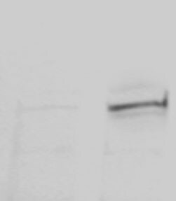
Western Blot: alpha-Smooth Muscle Actin Antibody [NB300-978]
Western Blot: alpha-Smooth Muscle Actin Antibody [NB300-978] - (1ug/ml) staining of Mouse Duodenum lysate (35ug protein in RIPA buffer). Detected by chemiluminescence.![Immunocytochemistry/ Immunofluorescence: alpha-Smooth Muscle Actin Antibody [NB300-978]](https://resources.rndsystems.com/images/products/alpha-Smooth-Muscle-Actin-Antibody-Immunocytochemistry-Immunofluorescence-NB300-978-img0006.jpg "Immunocytochemistry/ Immunofluorescence: alpha-Smooth Muscle Actin Antibody [NB300-978]")
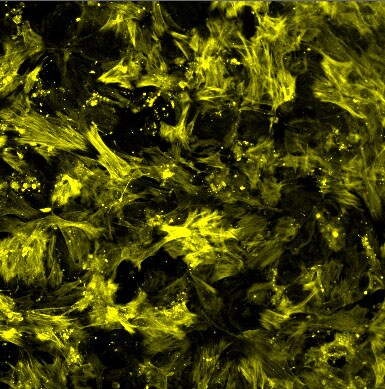
Immunocytochemistry/ Immunofluorescence: alpha-Smooth Muscle Actin Antibody [NB300-978]
Immunocytochemistry/Immunofluorescence: alpha-Smooth Muscle Actin Antibody [NB300-978] - SMA antibody staining on mouse fibroblasts. Primary antibody at 1:1000, incubation at 4C overnight. ICC/IF image submitted by a verified customer review.![Immunohistochemistry-Frozen: alpha-Smooth Muscle Actin Antibody [NB300-978]](https://resources.rndsystems.com/images/products/alpha-Smooth-Muscle-Actin-Antibody-Immunohistochemistry-Frozen-NB300-978-img0003.jpg "Immunohistochemistry-Frozen: alpha-Smooth Muscle Actin Antibody [NB300-978]")
Immunohistochemistry-Frozen: alpha-Smooth Muscle Actin Antibody [NB300-978]
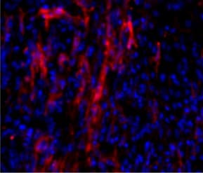
Immunohistochemistry-Frozen: alpha-Smooth Muscle Actin Antibody [NB300-978] - Infarcted mouse heart frozen tissue section immunostained with alpha-Smooth Muscle Actin Antibody. IHC-Fr image submitted by a verified customer review.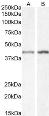
alpha-Smooth Muscle Actin Antibody [NB300-978] - (1ug/ml) staining of HeLa (A) and (2ug/ml) NIH3T3 (B) cell lysate (35ug protein in RIPA buffer). Detected by chemiluminescence.

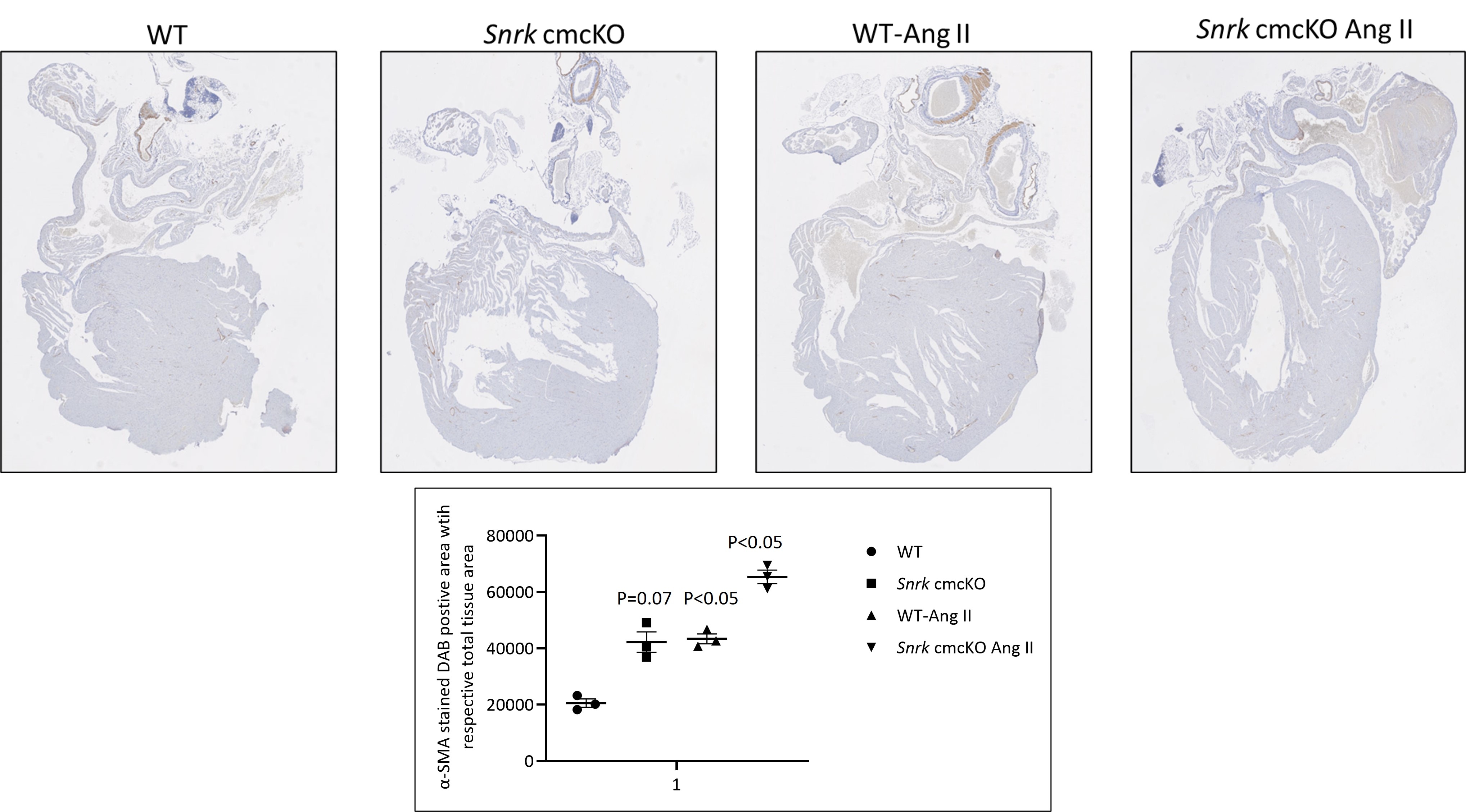
Immunohistochemistry-Paraffin: Goat Polyclonal alpha-Smooth Muscle Actin Antibody [NB300-978]
Immunohistochemistry-Paraffin: Goat Polyclonal alpha-Smooth Muscle Actin Antibody [NB300-978] - Analysis of alpha-Smooth Muscle Actin Antibody on mouse heart tissue. Image from a verified customer review.
Western Blot: alpha-Smooth Muscle Actin Antibody [NB300-978] -
Effect of Nebivolol on TGF-beta 1, p-Smad2/3, and alpha -SMA proteins expression. (A) representative western blot membranes of TGF-beta 1, Smad2/3, p-Smad2/3, alpha -SMA, and beta -actin proteins for all studied groups. (B–D) Expressions of TGF-beta 1, p-Smad2/3/t-Smad2/3, and alpha -SMA proteins were represented densitometrically using bands in (A). Bars are represented as mean +/- SEM. The Tukey-Kramar test was used to analyze the significant differences between groups after conducting a one-way ANOVA test, where ###p < 0.001, compared to the sham group, *p < 0.05,**p < 0.01, and ***p < 0.001, compared to CLP group, $$p < 0.01, and $$$p < 0.001, compared to CLP + Neb4 group. Image collected and cropped by CiteAb from the following open publication (https://www.nature.com/articles/s41598-024-64577-5), licensed under a CC-BY license. Not internally tested by Novus Biologicals.
Western Blot: alpha-Smooth Muscle Actin Antibody [NB300-978] -
Activation of multiple signaling pathways in TEC challenged by cytokines. Primary human renal TECs were treated with the cytokine cocktail. (A, B) Western blot of PI3K–Akt pathway, NFkb pathway, and Erk signaling pathway in cells treated with cytokine cocktail. (C) Densitometric quantitation of AktS473, pNFkB65, ErkTh202/Ty204, and pTenS380. All Western blots are representative of two independent experiments. Image collected and cropped by CiteAb from the following open publication (https://pubmed.ncbi.nlm.nih.gov/34305900), licensed under a CC-BY license. Not internally tested by Novus Biologicals.
Western Blot: alpha-Smooth Muscle Actin Antibody [NB300-978] -
Western blot analysis of function protein expression on primary human renal cortical epithelial cells challenged with cytokines. Primary human renal tubular epithelial cells are challenged with cytokine cocktail (15nM IFN gamma, 6nM TNF alpha, and 3nM IL1 beta ). (A, B) Expression of Snail, E-Cad, TJP1, SMA. AhR, IDO, KMO, KY, MHCI & II. (C, D) Densitometric quantitation of protein in (A, B). All Western blots are representative of at least three independent experiments. *P < 0.05, ***P < 0.0001 versus to cells without treatment, analysis is multiple t-tests, pairwise comparison of individual time-point to control indicated same p-value. Image collected and cropped by CiteAb from the following open publication (https://pubmed.ncbi.nlm.nih.gov/34305900), licensed under a CC-BY license. Not internally tested by Novus Biologicals.
Western Blot: alpha-Smooth Muscle Actin Antibody [NB300-978] -
Activation of multiple signaling pathways in TEC challenged by cytokines. Primary human renal TECs were treated with the cytokine cocktail. (A, B) Western blot of PI3K–Akt pathway, NFkb pathway, and Erk signaling pathway in cells treated with cytokine cocktail. (C) Densitometric quantitation of AktS473, pNFkB65, ErkTh202/Ty204, and pTenS380. All Western blots are representative of two independent experiments. Image collected and cropped by CiteAb from the following open publication (https://pubmed.ncbi.nlm.nih.gov/34305900), licensed under a CC-BY license. Not internally tested by Novus Biologicals.Applications for alpha-Smooth Muscle Actin Antibody
Application
Recommended Usage
Peptide ELISA
Detection limit 1:32000
Western Blot
0.3 - 2 ug/mL
Application Notes
Approx 45 kDa band observed in HeLa, NIH3T3, Mouse Duodenum and Human Heart lyates, and also in preliminary testing of Rat Duodenum lysates (calculated MW of 42 kDa according to NP_001604 and NP_031418.1). Alpha-Smooth Muscle Actin Antibody is validated for IHC-Fr, ICC/IF from verified customer reviews.
Reviewed Applications
Read 8 reviews rated 5 using NB300-978 in the following applications:
Formulation, Preparation, and Storage
Purification
Immunogen affinity purified
Formulation
Tris saline (20 mM Tris pH 7.3, 150 mM NaCl), 0.5% BSA
Preservative
0.02% Sodium Azide
Concentration
0.5 mg/ml
Shipping
The product is shipped with polar packs. Upon receipt, store it immediately at the temperature recommended below.
Stability & Storage
Store at -20C. Avoid freeze-thaw cycles.
Background: alpha-Smooth Muscle Actin
ACTA2 encodes alpha Smooth Muscle Actin and is also known as alpha-actin, alpha-actin-2, aortic smooth muscle or alpha smooth muscle actin (alpha-SMA, SMactin, alpha-SM-actin, ASMA). Alpha Smooth Muscle Actin is frequently used as a marker of smooth muscle differentiation and has a theoretical molecular weight of 42 kDa. Smooth muscle alpha actin is one of a few genes whose expression is predominantly expressed in vascular smooth muscle cells that is used to study myofibroblasts and fibrosis (1). Excessive accumulation of alpha smooth muscle actin-positive (ACTA2+) activated myofibroblasts is observed in Idiopathic pulmonary fibrosis (2). Expression of smooth muscle alpha actin is regulated by cell proliferation and is altered in pathological conditions including atherosclerosis and is common in metastatic cancers (1,3).
References
1. Chakraborty, R., Saddouk, F. Z., Carrao, A. C., Krause, D. S., Greif, D. M., & Martin, K. A. (2019). Promoters to Study Vascular Smooth Muscle. Arterioscler Thromb Vasc Biol, 39(4), 603-612. doi:10.1161/atvbaha.119.312449
2.El Agha, E., Moiseenko, A., Kheirollahi, V., De Langhe, S., Crnkovic, S., Kwapiszewska, G.,... Bellusci, S. (2017). Two-Way Conversion between Lipogenic and Myogenic Fibroblastic Phenotypes Marks the Progression and Resolution of Lung Fibrosis. Cell Stem Cell, 20(2), 261-273.e263. doi:10.1016/j.stem.2016.10.004
3. Chen, Y. C., Gonzalez, M. E., Burman, B., Zhao, X., Anwar, T., Tran, M.,... Kleer, C. G. (2019). Mesenchymal Stem/Stromal Cell Engulfment Reveals Metastatic Advantage in Breast Cancer. Cell Rep, 27(13), 3916-3926.e3915. doi:10.1016/j.celrep.2019.05.084
Long Name
Actin, Alpha 2, Smooth Muscle, Aorta
Alternate Names
AAT6, ACTA2, Actin alpha 2, ACTSA, ACTVS, alphaSmooth Muscle Actin, MYMY5, SMA
Entrez Gene IDs
59 (Human)
Gene Symbol
ACTA2
UniProt
Additional alpha-Smooth Muscle Actin Products
Product Documents for alpha-Smooth Muscle Actin Antibody
Certificate of Analysis
To download a Certificate of Analysis, please enter a lot or batch number in the search box below.
Product Specific Notices for alpha-Smooth Muscle Actin Antibody
This product is for research use only and is not approved for use in humans or in clinical diagnosis. Primary Antibodies are guaranteed for 1 year from date of receipt.
Related Research Areas
Citations for alpha-Smooth Muscle Actin Antibody
 Powered by Bioz
Customer Reviews for alpha-Smooth Muscle Actin Antibody (8)
5 out of 5
8 Customer Ratings
Have you used alpha-Smooth Muscle Actin Antibody?
Submit a review and receive an Amazon gift card!
$25/€18/£15/$25CAN/¥2500 Yen for a review with an image
$10/€7/£6/$10CAN/¥1110 Yen for a review without an image
Submit a review
Customer Images
Showing
1
-
5 of
8 reviews
Showing All
Filter By:
-
Application: Immunohistochemistry-ParaffinSample Tested: Mouse heartSpecies: MouseVerified Customer | Posted 12/20/2024Mouse heart IHC

-
Application: ImmunocytochemistrySample Tested: fibroblastsSpecies: MouseVerified Customer | Posted 09/15/2021SMA antibody staining on mouse fibroblasts.Primary antibody 1:1000 concentration, incubation overnight at 4C.
-
Application: Western BlotSample Tested: fibroblastsSpecies: HumanVerified Customer | Posted 03/31/2021left: control fibroblasts Right: SMA overexpression
-
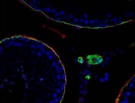
Application: ImmunocytochemistrySample Tested: Adult testisSpecies: MouseVerified Customer | Posted 01/14/2021Green channel is sma, colocalize with CD34(red).primary antibody 1:200 dilution.
-
Application: Immunohistochemistry-FrozenSample Tested: Mouse heart frozen sectionsSpecies: MouseVerified Customer | Posted 01/30/2020Infarcted mouse heart immunostained with SMA antibodyMouse frozen heart after myocardial infarction PFA fixed Primary antibody 1:200 Secondary antibody 1:500
-
Application: ImmunocytochemistrySample Tested: fibroblastsSpecies: HumanVerified Customer | Posted 10/09/2019Green color is SMA by using NB300-978 antibody in fibroblast tissue.
-
Application: Immunohistochemistry-FrozenSample Tested: corneal stromal cellsSpecies: RabbitVerified Customer | Posted 10/23/2018
-
Application: ImmunocytochemistrySample Tested: Eye lens tissueSpecies: RabbitVerified Customer | Posted 05/31/2018
There are no reviews that match your criteria.
Protocols
Find general support by application which include: protocols, troubleshooting, illustrated assays, videos and webinars.
- Antigen Retrieval Protocol (PIER)
- Antigen Retrieval for Frozen Sections Protocol
- Appropriate Fixation of IHC/ICC Samples
- Cellular Response to Hypoxia Protocols
- Chromogenic IHC Staining of Formalin-Fixed Paraffin-Embedded (FFPE) Tissue Protocol
- Chromogenic Immunohistochemistry Staining of Frozen Tissue
- ClariTSA™ Fluorophore Kits
- Detection & Visualization of Antibody Binding
- ELISA Sample Preparation & Collection Guide
- ELISA Troubleshooting Guide
- Fluorescent IHC Staining of Frozen Tissue Protocol
- Graphic Protocol for Heat-induced Epitope Retrieval
- Graphic Protocol for the Preparation and Fluorescent IHC Staining of Frozen Tissue Sections
- Graphic Protocol for the Preparation and Fluorescent IHC Staining of Paraffin-embedded Tissue Sections
- Graphic Protocol for the Preparation of Gelatin-coated Slides for Histological Tissue Sections
- How to Run an R&D Systems DuoSet ELISA
- How to Run an R&D Systems Quantikine ELISA
- How to Run an R&D Systems Quantikine™ QuicKit™ ELISA
- ICC Cell Smear Protocol for Suspension Cells
- ICC Immunocytochemistry Protocol Videos
- ICC for Adherent Cells
- IHC Sample Preparation (Frozen sections vs Paraffin)
- Immunocytochemistry (ICC) Protocol
- Immunocytochemistry Troubleshooting
- Immunofluorescence of Organoids Embedded in Cultrex Basement Membrane Extract
- Immunofluorescent IHC Staining of Formalin-Fixed Paraffin-Embedded (FFPE) Tissue Protocol
- Immunohistochemistry (IHC) and Immunocytochemistry (ICC) Protocols
- Immunohistochemistry Frozen Troubleshooting
- Immunohistochemistry Paraffin Troubleshooting
- Preparing Samples for IHC/ICC Experiments
- Preventing Non-Specific Staining (Non-Specific Binding)
- Primary Antibody Selection & Optimization
- Protocol for Heat-Induced Epitope Retrieval (HIER)
- Protocol for Making a 4% Formaldehyde Solution in PBS
- Protocol for VisUCyte™ HRP Polymer Detection Reagent
- Protocol for the Fluorescent ICC Staining of Cell Smears - Graphic
- Protocol for the Fluorescent ICC Staining of Cultured Cells on Coverslips - Graphic
- Protocol for the Preparation & Fixation of Cells on Coverslips
- Protocol for the Preparation and Chromogenic IHC Staining of Frozen Tissue Sections
- Protocol for the Preparation and Chromogenic IHC Staining of Frozen Tissue Sections - Graphic
- Protocol for the Preparation and Chromogenic IHC Staining of Paraffin-embedded Tissue Sections
- Protocol for the Preparation and Chromogenic IHC Staining of Paraffin-embedded Tissue Sections - Graphic
- Protocol for the Preparation and Fluorescent ICC Staining of Cells on Coverslips
- Protocol for the Preparation and Fluorescent ICC Staining of Non-adherent Cells
- Protocol for the Preparation and Fluorescent ICC Staining of Stem Cells on Coverslips
- Protocol for the Preparation and Fluorescent IHC Staining of Frozen Tissue Sections
- Protocol for the Preparation and Fluorescent IHC Staining of Paraffin-embedded Tissue Sections
- Protocol for the Preparation of Gelatin-coated Slides for Histological Tissue Sections
- Protocol for the Preparation of a Cell Smear for Non-adherent Cell ICC - Graphic
- Quantikine HS ELISA Kit Assay Principle, Alkaline Phosphatase
- Quantikine HS ELISA Kit Principle, Streptavidin-HRP Polymer
- R&D Systems Quality Control Western Blot Protocol
- Sandwich ELISA (Colorimetric) – Biotin/Streptavidin Detection Protocol
- Sandwich ELISA (Colorimetric) – Direct Detection Protocol
- TUNEL and Active Caspase-3 Detection by IHC/ICC Protocol
- The Importance of IHC/ICC Controls
- Troubleshooting Guide: ELISA
- Troubleshooting Guide: Immunohistochemistry
- Troubleshooting Guide: Western Blot Figures
- Western Blot Conditions
- Western Blot Protocol
- Western Blot Protocol for Cell Lysates
- Western Blot Troubleshooting
- Western Blot Troubleshooting Guide
- View all Protocols, Troubleshooting, Illustrated assays and Webinars
FAQs for alpha-Smooth Muscle Actin Antibody
Showing
1
-
2 of
2 FAQs
Showing All
-
Q: I do not see horse listed as a predicted or confirmed species. Would you be able to confirm if any of your alpha-SMA antibodies will react with equine samples?
A: We have not directly tested equine samples nor do we have cited publications that have tested equine samples. Since alpha-Smooth Muscle Actin is a highly conserved sequence, these antibodies should react with most mammal species.
-
Q: Which antibodies would you recommend for the best performance in IHC?
A: NB300-978, NBP1-30894, NBP2-22120, NBP2-32808, NBP2-33006, and NBP2-34522 have the broadest IHC applications, as they additionally list IHC-Paraffin and IHC-Frozen. To narrow the list down further, we would recommend using either NB300-978 or NBP2-33006 since these products have the most ratings and cited publications for use in IHC methods. NB300-978 has IHC-Free-Floating listed as an application as well.
-
Q: I do not see horse listed as a predicted or confirmed species. Would you be able to confirm if any of your alpha-SMA antibodies will react with equine samples?
A: We have not directly tested equine samples nor do we have cited publications that have tested equine samples. Since alpha-Smooth Muscle Actin is a highly conserved sequence, these antibodies should react with most mammal species.
-
Q: Which antibodies would you recommend for the best performance in IHC?
A: NB300-978, NBP1-30894, NBP2-22120, NBP2-32808, NBP2-33006, and NBP2-34522 have the broadest IHC applications, as they additionally list IHC-Paraffin and IHC-Frozen. To narrow the list down further, we would recommend using either NB300-978 or NBP2-33006 since these products have the most ratings and cited publications for use in IHC methods. NB300-978 has IHC-Free-Floating listed as an application as well.
Loading...
Associated Pathways
