Androgen R/NR3C4 Antibody
Novus Biologicals | Catalog # NB100-1446
![Western Blot: Androgen R/NR3C4 Antibody [NB100-1446]](https://resources.rndsystems.com/images/products/Androgen-R-NR3C4-Antibody-Western-Blot-NB100-1446-img0008.jpg "Western Blot: Androgen R/NR3C4 Antibody [NB100-1446]")
Loading...
Key Product Details
Species Reactivity
Validated:
Human, Equine, Feline
Predicted:
Bovine (100%), Canine (100%), Mouse (100%), Porcine (100%), Rat (100%). Backed by our 100% Guarantee.
Applications
Immunohistochemistry, Immunohistochemistry-Paraffin, Western Blot, Peptide ELISA, Immunocytochemistry/ Immunofluorescence, Chromatin Immunoprecipitation (ChIP)
Label
Unconjugated
Antibody Source
Polyclonal Goat IgG
Loading...
Product Specifications
Immunogen
Peptide with sequence EVQLGLGRVYPRPPSC corresponding to N-Terminus according to NP_000035.2.
Reactivity Notes
Feline and Equine reactivity reported from a verified customer review.
Localization
Nuclear
Specificity
This antibody is expected to recognize isoform 1 (NP_000035.2) only.
Clonality
Polyclonal
Host
Goat
Isotype
IgG
Theoretical MW
99 kDa.
Disclaimer note: The observed molecular weight of the protein may vary from the listed predicted molecular weight due to post translational modifications, post translation cleavages, relative charges, and other experimental factors.
Disclaimer note: The observed molecular weight of the protein may vary from the listed predicted molecular weight due to post translational modifications, post translation cleavages, relative charges, and other experimental factors.
Scientific Data Images for Androgen R/NR3C4 Antibody
Western Blot: Androgen R/NR3C4 Antibody [NB100-1446]
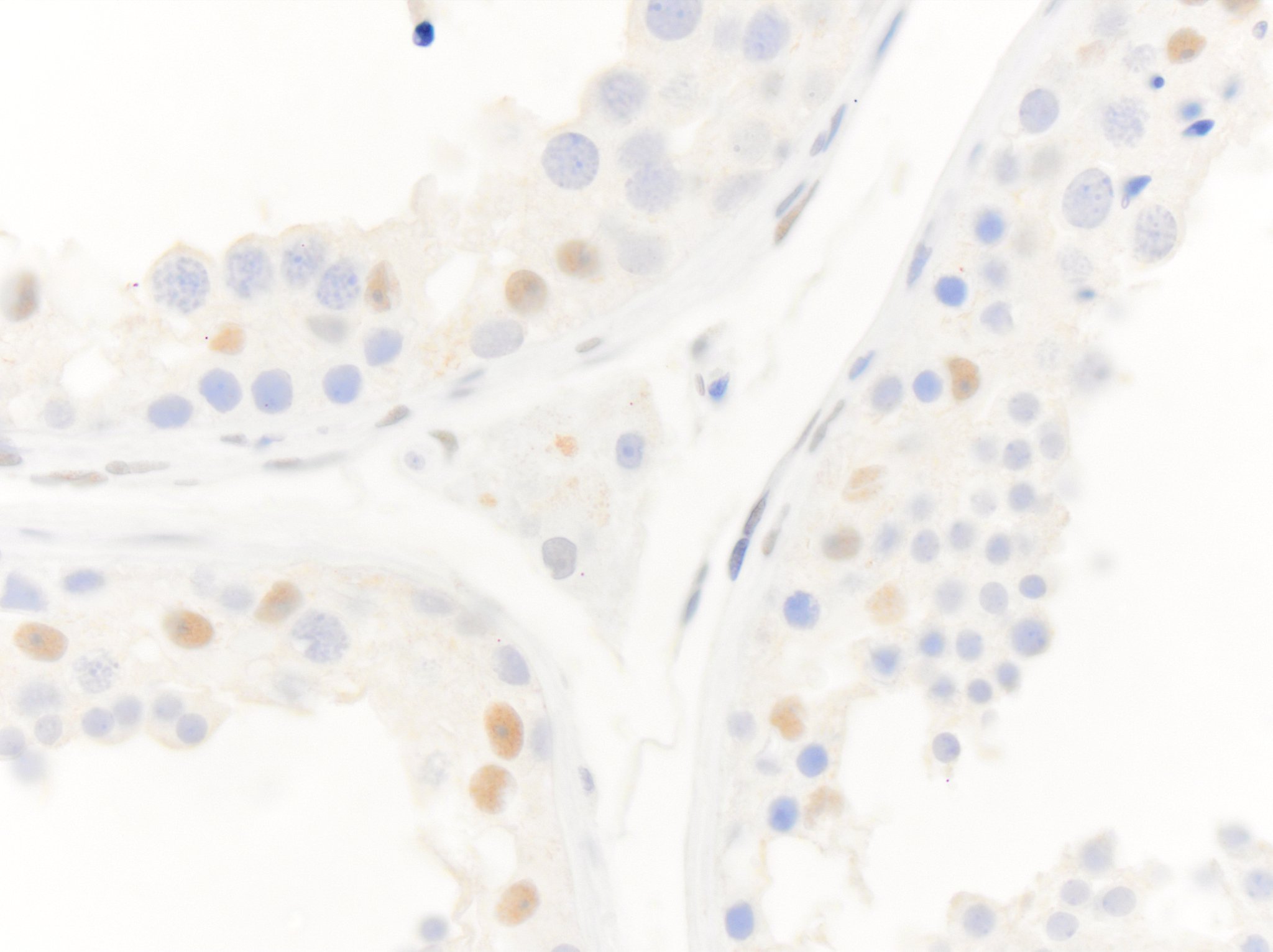
Western Blot: Androgen R/NR3C4 Antibody [NB100-1446] - Staining of Human Cerebellum (A) and (0.1ug/ml) negative control Pancreas (B) lysate (35ug protein in RIPA buffer). Detected by chemiluminescence.![Immunocytochemistry/ Immunofluorescence: Androgen R/NR3C4 Antibody [NB100-1446]](https://resources.rndsystems.com/images/products/Androgen-R-NR3C4-Antibody-Immunocytochemistry-Immunofluorescence-NB100-1446-img0010.jpg "Immunocytochemistry/ Immunofluorescence: Androgen R/NR3C4 Antibody [NB100-1446]")
Immunocytochemistry/ Immunofluorescence: Androgen R/NR3C4 Antibody [NB100-1446]
Immunocytochemistry/Immunofluorescence: Androgen R/NR3C4 Antibody [NB100-1446] - Analysis of paraformaldehyde fixed U2OS cells, permeabilized with 0.15% Triton. Primary incubation 1hr (10ug/ml) followed by Alexa Fluor 488 secondary antibody (2ug/ml), showing Mitochondrial/cytoplasmic staining. The nuclear stain is DAPI (blue). Negative control: Unimmunized goat IgG (10ug/ml) followed by Alexa Fluor 488 secondary antibody (2ug/ml).![Immunohistochemistry-Paraffin: Androgen R/NR3C4 Antibody [NB100-1446]](https://resources.rndsystems.com/images/products/Androgen-R-NR3C4-Antibody-Immunohistochemistry-Paraffin-NB100-1446-img0006.jpg "Immunohistochemistry-Paraffin: Androgen R/NR3C4 Antibody [NB100-1446]")
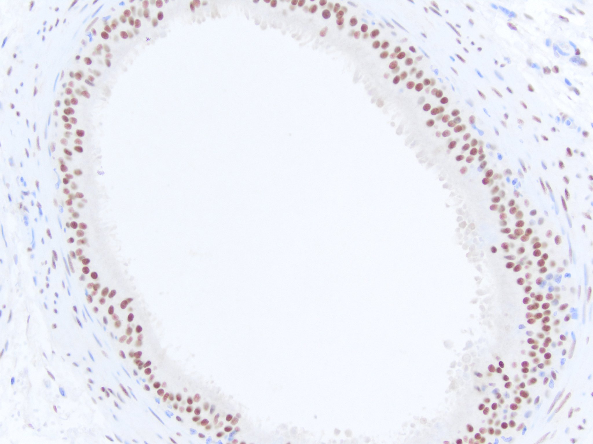
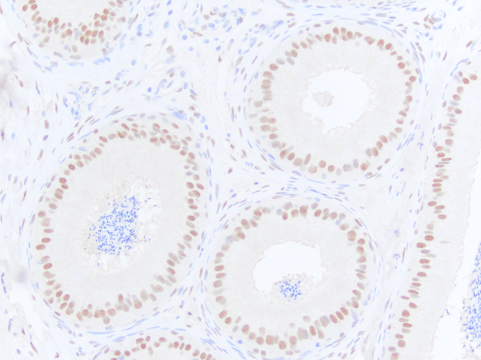
Immunohistochemistry-Paraffin: Androgen R/NR3C4 Antibody [NB100-1446]
Immunohistochemistry-Paraffin: Androgen R/NR3C4 Antibody [NB100-1446] - (2ug/ml) staining of paraffin embedded Human Prostate. Steamed antigen retrieval with citrate buffer pH 6, AP-staining.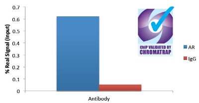
Chromatin Immunoprecipitation: Androgen R/NR3C4 Antibody [NB100-1446] - (Protein G) measuring FKBP5 enrichment.
![Immunocytochemistry/ Immunofluorescence: Androgen R/NR3C4 Antibody [NB100-1446]](https://resources.rndsystems.com/images/products/Androgen-R-NR3C4-Antibody-Immunocytochemistry-Immunofluorescence-NB100-1446-img0009.jpg "Immunocytochemistry/ Immunofluorescence: Androgen R/NR3C4 Antibody [NB100-1446]")
Immunocytochemistry/ Immunofluorescence: Androgen R/NR3C4 Antibody [NB100-1446]
Immunocytochemistry/Immunofluorescence: Androgen R/NR3C4 Antibody [NB100-1446] - Analysis of paraformaldehyde fixed MCF7 cells, permeabilized with 0.15% Triton. Primary incubation 1hr (10ug/ml) followed by Alexa Fluor 488 secondary antibody (2ug/ml), showing Mitochondrial/cytoplasmic staining. The nuclear stain is DAPI (blue). Negative control: Unimmunized goat IgG (10ug/ml) followed by Alexa Fluor 488 secondary antibody (2ug/ml).
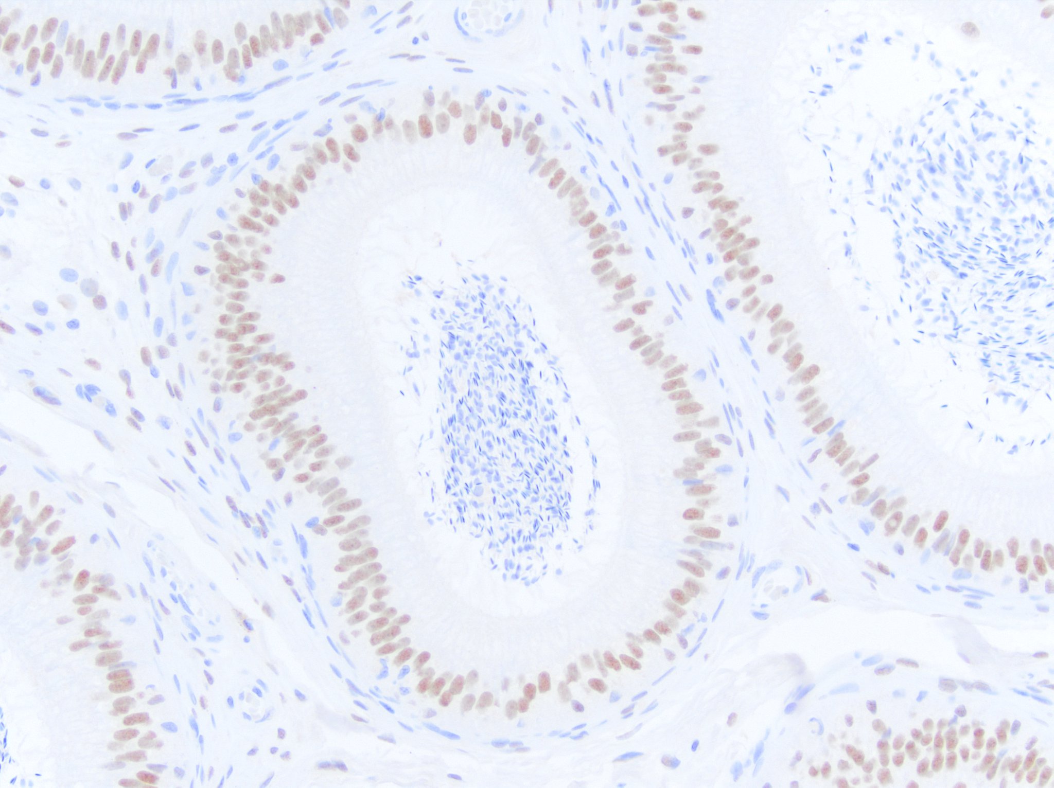
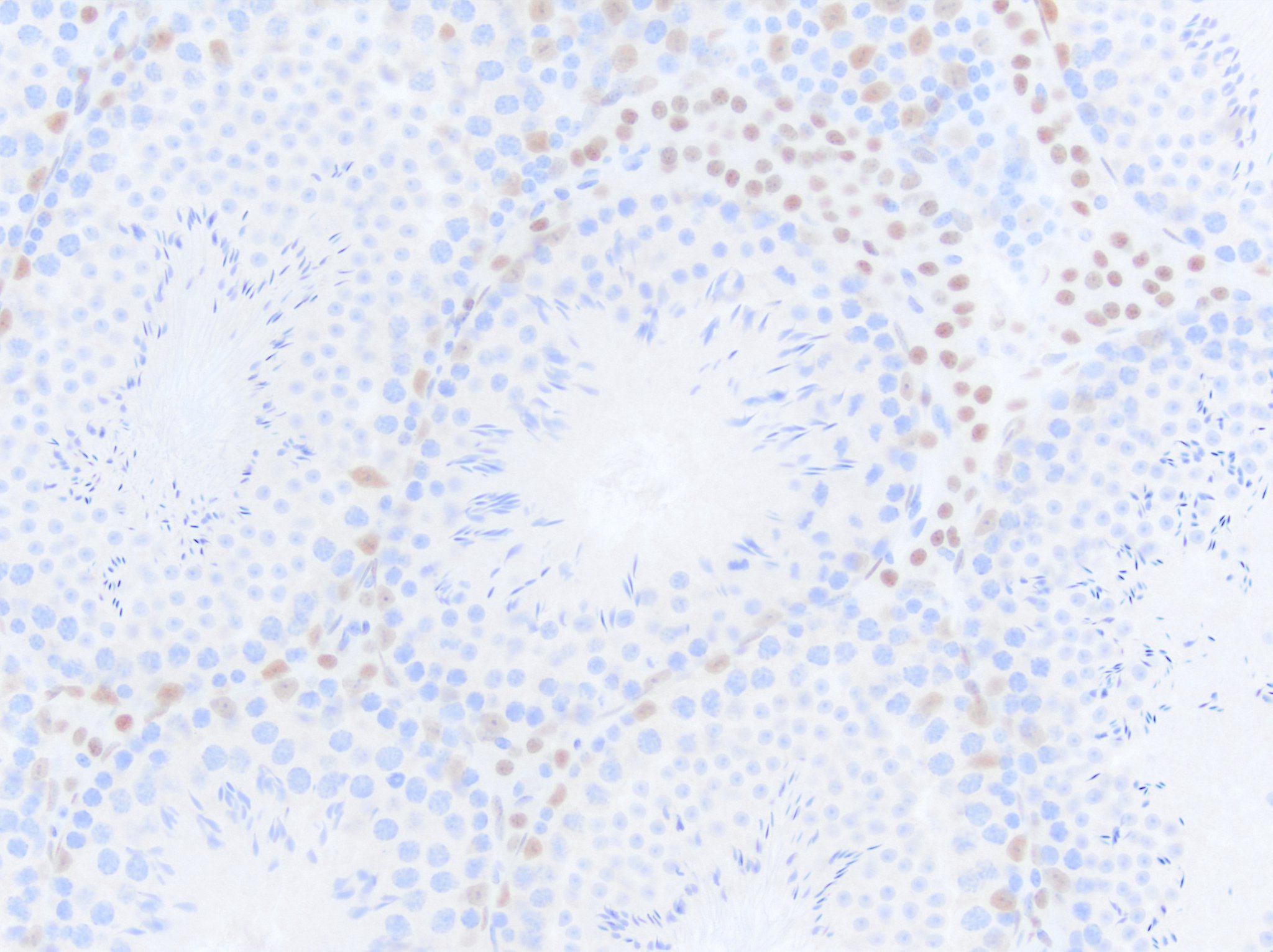
Immunohistochemistry-Paraffin: Goat Polyclonal Androgen R/NR3C4 Antibody [IMGENEX: IMG-3238] [NB100-1446] -
Immunohistochemistry-Paraffin: Goat Polyclonal Androgen R/NR3C4 Antibody [IMGENEX: IMG-3238] [NB100-1446] - Androgen receptor immunoreactivity in equine epididymis. NB100-1446 was diluted to 1 ug/mL and left on tissue sections for 30 min at room temperature. Immunoreactivity was best demonstrated with heat induced epitope retrieval using an EDTA based solution. Image from a verified customer review.
Immunohistochemistry-Paraffin: Goat Polyclonal Androgen R/NR3C4 Antibody [IMGENEX: IMG-3238] [NB100-1446] -
Immunohistochemistry-Paraffin: Goat Polyclonal Androgen R/NR3C4 Antibody [IMGENEX: IMG-3238] [NB100-1446] - Androgen receptor immunoreactivity in canine epididymis. NB100-1446 was diluted to 1 ug/mL and left on tissue sections for 30m at room temperature. Immunoreactivity was best demonstrated with heat induced epitope retrieval in an EDTA based buffer. Image from a verified customer review.
Immunohistochemistry-Paraffin: Goat Polyclonal Androgen R/NR3C4 Antibody [IMGENEX: IMG-3238] [NB100-1446] -
Immunohistochemistry-Paraffin: Goat Polyclonal Androgen R/NR3C4 Antibody [IMGENEX: IMG-3238] [NB100-1446] - Androgen receptor immunoreactivity in feline epididymis. NB100-1446 was diluted to 1 ug/mL and left on tissue sections for 30m at room temperature. Immunoreactivity was best demonstrated with heat induced epitope retrieval in an EDTA based buffer. Image from a verified customer review.
Immunohistochemistry-Paraffin: Goat Polyclonal Androgen R/NR3C4 Antibody [IMGENEX: IMG-3238] [NB100-1446] -
Immunohistochemistry-Paraffin: Goat Polyclonal Androgen R/NR3C4 Antibody [IMGENEX: IMG-3238] [NB100-1446] - Androgen receptor immunoreactivity in mouse testis. NB100-1446 was diluted to 1 ug/mL and left on tissue sections for 30m at room temperature. Immunoreactivity was best demonstrated with heat induced epitope retrieval in an EDTA based buffer. Image from a verified customer review.
Immunohistochemistry-Paraffin: Goat Polyclonal Androgen R/NR3C4 Antibody [IMGENEX: IMG-3238] [NB100-1446] -
Immunohistochemistry-Paraffin: Goat Polyclonal Androgen R/NR3C4 Antibody [IMGENEX: IMG-3238] [NB100-1446] - Androgen receptor immunoreactivity in human testis. NB100-1446 was diluted to 1ug per mL and left on tissue sections for 30m at room temperature. Immunoreactivity was best demonstrated with heat-induced epitope retrieval in an EDTA-based solution. Image from a verified customer review.Applications for Androgen R/NR3C4 Antibody
Application
Recommended Usage
Immunocytochemistry/ Immunofluorescence
10 ug/ml
Immunohistochemistry
2 - 3 ug/ml
Immunohistochemistry-Paraffin
2-3 ug/ml
Peptide ELISA
Detection limit 1:64000
Western Blot
1-0.5ug/ml
Application Notes
ChIP: A 6-fold increased presence of the FKBP5 locus observed when comparing the precipitations using Goat Anti-Androgen Receptor with non-specific goat IgG to bring down Androgen Receptor in a chromatin lysate from DHT-stimulated HEC50 cells.
WB: Approx. 95 kDa band observed in human brain and human heart lysates (calculated MW of 99 kDa band according to NP_000035).
IHC-P: Human prostate shows nuclear staining in the secretory cells of the gland.
WB: Approx. 95 kDa band observed in human brain and human heart lysates (calculated MW of 99 kDa band according to NP_000035).
IHC-P: Human prostate shows nuclear staining in the secretory cells of the gland.
Reviewed Applications
Read 5 reviews rated 4.8 using NB100-1446 in the following applications:
Formulation, Preparation, and Storage
Purification
Immunogen affinity purified
Formulation
Tris saline (20 mM Tris pH 7.3, 150 mM NaCl), 0.5% BSA
Preservative
0.02% Sodium Azide
Concentration
0.5 mg/ml
Shipping
The product is shipped with polar packs. Upon receipt, store it immediately at the temperature recommended below.
Stability & Storage
Store at -20C. Avoid freeze-thaw cycles.
Background: Androgen R/NR3C4
Long Name
Androgen Receptor
Alternate Names
AIS, AndrogenR, AR, DHTR, NR3C4, SBMA, SMAX1
Gene Symbol
AR
UniProt
Additional Androgen R/NR3C4 Products
Product Documents for Androgen R/NR3C4 Antibody
Certificate of Analysis
To download a Certificate of Analysis, please enter a lot or batch number in the search box below.
Product Specific Notices for Androgen R/NR3C4 Antibody
This product is for research use only and is not approved for use in humans or in clinical diagnosis. Primary Antibodies are guaranteed for 1 year from date of receipt.
Related Research Areas
Citations for Androgen R/NR3C4 Antibody
 Powered by Bioz
Customer Reviews for Androgen R/NR3C4 Antibody (5)
4.8 out of 5
5 Customer Ratings
Have you used Androgen R/NR3C4 Antibody?
Submit a review and receive an Amazon gift card!
$25/€18/£15/$25CAN/¥2500 Yen for a review with an image
$10/€7/£6/$10CAN/¥1110 Yen for a review without an image
Submit a review
Customer Images
Showing
1
-
5 of
5 reviews
Showing All
Filter By:
-
Application: Immunohistochemistry-ParaffinSample Tested: TestisSpecies: HumanVerified Customer | Posted 11/17/2023Androgen receptor immunoreactivity in human testis. NB100-1446 was diluted to 1ug per mL and left on tissue sections for 30m at room temperature. Immunoreactivity was best demonstrated with heat-induced epitope retrieval in an EDTA-based solution.

-
Application: Immunohistochemistry-ParaffinSample Tested: EpididymisSpecies: EquineVerified Customer | Posted 10/27/2023Androgen receptor immunoreactivity in horse epididymis. NB100 1446 was diluted to 1ug per mL and left on tissue sections for 30min at room temperature. Immunoreactivity was best demonstrated with heat induced epitope retrieval using an EDTA based solution
Bio-Techne ResponseThis review was submitted through the legacy Novus Innovators Program, reflecting a new species or application tested on a primary antibody.
-
Application: Immunohistochemistry-ParaffinSample Tested: EpididymisSpecies: CanineVerified Customer | Posted 10/27/2023Androgen receptor immunoreactivity in dog epididymis. NB100 1446 was diluted to 1ug per mL and left on tissue sections for 30m at room temperature. Immunoreactivity was best demonstrated with heat induced epitope retrieval in an EDTA based buffer.
-
Application: Immunohistochemistry-ParaffinSample Tested: EpididymisSpecies: FelineVerified Customer | Posted 10/27/2023Androgen receptor immunoreactivity in cat epididymis. NB100 1446 was diluted to 1ug per mL and left on tissue sections for 30m at room temperature. Immunoreactivity was best demonstrated with heat induced epitope retrieval in an EDTA based buffer.
Bio-Techne ResponseThis review was submitted through the legacy Novus Innovators Program, reflecting a new species or application tested on a primary antibody.
-
Application: Immunohistochemistry-ParaffinSample Tested: TestisSpecies: MouseVerified Customer | Posted 10/27/2023Androgen receptor immunoreactivity in mouse testis. NB100 1446 was diluted to 1ug per mL and left on tissue sections for 30m at room temperature. Immunoreactivity was best demonstrated with heat induced epitope retrieval in an EDTA based buffer
There are no reviews that match your criteria.
Protocols
Find general support by application which include: protocols, troubleshooting, illustrated assays, videos and webinars.
- Antigen Retrieval Protocol (PIER)
- Antigen Retrieval for Frozen Sections Protocol
- Appropriate Fixation of IHC/ICC Samples
- Cellular Response to Hypoxia Protocols
- ChIP Protocol Video
- Chromatin Immunoprecipitation (ChIP) Protocol
- Chromatin Immunoprecipitation Protocol
- Chromogenic IHC Staining of Formalin-Fixed Paraffin-Embedded (FFPE) Tissue Protocol
- Chromogenic Immunohistochemistry Staining of Frozen Tissue
- ClariTSA™ Fluorophore Kits
- Detection & Visualization of Antibody Binding
- ELISA Sample Preparation & Collection Guide
- ELISA Troubleshooting Guide
- Fluorescent IHC Staining of Frozen Tissue Protocol
- Graphic Protocol for Heat-induced Epitope Retrieval
- Graphic Protocol for the Preparation and Fluorescent IHC Staining of Frozen Tissue Sections
- Graphic Protocol for the Preparation and Fluorescent IHC Staining of Paraffin-embedded Tissue Sections
- Graphic Protocol for the Preparation of Gelatin-coated Slides for Histological Tissue Sections
- How to Run an R&D Systems DuoSet ELISA
- How to Run an R&D Systems Quantikine ELISA
- How to Run an R&D Systems Quantikine™ QuicKit™ ELISA
- ICC Cell Smear Protocol for Suspension Cells
- ICC Immunocytochemistry Protocol Videos
- ICC for Adherent Cells
- IHC Sample Preparation (Frozen sections vs Paraffin)
- Immunocytochemistry (ICC) Protocol
- Immunocytochemistry Troubleshooting
- Immunofluorescence of Organoids Embedded in Cultrex Basement Membrane Extract
- Immunofluorescent IHC Staining of Formalin-Fixed Paraffin-Embedded (FFPE) Tissue Protocol
- Immunohistochemistry (IHC) and Immunocytochemistry (ICC) Protocols
- Immunohistochemistry Frozen Troubleshooting
- Immunohistochemistry Paraffin Troubleshooting
- Preparing Samples for IHC/ICC Experiments
- Preventing Non-Specific Staining (Non-Specific Binding)
- Primary Antibody Selection & Optimization
- Protocol for Heat-Induced Epitope Retrieval (HIER)
- Protocol for Making a 4% Formaldehyde Solution in PBS
- Protocol for VisUCyte™ HRP Polymer Detection Reagent
- Protocol for the Fluorescent ICC Staining of Cell Smears - Graphic
- Protocol for the Fluorescent ICC Staining of Cultured Cells on Coverslips - Graphic
- Protocol for the Preparation & Fixation of Cells on Coverslips
- Protocol for the Preparation and Chromogenic IHC Staining of Frozen Tissue Sections
- Protocol for the Preparation and Chromogenic IHC Staining of Frozen Tissue Sections - Graphic
- Protocol for the Preparation and Chromogenic IHC Staining of Paraffin-embedded Tissue Sections
- Protocol for the Preparation and Chromogenic IHC Staining of Paraffin-embedded Tissue Sections - Graphic
- Protocol for the Preparation and Fluorescent ICC Staining of Cells on Coverslips
- Protocol for the Preparation and Fluorescent ICC Staining of Non-adherent Cells
- Protocol for the Preparation and Fluorescent ICC Staining of Stem Cells on Coverslips
- Protocol for the Preparation and Fluorescent IHC Staining of Frozen Tissue Sections
- Protocol for the Preparation and Fluorescent IHC Staining of Paraffin-embedded Tissue Sections
- Protocol for the Preparation of Gelatin-coated Slides for Histological Tissue Sections
- Protocol for the Preparation of a Cell Smear for Non-adherent Cell ICC - Graphic
- Quantikine HS ELISA Kit Assay Principle, Alkaline Phosphatase
- Quantikine HS ELISA Kit Principle, Streptavidin-HRP Polymer
- R&D Systems Quality Control Western Blot Protocol
- Sandwich ELISA (Colorimetric) – Biotin/Streptavidin Detection Protocol
- Sandwich ELISA (Colorimetric) – Direct Detection Protocol
- TUNEL and Active Caspase-3 Detection by IHC/ICC Protocol
- The Importance of IHC/ICC Controls
- Troubleshooting Guide: ELISA
- Troubleshooting Guide: Immunohistochemistry
- Troubleshooting Guide: Western Blot Figures
- Western Blot Conditions
- Western Blot Protocol
- Western Blot Protocol for Cell Lysates
- Western Blot Troubleshooting
- Western Blot Troubleshooting Guide
- View all Protocols, Troubleshooting, Illustrated assays and Webinars
Loading...
Associated Pathways
