B7-1/CD80 Antibody (62N3G8) - BSA Free
Novus Biologicals | Catalog # NBP2-25255
Key Product Details
Species Reactivity
Validated:
Human, Mouse
Cited:
Human, Mouse
Applications
Validated:
Multiplex Immunofluorescence, Immunohistochemistry, Immunohistochemistry-Paraffin, Western Blot, Flow Cytometry, COMET
Cited:
Immunohistochemistry-Paraffin, Western Blot
Label
Unconjugated
Antibody Source
Monoclonal Mouse IgG2b Kappa Clone # 62N3G8
Format
BSA Free
Loading...
Product Specifications
Immunogen
Amino acids 102-223 of human CD80 were used as the immunogen for the antibody.
Reactivity Notes
Mouse reactivity reported in scientific literature (PMID: 27440778).
Clonality
Monoclonal
Host
Mouse
Isotype
IgG2b Kappa
Scientific Data Images for B7-1/CD80 Antibody (62N3G8) - BSA Free

Detection of CD80/B7-1 in Human Hodgkin’s Lymphoma via seqIF™ staining on COMET™
CD80/B7-1 was detected in immersion fixed paraffin-embedded sections of human Hodgkin's Lymphoma using Mouse Anti-Human CD80/B7-1, Monoclonal Antibody (Catalog #NBP2-25255) at 20ug/mL at 37°Celsius for 4 minutes. Before incubation with the primary antibody, tissue underwent an all-in-one dewaxing and antigen retrieval preprocessing using PreTreatment Module (PT Module) and Dewax and HIER Buffer H (pH 9; Epredia Catalog # TA-999-DHBH). Tissue was stained using the Alexa Fluor™ 647 Goat anti-Mouse IgG Secondary Antibody at 1:200 at 37 ° Celsius for 2 minutes. (Yellow; Lunaphore Catalog # DR647MS) and counterstained with DAPI (blue; Lunaphore Catalog # DR100). Specific staining was localized to the membrane. Protocol available in COMET™ Panel Builder.![Western Blot: B7-1/CD80 Antibody (62N3G8)BSA Free [NBP2-25255]](https://resources.rndsystems.com/images/products/B7-1-CD80-Antibody-62N3G8-Western-Blot-NBP2-25255-img0010.jpg "Western Blot: B7-1/CD80 Antibody (62N3G8)BSA Free [NBP2-25255]")
Western Blot: B7-1/CD80 Antibody (62N3G8)BSA Free [NBP2-25255]
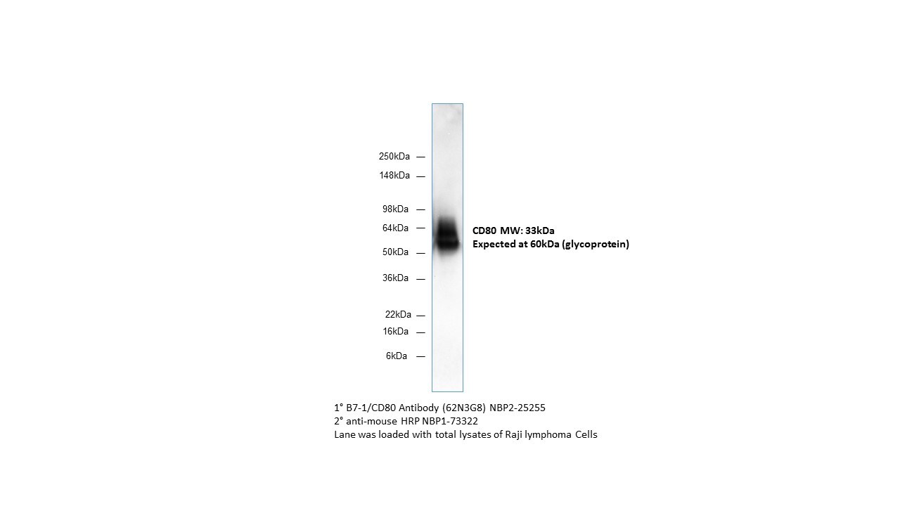
Western Blot: B7-1/CD80 Antibody (62N3G8) [NBP2-25255] - Analysis in human Raji lymphoma cells.![Immunohistochemistry-Paraffin: B7-1/CD80 Antibody (62N3G8) - BSA Free [NBP2-25255]](https://resources.rndsystems.com/images/products/B7-1-CD80-Antibody-62N3G8-Immunohistochemistry-Paraffin-NBP2-25255-img0002.jpg "Immunohistochemistry-Paraffin: B7-1/CD80 Antibody (62N3G8) - BSA Free [NBP2-25255]")
Immunohistochemistry-Paraffin: B7-1/CD80 Antibody (62N3G8) - BSA Free [NBP2-25255]
Immunohistochemistry-Paraffin: B7-1/CD80 Antibody (62N3G8) [NBP2-25255] - Human thymus stained with CD80 antibody at 5 ug/ml using peroxidase-conjugate and DAB chromogen. Note membrane and cytoplasmic staining.![Flow Cytometry: B7-1/CD80 Antibody (62N3G8) - BSA Free [NBP2-25255]](https://resources.rndsystems.com/images/products/B7-1-CD80-Antibody-62N3G8-Flow-Cytometry-NBP2-25255-img0009.jpg "Flow Cytometry: B7-1/CD80 Antibody (62N3G8) - BSA Free [NBP2-25255]")
Flow Cytometry: B7-1/CD80 Antibody (62N3G8) - BSA Free [NBP2-25255]
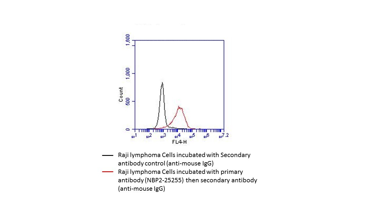
Flow Cytometry: B7-1/CD80 Antibody (62N3G8) [NBP2-25255] - Raji lymphoma cells. Image from verified customer review.![Western Blot: B7-1/CD80 Antibody (62N3G8)BSA Free [NBP2-25255]](https://resources.rndsystems.com/images/products/B7-1-CD80-Antibody-62N3G8-Western-Blot-NBP2-25255-img0003.jpg "Western Blot: B7-1/CD80 Antibody (62N3G8)BSA Free [NBP2-25255]")
Western Blot: B7-1/CD80 Antibody (62N3G8)BSA Free [NBP2-25255]
Western Blot: B7-1/CD80 Antibody (62N3G8) [NBP2-25255] - Analysis of CD80 in A) a partial recombinant protein and B) Daudi lysate using this antibody at 0.1 and 1 g/ml, respectively. Goat anti-mouse Ig HRP secondary antibody and PicoTect ECL substrate solution were used for this test.![Flow Cytometry: B7-1/CD80 Antibody (62N3G8) - BSA Free [NBP2-25255]](https://resources.rndsystems.com/images/products/B7-1-CD80-Antibody-62N3G8-Flow-Cytometry-NBP2-25255-img0008.jpg "Flow Cytometry: B7-1/CD80 Antibody (62N3G8) - BSA Free [NBP2-25255]")
Flow Cytometry: B7-1/CD80 Antibody (62N3G8) - BSA Free [NBP2-25255]
Flow Cytometry: B7-1/CD80 Antibody (62N3G8) [NBP2-25255] - Analysis using the Alexa Fluor (R) 700 conjugate of NBP2-25255. Staining of CD80 in human PBMC (monocyte depleted) using anti-CD80 antibody. Image from verified customer review.Applications for B7-1/CD80 Antibody (62N3G8) - BSA Free
Application
Recommended Usage
Flow Cytometry
1:10-1:1000. Use reported by customer review
Immunohistochemistry
1:10-1:500
Immunohistochemistry-Paraffin
1:10-1:500
Multiplex Immunofluorescence
20ug/mL
Western Blot
reported by customer review
Application Notes
Staining of formalin-fixed tissues is enhanced by boiling tissue sections in 10 mM sodium citrate buffer, pH 6.0 for 10-20 min followed by cooling at RT for 20 min.
Reviewed Applications
Read 2 reviews rated 5 using NBP2-25255 in the following applications:
Flow Cytometry Panel Builder
Bio-Techne Knows Flow Cytometry
Save time and reduce costly mistakes by quickly finding compatible reagents using the Panel Builder Tool.
Advanced Features
- Spectra Viewer - Custom analysis of spectra from multiple fluorochromes
- Spillover Popups - Visualize the spectra of individual fluorochromes
- Antigen Density Selector - Match fluorochrome brightness with antigen density

Formulation, Preparation, and Storage
Purification
Protein G purified
Formulation
PBS
Format
BSA Free
Preservative
0.05% Sodium Azide
Concentration
1.0 mg/ml
Shipping
The product is shipped with polar packs. Upon receipt, store it immediately at the temperature recommended below.
Stability & Storage
Store at 4C short term. Aliquot and store at -20C long term. Avoid freeze-thaw cycles.
Background: B7-1/CD80
Additional B7-1/CD80 Products
Product Documents for B7-1/CD80 Antibody (62N3G8) - BSA Free
Certificate of Analysis
To download a Certificate of Analysis, please enter a lot or batch number in the search box below.
Product Specific Notices for B7-1/CD80 Antibody (62N3G8) - BSA Free
This product is for research use only and is not approved for use in humans or in clinical diagnosis. Primary Antibodies are guaranteed for 1 year from date of receipt.
Related Research Areas
Citations for B7-1/CD80 Antibody (62N3G8) - BSA Free
 Powered by Bioz
Customer Reviews for B7-1/CD80 Antibody (62N3G8) - BSA Free (2)
5 out of 5
2 Customer Ratings
Have you used B7-1/CD80 Antibody (62N3G8) - BSA Free?
Submit a review and receive an Amazon gift card!
$25/€18/£15/$25CAN/¥2500 Yen for a review with an image
$10/€7/£6/$10CAN/¥1110 Yen for a review without an image
Submit a review
Customer Images
Showing
1
-
2 of
2 reviews
Showing All
Filter By:
-
Application: Western BlotSample Tested: Raji whole cell lysateSpecies: HumanVerified Customer | Posted 06/28/2018NBP2-25255 works both in flow assay and western blot.

-
Application: Flow CytometrySample Tested: Raji lymphoma cellsSpecies: HumanVerified Customer | Posted 06/28/2018
There are no reviews that match your criteria.
Protocols
Find general support by application which include: protocols, troubleshooting, illustrated assays, videos and webinars.
- 7-Amino Actinomycin D (7-AAD) Cell Viability Flow Cytometry Protocol
- Antigen Retrieval Protocol (PIER)
- Antigen Retrieval for Frozen Sections Protocol
- Appropriate Fixation of IHC/ICC Samples
- Cellular Response to Hypoxia Protocols
- Chromogenic IHC Staining of Formalin-Fixed Paraffin-Embedded (FFPE) Tissue Protocol
- Chromogenic Immunohistochemistry Staining of Frozen Tissue
- ClariTSA™ Fluorophore Kits
- Detection & Visualization of Antibody Binding
- Extracellular Membrane Flow Cytometry Protocol
- Flow Cytometry Protocol for Cell Surface Markers
- Flow Cytometry Protocol for Staining Membrane Associated Proteins
- Flow Cytometry Staining Protocols
- Flow Cytometry Troubleshooting Guide
- Fluorescent IHC Staining of Frozen Tissue Protocol
- Graphic Protocol for Heat-induced Epitope Retrieval
- Graphic Protocol for the Preparation and Fluorescent IHC Staining of Frozen Tissue Sections
- Graphic Protocol for the Preparation and Fluorescent IHC Staining of Paraffin-embedded Tissue Sections
- Graphic Protocol for the Preparation of Gelatin-coated Slides for Histological Tissue Sections
- IHC Sample Preparation (Frozen sections vs Paraffin)
- Immunofluorescent IHC Staining of Formalin-Fixed Paraffin-Embedded (FFPE) Tissue Protocol
- Immunohistochemistry (IHC) and Immunocytochemistry (ICC) Protocols
- Immunohistochemistry Frozen Troubleshooting
- Immunohistochemistry Paraffin Troubleshooting
- Intracellular Flow Cytometry Protocol Using Alcohol (Methanol)
- Intracellular Flow Cytometry Protocol Using Detergents
- Intracellular Nuclear Staining Flow Cytometry Protocol Using Detergents
- Intracellular Staining Flow Cytometry Protocol Using Alcohol Permeabilization
- Intracellular Staining Flow Cytometry Protocol Using Detergents to Permeabilize Cells
- Preparing Samples for IHC/ICC Experiments
- Preventing Non-Specific Staining (Non-Specific Binding)
- Primary Antibody Selection & Optimization
- Propidium Iodide Cell Viability Flow Cytometry Protocol
- Protocol for Heat-Induced Epitope Retrieval (HIER)
- Protocol for Liperfluo
- Protocol for Making a 4% Formaldehyde Solution in PBS
- Protocol for VisUCyte™ HRP Polymer Detection Reagent
- Protocol for the Characterization of Human Th22 Cells
- Protocol for the Characterization of Human Th9 Cells
- Protocol for the Preparation & Fixation of Cells on Coverslips
- Protocol for the Preparation and Chromogenic IHC Staining of Frozen Tissue Sections
- Protocol for the Preparation and Chromogenic IHC Staining of Frozen Tissue Sections - Graphic
- Protocol for the Preparation and Chromogenic IHC Staining of Paraffin-embedded Tissue Sections
- Protocol for the Preparation and Chromogenic IHC Staining of Paraffin-embedded Tissue Sections - Graphic
- Protocol for the Preparation and Fluorescent IHC Staining of Frozen Tissue Sections
- Protocol for the Preparation and Fluorescent IHC Staining of Paraffin-embedded Tissue Sections
- Protocol for the Preparation of Gelatin-coated Slides for Histological Tissue Sections
- Protocol: Annexin V and PI Staining by Flow Cytometry
- Protocol: Annexin V and PI Staining for Apoptosis by Flow Cytometry
- R&D Systems Quality Control Western Blot Protocol
- TUNEL and Active Caspase-3 Detection by IHC/ICC Protocol
- The Importance of IHC/ICC Controls
- Troubleshooting Guide: Fluorokine Flow Cytometry Kits
- Troubleshooting Guide: Immunohistochemistry
- Troubleshooting Guide: Western Blot Figures
- Western Blot Conditions
- Western Blot Protocol
- Western Blot Protocol for Cell Lysates
- Western Blot Troubleshooting
- Western Blot Troubleshooting Guide
- View all Protocols, Troubleshooting, Illustrated assays and Webinars


