Caspase-11 Antibody (17D9) - BSA Free
Novus Biologicals | Catalog # NB120-10454
Key Product Details
Validated by
Species Reactivity
Validated:
Cited:
Applications
Validated:
Cited:
Label
Antibody Source
Format
Product Specifications
Immunogen
Reactivity Notes
Localization
Clonality
Host
Isotype
Theoretical MW
Disclaimer note: The observed molecular weight of the protein may vary from the listed predicted molecular weight due to post translational modifications, post translation cleavages, relative charges, and other experimental factors.
Scientific Data Images for Caspase-11 Antibody (17D9) - BSA Free
![Western Blot: Caspase-11 Antibody (17D9)BSA Free [NB120-10454]](https://resources.rndsystems.com/images/products/Caspase-11-Antibody-17D9-Western-Blot-NB120-10454-img0003.jpg "Western Blot: Caspase-11 Antibody (17D9)BSA Free [NB120-10454]")
Western Blot: Caspase-11 Antibody (17D9)BSA Free [NB120-10454]
Western Blot: Caspase-11 Antibody (17D9) [NB120-10454] - Mouse liver extract was separated on a 4-15% gel and transfered to PVDF membrane. The membrane was probed with anti-caspase 11 antibody and detected with an anti-rat HRP secondary antibody. Both procaspase 11 and fully cleaved caspase 11 are detected.![Immunohistochemistry-Paraffin: Caspase-11 Antibody (17D9) - BSA Free [NB120-10454]](https://resources.rndsystems.com/images/products/Caspase-11-Antibody-17D9-Immunohistochemistry-Paraffin-NB120-10454-img0006.jpg "Immunohistochemistry-Paraffin: Caspase-11 Antibody (17D9) - BSA Free [NB120-10454]")
Immunohistochemistry-Paraffin: Caspase-11 Antibody (17D9) - BSA Free [NB120-10454]
Immunohistochemistry-Paraffin: Caspase-11 Antibody (17D9) [NB120-10454] - IHC-P analysis of a formalin fixed paraffin embedded tissue section of mouse lung using Caspase 11 antibody (clone 17D9) at 1:100 diluton. The antibody generated cytoplasmic signal with more intense staining in the bronchiolar epithelium while the staining signal in alveolar cells was relatively low.![Western Blot: Caspase-11 Antibody (17D9)BSA Free [NB120-10454]](https://resources.rndsystems.com/images/products/Caspase-11-Antibody-17D9-Western-Blot-NB120-10454-img0007.jpg "Western Blot: Caspase-11 Antibody (17D9)BSA Free [NB120-10454]")
![Immunocytochemistry/ Immunofluorescence: Caspase-11 Antibody (17D9) - BSA Free [NB120-10454]](https://resources.rndsystems.com/images/products/Caspase-11-Antibody-17D9-Immunocytochemistry-Immunofluorescence-NB120-10454-img0004.jpg "Immunocytochemistry/ Immunofluorescence: Caspase-11 Antibody (17D9) - BSA Free [NB120-10454]")
Immunocytochemistry/ Immunofluorescence: Caspase-11 Antibody (17D9) - BSA Free [NB120-10454]
Immunocytochemistry/Immunofluorescence: Caspase-11 Antibody (17D9) [NB120-10454] - Caspase 11 antibody was tested in NIH-3T3 cells at a 1:100 dilution using a Dylight 488 conjugated secondary antibody (Green). Actin (Red) and DNA (Blue) were counterstained using Phalloidin 568 and DAPI.![Western Blot: Caspase-11 Antibody (17D9)BSA Free [NB120-10454]](https://resources.rndsystems.com/images/products/Caspase-11-Antibody-17D9-Western-Blot-NB120-10454-img0005.jpg "Western Blot: Caspase-11 Antibody (17D9)BSA Free [NB120-10454]")
Western Blot: Caspase-11 Antibody (17D9)BSA Free [NB120-10454]
Western Blot: Caspase-11 Antibody (17D9) [NB120-10454] - Caspase 11 Antibody (17D9) [NB120-10454] - analysis of Caspase 11 in LPS stimulated bone marrow derived macrophages using anti-Caspase 11 antibody. Image from verified customer review.![Knockout Validated: Caspase-11 Antibody (17D9) - BSA Free [NB120-10454]](https://resources.rndsystems.com/images/products/Caspase-11-Antibody-17D9-Knockout-Validated-NB120-10454-img0008.jpg "Western Blot: Caspase-11 Antibody (17D9) - BSA Free [NB120-10454]")
Western Blot: Caspase-11 Antibody (17D9) - BSA Free [NB120-10454]
Western Blot: Caspase-11 Antibody (17D9) [NB120-10454] - Casp11fl/fl and Casp11EC-/- mice (n = 3) were challenged with LPS (40 mg/kg i.p.) for 6 hours. Endothelial Cells were isolated from lungs, and western blot analysis was performed for pro-IL-1Beta, mature IL-1Beta, and caspase-1 cleavage. This image is from PMID: 28990935.![Knockout Validated: Caspase-11 Antibody (17D9) - BSA Free [NB120-10454]](https://resources.rndsystems.com/images/products/Caspase-11-Antibody-17D9-Knockout-Validated-NB120-10454-img0009.jpg "Western Blot: Caspase-11 Antibody (17D9) - BSA Free [NB120-10454]")
![Knockout Validated: Caspase-11 Antibody (17D9) - BSA Free [NB120-10454]](https://resources.rndsystems.com/images/products/Caspase-11-Antibody-17D9-Knockout-Validated-NB120-10454-img0010.jpg "Western Blot: Caspase-11 Antibody (17D9) - BSA Free [NB120-10454]")
![Knockout Validated: Caspase-11 Antibody (17D9) - BSA Free [NB120-10454]](https://resources.rndsystems.com/images/products/Caspase-11-Antibody-17D9-Knockout-Validated-NB120-10454-img0011.jpg "Western Blot: Caspase-11 Antibody (17D9) - BSA Free [NB120-10454]")
 - BSA Free [NB120-10454] -")
Western Blot: Caspase-11 Antibody (17D9) - BSA Free [NB120-10454] -
Western Blot: Caspase-11 Antibody (17D9) - BSA Free [NB120-10454] - Histone induce BMECs pyroptosis is dependent on activations of caspase 1, caspase 3 & NLRP3. BMECs were incubated with stimulated histone for 16 h, & the activities of these proteins were determined by western blotting. Date are presented as mean ± SEM (n = 5). P-values of < 0.05 were considered significant (**P < 0.01, ***P < 0.001, & “ns” means not significant). Image collected & cropped by CiteAb from the following publication (https://pubmed.ncbi.nlm.nih.gov/31156617), licensed under a CC-BY license. Not internally tested by Novus Biologicals. - BSA Free [NB120-10454] -")
Western Blot: Caspase-11 Antibody (17D9) - BSA Free [NB120-10454] -
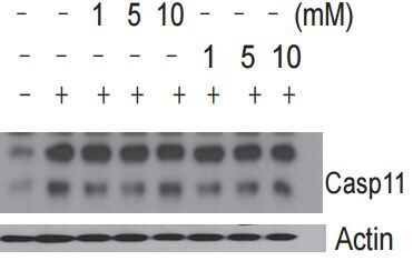
Western Blot: Caspase-11 Antibody (17D9) - BSA Free [NB120-10454] - Caspase-11 is expressed in Casp1Null bone marrow-derived macrophages.Immunoblot analysis of caspase-1, caspase-11 & GAPDH (loading control) in unprimed WT or mutant BMDMs at various times (above lane) after stimulation with IFN-beta (250U/ml), IFN-gamma (100 ng/ml) or LPS (100 ng/ml). Data are representative of two independent experiments. Image collected & cropped by CiteAb from the following publication (https://pubmed.ncbi.nlm.nih.gov/28345580), licensed under a CC-BY license. Not internally tested by Novus Biologicals. - BSA Free [NB120-10454] -")
Immunohistochemistry: Caspase-11 Antibody (17D9) - BSA Free [NB120-10454] -
Immunohistochemistry: Caspase-11 Antibody (17D9) - BSA Free [NB120-10454] - Immunocytochemistry analysis of histone induced activations of caspase 1, caspase 3, & NLRP3 (400×). BMECs were cultured briefly on cover glasses (pre-treated with poly-L –lysine, 0.1 mg/mL, Sigma-Aldrich) & incubated with histone (50 & 100 μg/mL) for 16 h. The samples were visualized with DAB, counterstained with hematoxylin & observed by inverted microscope. Three independent experiments were carried out by light microscope analyses. Image collected & cropped by CiteAb from the following publication (https://pubmed.ncbi.nlm.nih.gov/31156617), licensed under a CC-BY license. Not internally tested by Novus Biologicals. - BSA Free [NB120-10454] -")
Western Blot: Caspase-11 Antibody (17D9) - BSA Free [NB120-10454] -
Western Blot: Caspase-11 Antibody (17D9) - BSA Free [NB120-10454] - Differential roles of caspase-1 & caspase-11 in response to infection with Aspergillus fumigatus.(A) Immunoblot analysis of pro-caspase-1 (Pro-Casp-1) & the caspase-1 subunit p20 (Casp-1 p20) & GAPDH (loading control) in unprimed WT or mutant bone marrow-derived dendritic cells left untreated (medium alone [Med]) or assessed 20 h after infection with A. fumigatus (MOI, 10). (B) Release of IL-1 beta & IL-18 after treatment as in (A). (C) Survival of 8-week-old WT & mutant mice infected with 5 × 10⁵ A. fumigatus conidia after immunosuppression with cyclophosphamide & cortisone acetate. *P < 0.05, ****P < 0.0001 (log-rank test). Data are representative of two (C) or three independent experiments (A & B; mean & s.e.m. are representative of values from three independent experiments in B). Image collected & cropped by CiteAb from the following publication (https://pubmed.ncbi.nlm.nih.gov/28345580), licensed under a CC-BY license. Not internally tested by Novus Biologicals. - BSA Free [NB120-10454] -")
Western Blot: Caspase-11 Antibody (17D9) - BSA Free [NB120-10454] -
Western Blot: Caspase-11 Antibody (17D9) - BSA Free [NB120-10454] - GBP2 & IRGB10 regulates caspase-11 mediated non-canonical NLRP3 activation & pyroptosis.a Microscopic analysis of cell death in unprimed WT, Gbp2−/−, Gbp5−/−, Gbpchr3-KO,Irgb10−/−, Irgb10−/−Gbpchr3-KO, Caspase11−/− & Nlrp3−/− BMDMs infected with popB (MOI 10) for 16 h. (Scale, 15 μm). Arrow heads indicate pyroptotic cells. b–e Immunoblot analysis of caspase-1, IL-1 beta, IL-18, & IL-6 release in WT, Gbp2−/−, Gbp5−/−, Gbpchr3-KO, Irgb10−/−, Irgb10−/−Gbpchr3-KO, Caspase11−/− & Nlrp3−/− BMDMs infected with popB (MOI10). f Bacterial CFU in unprimed WT, Gbp2−/−, Gbp5−/−, Gbpchr3-KO, Irgb10−/−, Irgb10−/−Gbpchr3-KO, Caspase11−/− & Nlrp3−/− BMDMs infected with popB (MOI10), 4 h & 16 h. Data are representative of three independent experiments. ns-not significant, *p < 0.05, **p < 0.01, ***p < 0.001,****p < 0.0001 (two-tailed T test) Image collected & cropped by CiteAb from the following publication (https://pubmed.ncbi.nlm.nih.gov/30062052), licensed under a CC-BY license. Not internally tested by Novus Biologicals. - BSA Free [NB120-10454] -")
Flow Cytometry: Caspase-11 Antibody (17D9) - BSA Free [NB120-10454] -
Flow Cytometry: Caspase-11 Antibody (17D9) - BSA Free [NB120-10454] - The expression of circHIPK3 in serum samples of patients with AP & in caerulein-stimulated pancreatic acinar cells. (A) QPCR was performed to detect circHIPK3 expression in serum samples of patients with AP & healthy subjects. MAP, mild acute pancreatitis; SAP, severe Acute Pancreatitis. (B) CCK8 assay was performed to measure the cell viability in AR42J cells after caerulein treatment. (C) Levels of inflammatory cytokines IL-1 beta, IL-6, IL-8, & TNF-alpha were measured by ELISA kits in culture medium after caerulein treatment. (D) Amylase activity was measured by ELISA kit in AR42J cells after caerulein treatment. (E) The pyroptosis-related proteins caspase 1 & caspase 11 were analyzed by immunoblot in AR42J cells after caerulein treatment. (F) PI staining was performed in AR42J cells after caerulein treatment, & the PI-positive cells were counted. (G) Flow-cytometric analysis of caspase-1/11+PI+ cells in AR42J cells after caerulein treatment. Data are presented as a representative plot (upper) & quantified percentages (lower). (H) circHIPK3 expression was determined by qPCR in AR42J cells after caerulein treatment. *p < 0.05. Image collected & cropped by CiteAb from the following publication (https://pubmed.ncbi.nlm.nih.gov/32318575), licensed under a CC-BY license. Not internally tested by Novus Biologicals.Applications for Caspase-11 Antibody (17D9) - BSA Free
Flow Cytometry
Immunoblotting
Immunocytochemistry/ Immunofluorescence
Immunohistochemistry
Immunohistochemistry-Frozen
Immunohistochemistry-Paraffin
Immunoprecipitation
Western Blot
Reviewed Applications
Read 6 reviews rated 4 using NB120-10454 in the following applications:
Flow Cytometry Panel Builder
Bio-Techne Knows Flow Cytometry
Save time and reduce costly mistakes by quickly finding compatible reagents using the Panel Builder Tool.
Advanced Features
- Spectra Viewer - Custom analysis of spectra from multiple fluorochromes
- Spillover Popups - Visualize the spectra of individual fluorochromes
- Antigen Density Selector - Match fluorochrome brightness with antigen density

Formulation, Preparation, and Storage
Purification
Formulation
Format
Preservative
Concentration
Shipping
Stability & Storage
Background: Caspase-11
Alternate Names
Gene Symbol
UniProt
Additional Caspase-11 Products
Product Documents for Caspase-11 Antibody (17D9) - BSA Free
Certificate of Analysis
To download a Certificate of Analysis, please enter a lot or batch number in the search box below.
Product Specific Notices for Caspase-11 Antibody (17D9) - BSA Free
This product is for research use only and is not approved for use in humans or in clinical diagnosis. Primary Antibodies are guaranteed for 1 year from date of receipt.
Citations for Caspase-11 Antibody (17D9) - BSA Free
 Powered by Bioz
Customer Reviews for Caspase-11 Antibody (17D9) - BSA Free (6)
Have you used Caspase-11 Antibody (17D9) - BSA Free?
Submit a review and receive an Amazon gift card!
$25/€18/£15/$25CAN/¥2500 Yen for a review with an image
$10/€7/£6/$10CAN/¥1110 Yen for a review without an image
Submit a review
Customer Images

-(01-ml)_NB120-10454_8586.jpg)
-
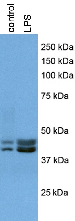
Application: Western BlotSample Tested: Mouse ColonSpecies: MouseVerified Customer | Posted 03/31/2018
-
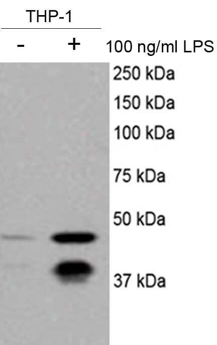
Application: Western BlotSample Tested: mouse primary hepatocytesSpecies: MouseVerified Customer | Posted 12/18/2017Review for anti-caspase 11 antibody (NB120-10454)100 ng/ml LPS treated for 1 hour Name: Anti-caspase 11 antibody (NB120-10454) Catalog #: Anti-caspase 11 antibody (NB120-10454) Lot Number: Anti-caspase 11 antibody (NB120-10454, Lot # A-6) PO/Order Number: Click here to enter text.. WB Image Description (Please provide labels for all lanes): mouse primary hepatocytes treated without (lane 1), or with 100 ng/ml LPS (lane 2) Sample Information: Cell Line or Tissue: mouse primary hepatocytes Species: mouse Treatment: 100 ng/ml LPS Lysate Preparation: Date of lysate preparation: December 4, 2017 Lysis buffer used: 1X lysis buffer from Cell Signaling by adding PMSF Reducing agent: beta-mercaptoethanol, DTT If boiled (temperature/time): Yes Controls: Positive Control: No Negative Control: No Loading Control (please attach additional images if applicable): No Protein Amount Loaded per lane: 20 ug Antibody Storage Conditions: -20℃ Electrophoresis: Gel Percentage: 12% Electrophoresis Conditions: Tris-Glycine-SDS at room temperature Voltage: 120V Time: 2 hours Membrane Transfer: Method (Submersion/Semi-dry): wet transfer Membrane Type (PVDF/Nitrocellulose): Nitrocellulose Time: 2 hours Voltage: 100V Blocking: Blocking Solution: 5% milk in 1X TBST Time: 1 hour at room temperature Primary Antibody: Dilution: 1/200 Diluent Buffer: 2.5% BSA Incubation Time: overnight Incubation Temperature: 4℃ Washing Conditions: Wash Solution: 1X TBST Time and Repetitions: 5 min each for 3 times Secondary Antibody Manufacturer and Catalog #: Thermo Scientific, cat # 34095, Lot # SG251180 Secondary description: goat anti-rat secondary antibody Dilution: 1/5000 Diluent Buffer: 3% milk Incubation Time: 1 hour Incubation Temperature: room temperature Detection Method: Detection: ECL (GE, cat # RPN2209, lot #n9838243) Procedure: Add equal volume of A and B, mix and apply on the membranes for 3-5 min before exposure Development Time: 25 seconds Molecular weight of band(s): ~ 38, 43 kDa Experimental Concerns and Observations: Specific bands around 38, 43 kDa were observed

-
Application: Western BlotSample Tested:Species: HumanVerified Customer | Posted 08/06/2016Review for Caspase-11 Antibody (NB120-10454)
-
Application: Western BlotSample Tested: mouse macrophageSpecies: MouseVerified Customer | Posted 02/02/2016pro-Caspase 11 western blot
-
Application: Western BlotSample Tested: Bone Marrrow Derived MacrophagesSpecies: MouseVerified Customer | Posted 03/19/2015Caspase 11 with LPS Stimulation
-
Application: Western BlotSample Tested:Species: MouseVerified Customer | Posted 07/01/2014Mouse B cell
There are no reviews that match your criteria.
Protocols
View specific protocols for Caspase-11 Antibody (17D9) - BSA Free (NB120-10454):
Culture cells to appropriate density in 35 mm culture dishes or 6-well plates.
1. Remove culture medium and wash the cells briefly in PBS. Add 10% formalin to the dish and fix at room temperature for 10 minutes.
2. Remove the formalin and wash the cells in PBS.
3. Permeablize the cells with 0.1% Triton X100 or other suitable detergent for 10 min.
4. Remove the permeablization buffer and wash three times for 10 minutes each in PBS. Be sure to not let the specimen dry out.
5. To block nonspecific antibody binding, incubate in 10% normal goat serum from 1 hour to overnight at room temperature.
6. Add primary antibody at appropriate dilution and incubate overnight at 4C.
7. Remove primary antibody and replace with PBS. Wash three times for 10 minutes each.
8. Add secondary antibody at appropriate dilution. Incubate for 1 hour at room temperature.
9. Remove secondary antibody and replace with PBS. Wash three times for 10 minutes each.
10. Counter stain DNA with DAPi if required.
1. Perform SDS-PAGE on samples to be analyzed, loading 10-25 ug of total protein per lane.
2. Transfer proteins to PVDF membrane according to the instructions provided by the manufacturer of the membrane and transfer apparatus.
3. Stain the membrane with Ponceau S (or similar product) to assess transfer success, and mark molecular weight standards where appropriate.
4. Rinse the blot TBS -0.05% Tween 20 (TBST).
5. Block the membrane in 5% Non-fat milk in TBST (blocking buffer) for at least 1 hour.
6. Wash the membrane in TBST three times for 10 minutes each.
7. Dilute primary antibody in 1% BSA and incubate overnight at 4C with gentle rocking.
8. Wash the membrane in TBST three times for 10 minutes each.
9. Incubate the membrane in diluted HRP conjugated secondary antibody in blocking buffer (as per manufacturer's instructions) for 1 hour at room temperature.
10. Wash the blot in TBST three times for 10 minutes each (this step can be repeated as required to reduce background).
11. Apply the detection reagent of choice in accordance with the manufacturer's instructions.
Find general support by application which include: protocols, troubleshooting, illustrated assays, videos and webinars.
- 7-Amino Actinomycin D (7-AAD) Cell Viability Flow Cytometry Protocol
- Antigen Retrieval Protocol (PIER)
- Antigen Retrieval for Frozen Sections Protocol
- Appropriate Fixation of IHC/ICC Samples
- Cellular Response to Hypoxia Protocols
- Chromogenic IHC Staining of Formalin-Fixed Paraffin-Embedded (FFPE) Tissue Protocol
- Chromogenic Immunohistochemistry Staining of Frozen Tissue
- ClariTSA™ Fluorophore Kits
- Detection & Visualization of Antibody Binding
- ELISA Sample Preparation & Collection Guide
- ELISA Troubleshooting Guide
- Extracellular Membrane Flow Cytometry Protocol
- Flow Cytometry Protocol for Cell Surface Markers
- Flow Cytometry Protocol for Staining Membrane Associated Proteins
- Flow Cytometry Staining Protocols
- Flow Cytometry Troubleshooting Guide
- Fluorescent IHC Staining of Frozen Tissue Protocol
- Graphic Protocol for Heat-induced Epitope Retrieval
- Graphic Protocol for the Preparation and Fluorescent IHC Staining of Frozen Tissue Sections
- Graphic Protocol for the Preparation and Fluorescent IHC Staining of Paraffin-embedded Tissue Sections
- Graphic Protocol for the Preparation of Gelatin-coated Slides for Histological Tissue Sections
- How to Run an R&D Systems DuoSet ELISA
- How to Run an R&D Systems Quantikine ELISA
- How to Run an R&D Systems Quantikine™ QuicKit™ ELISA
- ICC Cell Smear Protocol for Suspension Cells
- ICC Immunocytochemistry Protocol Videos
- ICC for Adherent Cells
- IHC Sample Preparation (Frozen sections vs Paraffin)
- Immunocytochemistry (ICC) Protocol
- Immunocytochemistry Troubleshooting
- Immunofluorescence of Organoids Embedded in Cultrex Basement Membrane Extract
- Immunofluorescent IHC Staining of Formalin-Fixed Paraffin-Embedded (FFPE) Tissue Protocol
- Immunohistochemistry (IHC) and Immunocytochemistry (ICC) Protocols
- Immunohistochemistry Frozen Troubleshooting
- Immunohistochemistry Paraffin Troubleshooting
- Immunoprecipitation Protocol
- Intracellular Flow Cytometry Protocol Using Alcohol (Methanol)
- Intracellular Flow Cytometry Protocol Using Detergents
- Intracellular Nuclear Staining Flow Cytometry Protocol Using Detergents
- Intracellular Staining Flow Cytometry Protocol Using Alcohol Permeabilization
- Intracellular Staining Flow Cytometry Protocol Using Detergents to Permeabilize Cells
- Preparing Samples for IHC/ICC Experiments
- Preventing Non-Specific Staining (Non-Specific Binding)
- Primary Antibody Selection & Optimization
- Propidium Iodide Cell Viability Flow Cytometry Protocol
- Protocol for Heat-Induced Epitope Retrieval (HIER)
- Protocol for Liperfluo
- Protocol for Making a 4% Formaldehyde Solution in PBS
- Protocol for VisUCyte™ HRP Polymer Detection Reagent
- Protocol for the Characterization of Human Th22 Cells
- Protocol for the Characterization of Human Th9 Cells
- Protocol for the Fluorescent ICC Staining of Cell Smears - Graphic
- Protocol for the Fluorescent ICC Staining of Cultured Cells on Coverslips - Graphic
- Protocol for the Preparation & Fixation of Cells on Coverslips
- Protocol for the Preparation and Chromogenic IHC Staining of Frozen Tissue Sections
- Protocol for the Preparation and Chromogenic IHC Staining of Frozen Tissue Sections - Graphic
- Protocol for the Preparation and Chromogenic IHC Staining of Paraffin-embedded Tissue Sections
- Protocol for the Preparation and Chromogenic IHC Staining of Paraffin-embedded Tissue Sections - Graphic
- Protocol for the Preparation and Fluorescent ICC Staining of Cells on Coverslips
- Protocol for the Preparation and Fluorescent ICC Staining of Non-adherent Cells
- Protocol for the Preparation and Fluorescent ICC Staining of Stem Cells on Coverslips
- Protocol for the Preparation and Fluorescent IHC Staining of Frozen Tissue Sections
- Protocol for the Preparation and Fluorescent IHC Staining of Paraffin-embedded Tissue Sections
- Protocol for the Preparation of Gelatin-coated Slides for Histological Tissue Sections
- Protocol for the Preparation of a Cell Smear for Non-adherent Cell ICC - Graphic
- Protocol: Annexin V and PI Staining by Flow Cytometry
- Protocol: Annexin V and PI Staining for Apoptosis by Flow Cytometry
- Quantikine HS ELISA Kit Assay Principle, Alkaline Phosphatase
- Quantikine HS ELISA Kit Principle, Streptavidin-HRP Polymer
- R&D Systems Quality Control Western Blot Protocol
- Sandwich ELISA (Colorimetric) – Biotin/Streptavidin Detection Protocol
- Sandwich ELISA (Colorimetric) – Direct Detection Protocol
- TUNEL and Active Caspase-3 Detection by IHC/ICC Protocol
- The Importance of IHC/ICC Controls
- Troubleshooting Guide: ELISA
- Troubleshooting Guide: Fluorokine Flow Cytometry Kits
- Troubleshooting Guide: Immunohistochemistry
- Troubleshooting Guide: Western Blot Figures
- Western Blot Conditions
- Western Blot Protocol
- Western Blot Protocol for Cell Lysates
- Western Blot Troubleshooting
- Western Blot Troubleshooting Guide
- View all Protocols, Troubleshooting, Illustrated assays and Webinars
FAQs for Caspase-11 Antibody (17D9) - BSA Free
-
Q: If I dilute the 100 uL of antibody (the whole delivered content) in 10 mL of buffer to incubate my blottet membrane in (and I need this volume of about 10 mL for the membrane to float in it on the shaker). I will be able to re-use this one dilution (which is 10 ug/mL) for 100 western blots? So that means for 100 samples? Or even for 100 membranes? Usually we use a antibody-dilution of 1:1000 (around 1ug/mL), this would be 1:100 (10ug/mL).
A: I am hoping I can assist you in determining usage of this antibody in your assay. The antibody comes at a starting concentration of 1 mg/mL. Since you get 100uL in the vial you will have 100 ug of antibody. The lab recommends using a dilution of 1:100-1:200 for WB. Therefore if you wished to use 10 mL volume on your incubation you would need to add 100 uL of antibody into 9.9mL of buffer to give you a final concentration of 10 ug/uL. Since you are using such a large final volume you will only get 1-2 blots per Western ran since we do not recommend reusing your antibody. If you can run the blots with much lower final volume such as 1 mL instead you can get away with a lot more Westerns.
-
Q: If it has been determined, can you let me know as specifically as possible the epitope that the Caspase-11 antibody recognizes, eg. peptide sequence or approximate region of the p30 subunit?
A: This antibody has not been epitope mapped and was generated using full-length p30. The only information we have is that the antibody may be used for immunoblotting [to detect the full length caspase 11 (apparent size of 35-43 kDa), and the p10 and p30fragments], so it must recognize a region similar in both subunits.
-
Q: If I dilute the 100 uL of antibody (the whole delivered content) in 10 mL of buffer to incubate my blottet membrane in (and I need this volume of about 10 mL for the membrane to float in it on the shaker). I will be able to re-use this one dilution (which is 10 ug/mL) for 100 western blots? So that means for 100 samples? Or even for 100 membranes? Usually we use a antibody-dilution of 1:1000 (around 1ug/mL), this would be 1:100 (10ug/mL).
A: I am hoping I can assist you in determining usage of this antibody in your assay. The antibody comes at a starting concentration of 1 mg/mL. Since you get 100uL in the vial you will have 100 ug of antibody. The lab recommends using a dilution of 1:100-1:200 for WB. Therefore if you wished to use 10 mL volume on your incubation you would need to add 100 uL of antibody into 9.9mL of buffer to give you a final concentration of 10 ug/uL. Since you are using such a large final volume you will only get 1-2 blots per Western ran since we do not recommend reusing your antibody. If you can run the blots with much lower final volume such as 1 mL instead you can get away with a lot more Westerns.
-
Q: If it has been determined, can you let me know as specifically as possible the epitope that the Caspase-11 antibody recognizes, eg. peptide sequence or approximate region of the p30 subunit?
A: This antibody has not been epitope mapped and was generated using full-length p30. The only information we have is that the antibody may be used for immunoblotting [to detect the full length caspase 11 (apparent size of 35-43 kDa), and the p10 and p30fragments], so it must recognize a region similar in both subunits.