CD11b Antibody (M1/70.15) - BSA Free
Novus Biologicals | Catalog # NB600-1327
Key Product Details
Validated by
Biological Validation
Species Reactivity
Validated:
Human, Mouse, Rabbit
Cited:
Human, Mouse, Rat, Rabbit
Applications
Validated:
Immunohistochemistry, Immunohistochemistry-Paraffin, Immunohistochemistry-Frozen, Flow Cytometry, Immunocytochemistry/ Immunofluorescence, Immunoprecipitation, CyTOF-ready
Cited:
Immunohistochemistry-Paraffin, Immunohistochemistry-Frozen, Flow Cytometry, Immunocytochemistry/ Immunofluorescence, IF/IHC
Label
Unconjugated
Antibody Source
Monoclonal Rat IgG2b Lambda Clone # M1/70.15
Format
BSA Free
Loading...
Product Specifications
Immunogen
Rat Monoclonal CD11b Antibody (M1/70.15) was made to T cell enriched splenocytes from B10 mice.
Reactivity Notes
Mouse reactivity reported in a verified customer review. Rabbit reactivity reported in scientific literature (PMID: 28700779). Human reactivity reported in scientific literature (PMID: 28683563).
Marker
Microglia Marker, Myeloid Marker
Clonality
Monoclonal
Host
Rat
Isotype
IgG2b Lambda
Theoretical MW
127.2 kDa.
Disclaimer note: The observed molecular weight of the protein may vary from the listed predicted molecular weight due to post translational modifications, post translation cleavages, relative charges, and other experimental factors.
Disclaimer note: The observed molecular weight of the protein may vary from the listed predicted molecular weight due to post translational modifications, post translation cleavages, relative charges, and other experimental factors.
Scientific Data Images for CD11b Antibody (M1/70.15) - BSA Free
![Immunohistochemistry: CD11b Antibody (M1/70.15) - BSA Free [NB600-1327]](https://resources.rndsystems.com/images/products/CD11b-Antibody-M1-70-15-Immunohistochemistry-NB600-1327-img0012.jpg "Immunohistochemistry: CD11b Antibody (M1/70.15) - BSA Free [NB600-1327]")
Immunohistochemistry: CD11b Antibody (M1/70.15) - BSA Free [NB600-1327]
Immunohistochemistry: CD11b Antibody (M1/70.15) [NB600-1327] - Immunohistochemical analysis of frozen lymph node section using CD11b Antibody (M1/70.15) [NB600-1327] followed by staining with HRP-conjugated secondary antibody.![Immunocytochemistry/ Immunofluorescence: CD11b Antibody (M1/70.15) - BSA Free [NB600-1327]](https://resources.rndsystems.com/images/products/CD11b-Antibody-M1-70-15-Immunocytochemistry-Immunofluorescence-NB600-1327-img0013.jpg "Immunocytochemistry/ Immunofluorescence: CD11b Antibody (M1/70.15) - BSA Free [NB600-1327]")
Immunocytochemistry/ Immunofluorescence: CD11b Antibody (M1/70.15) - BSA Free [NB600-1327]
Immunocytochemistry/Immunofluorescence: CD11b Antibody (M1/70.15) [NB600-1327] - Characterization of microglia cultures by multiple staining with CD11b Antibody (M1/70.15) [NB600-1327] (panels a. & c.) and anti GFAP (panels b & d). Nuclei appear blue due to 4',6-diamidino-2-phenylindole (DAPI) counter staining.![Flow Cytometry: CD11b Antibody (M1/70.15) - BSA Free [NB600-1327]](https://resources.rndsystems.com/images/products/CD11b-Antibody-M1-70-15-Flow-Cytometry-NB600-1327-img0017.jpg "Flow Cytometry: CD11b Antibody (M1/70.15) - BSA Free [NB600-1327]")
Flow Cytometry: CD11b Antibody (M1/70.15) - BSA Free [NB600-1327]
Flow Cytometry: CD11b Antibody (M1/70.15) [NB600-1327] - A surface stain was performed on Raw264.7 cells with CD11b (M1/70.15) Antibody [NB600-1327R] (blue) and a matched isotype control (orange), both conjugated to DyLight 550. Cells were incubated in an antibody dilution of 5 ug/mL for 20 minutes at room temperature.![Immunocytochemistry/ Immunofluorescence: CD11b Antibody (M1/70.15) - BSA Free [NB600-1327]](https://resources.rndsystems.com/images/products/CD11b-Antibody-M1-70-15-BSA-Free-Immunocytochemistry-Immunofluorescence-NB600-1327-img0020.jpg "Immunocytochemistry/ Immunofluorescence: CD11b Antibody (M1/70.15) - BSA Free [NB600-1327]")
Immunocytochemistry/ Immunofluorescence: CD11b Antibody (M1/70.15) - BSA Free [NB600-1327]
Immunocytochemistry/Immunofluorescence: CD11b Antibody (M1/70.15) - BSA Free [NB600-1327] - Raw264.7 cells were fixed in 4% paraformaldehyde for 10 minutes and permeabilized in 0.05% Triton X-100 in PBS for 5 minutes. The cells were incubated with CD11b Antibody [M1/70.15] (NB600-1327) at 1ug/ml overnight at 4C and detected with an anti-rat DyLight 488 (Green) at a 1:1000 dilution for 60 minutes. Nuclei were counterstained with DAPI (Blue). Cells were imaged using a 100X objective and digitally deconvolved.![Immunocytochemistry/ Immunofluorescence: CD11b Antibody (M1/70.15) - BSA Free [NB600-1327]](https://resources.rndsystems.com/images/products/CD11b-Antibody-M1-70-15-BSA-Free-Immunocytochemistry-Immunofluorescence-NB600-1327-img0018.jpg "Immunocytochemistry/ Immunofluorescence: CD11b Antibody (M1/70.15) - BSA Free [NB600-1327]")
Immunocytochemistry/ Immunofluorescence: CD11b Antibody (M1/70.15) - BSA Free [NB600-1327]
Immunocytochemistry/Immunofluorescence: CD11b Antibody (M1/70.15) - BSA Free [NB600-1327] - Raw264.7 cells were fixed in 4% paraformaldehyde for 10 minutes and permeabilized in 0.05% Triton X-100 in PBS for 5 minutes. The cells were incubated with CD11b Antibody [M1/70.15] conjugated to Alexa Fluor 488 (NB600-1327AF488) at 5 ug/ml for 1 hour at room temperature. Nuclei were counterstained with DAPI (Blue). Cells were imaged using a 100X objective and digitally deconvolved.![Immunocytochemistry/ Immunofluorescence: CD11b Antibody (M1/70.15) - BSA Free [NB600-1327]](https://resources.rndsystems.com/images/products/CD11b-Antibody-M1-70-15-Immunocytochemistry-Immunofluorescence-NB600-1327-img0010.jpg "Immunocytochemistry/ Immunofluorescence: CD11b Antibody (M1/70.15) - BSA Free [NB600-1327]")
Immunocytochemistry/ Immunofluorescence: CD11b Antibody (M1/70.15) - BSA Free [NB600-1327]
Immunocytochemistry/Immunofluorescence: CD11b Antibody (M1/70.15) [NB600-1327] - Immunocytochemistry/Immunofluorescence (ICC/IF) analysis of RAW264.7 cells using CD11b antibody (M1/70.15) [NB600-1327] with Dylight 488 conjugated secondary (green). Nuclei and tubulin were stained with 4',6-diamidino-2-phenylindole (DAPI) (blue) and Dylight 550 (red).![Immunocytochemistry/ Immunofluorescence: CD11b Antibody (M1/70.15) - BSA Free [NB600-1327]](https://resources.rndsystems.com/images/products/CD11b-Antibody-M1-70-15-Immunocytochemistry-Immunofluorescence-NB600-1327-img0015.jpg "Immunocytochemistry/ Immunofluorescence: CD11b Antibody (M1/70.15) - BSA Free [NB600-1327]")
Immunocytochemistry/ Immunofluorescence: CD11b Antibody (M1/70.15) - BSA Free [NB600-1327]
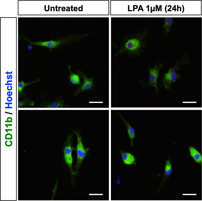
Immunocytochemistry/Immunofluorescence: CD11b Antibody (M1/70.15) [NB600-1327] - Immunocytochemical/Immunofluorescent staining in untreated and treated mouse microglia cells using CD11b Antibody (M1/70.15) [NB600-1327]. Image from a verified customer review.![Immunocytochemistry/ Immunofluorescence: CD11b Antibody (M1/70.15) - BSA Free [NB600-1327]](https://resources.rndsystems.com/images/products/CD11b-Antibody-M1-70-15-Immunocytochemistry-Immunofluorescence-NB600-1327-img0016.jpg "Immunocytochemistry/ Immunofluorescence: CD11b Antibody (M1/70.15) - BSA Free [NB600-1327]")
Immunocytochemistry/ Immunofluorescence: CD11b Antibody (M1/70.15) - BSA Free [NB600-1327]
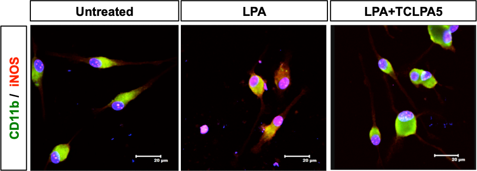
Immunocytochemistry/Immunofluorescence: CD11b Antibody (M1/70.15) [NB600-1327] - Immunocytochemical/Immunofluorescent analysis in mouse primary microglia. Cells were treated with lysophosphatidic acid (LPA) or LPA and an LPAR5 inhibitor in order to investigate the expression of iNOS after 24 hours. CD11b Antibody (M1/70.15) [NB600-1327] was used in a dilution 1:100 in order to stain primary murine microglia. Cy2 (green) and Cy3 (red) were used as secondary antibodies.![Immunocytochemistry/ Immunofluorescence: CD11b Antibody (M1/70.15) - BSA Free [NB600-1327]](https://resources.rndsystems.com/images/products/CD11b-Antibody-M1-70-15-BSA-Free-Immunocytochemistry-Immunofluorescence-NB600-1327-img0019.jpg "Immunocytochemistry/ Immunofluorescence: CD11b Antibody (M1/70.15) - BSA Free [NB600-1327]")
Immunocytochemistry/ Immunofluorescence: CD11b Antibody (M1/70.15) - BSA Free [NB600-1327]
Immunocytochemistry/Immunofluorescence: CD11b Antibody (M1/70.15) - BSA Free [NB600-1327] - Raw264.7 cells were fixed in 4% paraformaldehyde for 10 minutes and permeabilized in 0.05% Triton X-100 in PBS for 5 minutes. The cells were incubated with CD11b Antibody [M1/70.15] conjugated to Alexa Fluor 647 (NB600-1327AF647) at 5 ug/ml for 1 hour at room temperature. Nuclei were counterstained with DAPI (Blue). Cells were imaged using a 100X objective and digitally deconvolved.![Flow Cytometry: CD11b Antibody (M1/70.15) - BSA Free [NB600-1327]](https://resources.rndsystems.com/images/products/CD11b-Antibody-M1-70-15-Flow-Cytometry-NB600-1327-img0004.jpg "Flow Cytometry: CD11b Antibody (M1/70.15) - BSA Free [NB600-1327]")
Flow Cytometry: CD11b Antibody (M1/70.15) - BSA Free [NB600-1327]
Flow Cytometry: CD11b Antibody (M1/70.15) [NB600-1327] - Staining of mouse peritoneal macrophages with Rat anti Mouse CD11b Antibody (M1/70.15) [NB600-1327].![Flow Cytometry: CD11b Antibody (M1/70.15) - BSA Free [NB600-1327]](https://resources.rndsystems.com/images/products/CD11b-Antibody-M1-70-15-Flow-Cytometry-NB600-1327-img0007.jpg "Flow Cytometry: CD11b Antibody (M1/70.15) - BSA Free [NB600-1327]")
Flow Cytometry: CD11b Antibody (M1/70.15) - BSA Free [NB600-1327]
Flow Cytometry: CD11b Antibody (M1/70.15) [NB600-1327] - Staining of mouse peritoneal macrophages with Rat anti Mouse CD11b Antibody (M1/70.15) [NB600-1327]: RPE - Alexa Fluor 750![Flow Cytometry: CD11b Antibody (M1/70.15) - BSA Free [NB600-1327]](https://resources.rndsystems.com/images/products/CD11b-Antibody-M1-70-15-Flow-Cytometry-NB600-1327-img0014.jpg "Flow Cytometry: CD11b Antibody (M1/70.15) - BSA Free [NB600-1327]")
Flow Cytometry: CD11b Antibody (M1/70.15) - BSA Free [NB600-1327]
Flow Cytometry: CD11b Antibody (M1/70.15) [NB600-1327] - Staining of mouse peritoneal macrophages using the Alexa Fluor (R) 488 conjugate of CD11b Antibody (M1/70.15) [NB600-1327AF488]. in Raw 264.7 Mouse Cell Line.")
CD11b (M1/70.15) in Raw 264.7 Mouse Cell Line.
CD11b (M1/70.15) was detected in immersion fixed Raw 264.7 mouse macrophage cell line using Rat anti-CD11b (M1/70.15) Protein-G purified Monoclonal Antibody conjugated to DyLight 550 (Catalog # NB600-1327R) (red) at 10 µg/mL overnight at 4C. Cells were counterstained with DAPI (blue). Cells were imaged using a 100X objective and digitally deconvolved.Applications for CD11b Antibody (M1/70.15) - BSA Free
Application
Recommended Usage
Flow Cytometry
1:50. Use reported in scientific literature (PMID 28683563)
Immunocytochemistry/ Immunofluorescence
1:10-1:500. Use reported by customer review
Immunohistochemistry
1:10-1:500
Immunohistochemistry-Frozen
1:10-1:500. Use reported in scientific literature
Immunohistochemistry-Paraffin
1:10-1:500. Use reported in scientific literature (PMID 28700779)
Immunoprecipitation
1:10-1:500
Application Notes
IHC-P: Perform enzymatic antigen retrieval before commencing with IHC staining protocol. NB600-1327 has been reported to as being suitable for use on PLP fixed paraffin embedded tissue but has not been tested for use on formalin fixed tissue. This antibody is CyTOF ready.
Reviewed Applications
Read 2 reviews rated 5 using NB600-1327 in the following applications:
Flow Cytometry Panel Builder
Bio-Techne Knows Flow Cytometry
Save time and reduce costly mistakes by quickly finding compatible reagents using the Panel Builder Tool.
Advanced Features
- Spectra Viewer - Custom analysis of spectra from multiple fluorochromes
- Spillover Popups - Visualize the spectra of individual fluorochromes
- Antigen Density Selector - Match fluorochrome brightness with antigen density

Formulation, Preparation, and Storage
Purification
Protein G purified
Formulation
PBS
Format
BSA Free
Preservative
0.02% Sodium Azide
Concentration
1.0 mg/ml
Shipping
The product is shipped with polar packs. Upon receipt, store it immediately at the temperature recommended below.
Stability & Storage
Store at 4C short term. Aliquot and store at -20C long term. Avoid freeze-thaw cycles.
Background: CD11b/Integrin alpha M
References
1. Rosetti, F., & Mayadas, T. N. (2016). The many faces of Mac-1 in autoimmune disease. Immunol Rev, 269(1), 175-193. doi:10.1111/imr.12373
2. Kim, D., Kim, T. H., Wu, G., Park, B. K., Ha, J. H., Kim, Y. S.,... Kwon, H. J. (2016). Extracellular Release of CD11b by TLR9 Stimulation in Macrophages. PLoS One, 11(3), e0150677. doi:10.1371/journal.pone.0150677
3. Christensen, J. E., Andreasen, S. O., Christensen, J. P., & Thomsen, A. R. (2001). CD11b expression as a marker to distinguish between recently activated effector CD8(+) T cells and memory cells. Int Immunol, 13(4), 593-600. doi:10.1093/intimm/13.4.593
4. Nath, S. K., Han, S., Kim-Howard, X., Kelly, J. A., Viswanathan, P., Gilkeson, G. S.,... Harley, J. B. (2008). A nonsynonymous functional variant in integrin-alpha(M) (encoded by ITGAM) is associated with systemic lupus erythematosus. Nat Genet, 40(2), 152-154. doi:10.1038/ng.71
Alternate Names
CD11b, Integrin alpha M, ITGAM, cd11b flow antibody, M1/70, M1/70 cd11b, m1/70 clone, M1/70.15 Antibody, M1/70.15 CD11b, M1/70.15 CD11b Antibody, M1/70.15 Clone, M1/70.15 Conjugated, M1/70.15 ICC Antibody, M1/70.15 IHC Antibody, M1/70.15 Immunocytochemistry Antibody, M1/70.15 Immunohistochemistry Antibody, M1/70.15 Microglia Marker, M1/70.15 Myeloid Marker
Gene Symbol
ITGAM
UniProt
Additional CD11b/Integrin alpha M Products
Product Documents for CD11b Antibody (M1/70.15) - BSA Free
Certificate of Analysis
To download a Certificate of Analysis, please enter a lot or batch number in the search box below.
Product Specific Notices for CD11b Antibody (M1/70.15) - BSA Free
This product is for research use only and is not approved for use in humans or in clinical diagnosis. Primary Antibodies are guaranteed for 1 year from date of receipt.
Related Research Areas
Citations for CD11b Antibody (M1/70.15) - BSA Free
 Powered by Bioz
Customer Reviews for CD11b Antibody (M1/70.15) - BSA Free (2)
5 out of 5
2 Customer Ratings
Have you used CD11b Antibody (M1/70.15) - BSA Free?
Submit a review and receive an Amazon gift card!
$25/€18/£15/$25CAN/¥2500 Yen for a review with an image
$10/€7/£6/$10CAN/¥1110 Yen for a review without an image
Submit a review
Customer Images
Showing
1
-
2 of
2 reviews
Showing All
Filter By:
-
Application: ImmunocytochemistrySample Tested: Murine primary microgliaSpecies: MouseVerified Customer | Posted 06/10/2019Cells were treated with LPA or LPA and an LPAR5 inhibitor in order to investigate the expression of iNOS after 24 hours. CD11b antibody was used in a dilution 1:100 in order to stain primary murine microglia.Cy2 (green) and Cy3 (red) were used as secondary antibodies.

-
Application: ImmunocytochemistrySample Tested: Microglia cellsSpecies: MouseVerified Customer | Posted 06/07/2019
There are no reviews that match your criteria.
Protocols
Find general support by application which include: protocols, troubleshooting, illustrated assays, videos and webinars.
- 7-Amino Actinomycin D (7-AAD) Cell Viability Flow Cytometry Protocol
- Antigen Retrieval Protocol (PIER)
- Antigen Retrieval for Frozen Sections Protocol
- Appropriate Fixation of IHC/ICC Samples
- Cellular Response to Hypoxia Protocols
- Chromogenic IHC Staining of Formalin-Fixed Paraffin-Embedded (FFPE) Tissue Protocol
- Chromogenic Immunohistochemistry Staining of Frozen Tissue
- ClariTSA™ Fluorophore Kits
- Detection & Visualization of Antibody Binding
- Extracellular Membrane Flow Cytometry Protocol
- Flow Cytometry Protocol for Cell Surface Markers
- Flow Cytometry Protocol for Staining Membrane Associated Proteins
- Flow Cytometry Staining Protocols
- Flow Cytometry Troubleshooting Guide
- Fluorescent IHC Staining of Frozen Tissue Protocol
- Graphic Protocol for Heat-induced Epitope Retrieval
- Graphic Protocol for the Preparation and Fluorescent IHC Staining of Frozen Tissue Sections
- Graphic Protocol for the Preparation and Fluorescent IHC Staining of Paraffin-embedded Tissue Sections
- Graphic Protocol for the Preparation of Gelatin-coated Slides for Histological Tissue Sections
- ICC Cell Smear Protocol for Suspension Cells
- ICC Immunocytochemistry Protocol Videos
- ICC for Adherent Cells
- IHC Sample Preparation (Frozen sections vs Paraffin)
- Immunocytochemistry (ICC) Protocol
- Immunocytochemistry Troubleshooting
- Immunofluorescence of Organoids Embedded in Cultrex Basement Membrane Extract
- Immunofluorescent IHC Staining of Formalin-Fixed Paraffin-Embedded (FFPE) Tissue Protocol
- Immunohistochemistry (IHC) and Immunocytochemistry (ICC) Protocols
- Immunohistochemistry Frozen Troubleshooting
- Immunohistochemistry Paraffin Troubleshooting
- Immunoprecipitation Protocol
- Intracellular Flow Cytometry Protocol Using Alcohol (Methanol)
- Intracellular Flow Cytometry Protocol Using Detergents
- Intracellular Nuclear Staining Flow Cytometry Protocol Using Detergents
- Intracellular Staining Flow Cytometry Protocol Using Alcohol Permeabilization
- Intracellular Staining Flow Cytometry Protocol Using Detergents to Permeabilize Cells
- Preparing Samples for IHC/ICC Experiments
- Preventing Non-Specific Staining (Non-Specific Binding)
- Primary Antibody Selection & Optimization
- Propidium Iodide Cell Viability Flow Cytometry Protocol
- Protocol for Heat-Induced Epitope Retrieval (HIER)
- Protocol for Liperfluo
- Protocol for Making a 4% Formaldehyde Solution in PBS
- Protocol for VisUCyte™ HRP Polymer Detection Reagent
- Protocol for the Characterization of Human Th22 Cells
- Protocol for the Characterization of Human Th9 Cells
- Protocol for the Fluorescent ICC Staining of Cell Smears - Graphic
- Protocol for the Fluorescent ICC Staining of Cultured Cells on Coverslips - Graphic
- Protocol for the Preparation & Fixation of Cells on Coverslips
- Protocol for the Preparation and Chromogenic IHC Staining of Frozen Tissue Sections
- Protocol for the Preparation and Chromogenic IHC Staining of Frozen Tissue Sections - Graphic
- Protocol for the Preparation and Chromogenic IHC Staining of Paraffin-embedded Tissue Sections
- Protocol for the Preparation and Chromogenic IHC Staining of Paraffin-embedded Tissue Sections - Graphic
- Protocol for the Preparation and Fluorescent ICC Staining of Cells on Coverslips
- Protocol for the Preparation and Fluorescent ICC Staining of Non-adherent Cells
- Protocol for the Preparation and Fluorescent ICC Staining of Stem Cells on Coverslips
- Protocol for the Preparation and Fluorescent IHC Staining of Frozen Tissue Sections
- Protocol for the Preparation and Fluorescent IHC Staining of Paraffin-embedded Tissue Sections
- Protocol for the Preparation of Gelatin-coated Slides for Histological Tissue Sections
- Protocol for the Preparation of a Cell Smear for Non-adherent Cell ICC - Graphic
- Protocol: Annexin V and PI Staining by Flow Cytometry
- Protocol: Annexin V and PI Staining for Apoptosis by Flow Cytometry
- TUNEL and Active Caspase-3 Detection by IHC/ICC Protocol
- The Importance of IHC/ICC Controls
- Troubleshooting Guide: Fluorokine Flow Cytometry Kits
- Troubleshooting Guide: Immunohistochemistry
- View all Protocols, Troubleshooting, Illustrated assays and Webinars
FAQs for CD11b Antibody (M1/70.15) - BSA Free
Showing
1
-
5 of
8 FAQs
Showing All
-
Q: Do you know if this CD11b antibody can be used in CD11b positive cell depletion?
A: This particular CD11b antibody is not reccomended for depletion studies due to the preservative used; however, our CD11b antibody NBP1-06650 is offered in a preservative free format and has been validated for blocking.
-
Q: I am looking for a Mac-1 (microglia cell marker) antibody to do immunostaining with my mouse brain and I checked, you have a lot of antibodies for this biomarker. I don't know which one is the best to be used for immunohistology on mouse brain tissue. Could you please help with this?
A:
I would recommend NB110-89474. This antibody has been published in 11 scientific articles, including several times in IHC in mouse. As long as CD11b is expressed in your sample, it should work just fine and is backed by our 100% Novus Guarantee.
-
Q: I'm not sure if this will work for me, can I get a free sample of your CD11b antibody to test?
A: We don't offer free samples but we do offer this CD11b antibody in a trial size of 0.025 ml at a reduced cost, all antibodies are backed by our 100% guarantee to work in all validated species and applications on the product's web page.
-
Q: Is CD11b antibody also staining neutrophils in the brain
A: CD11b is typically used as a marker for granulocytes and macrophages. You may need a combination of markers to specifically identify neutrophils
-
Q: Looking for a bone marrow macrophage marker can you recommend a CD11b antibody for ICC?
A: I would recommend NB110-89474 which is our most popular CD11b antibody and has been validated in ICC and cited in 18 publications.
-
Q: The protocol says to do enzymatic antigen retrieval before incubating with the CD11b antibody, which enzyme is used?
A: The protocol is referencting the PIER method using Proteinase K as the enzyme.
-
Q: We need a CD11b antibody for IHC-P on mouse tissue conjugated to FITC, do you have one that will work?
A: We have 3 CD11b antibodies for IHC-P and just one CD11 b antibody we offer conjugated to FITC, NB110-89474F.
-
Q: What types of cells can this CD11b antibody be used as a marker for?
A: A study used this CD11b antibody to stain granulocytes in mouse uterine tissue in IHC. CD11b is also expressed on other leukocytes such as monocytes, macrophages, and natural killer cells.
-
Q: Do you know if this CD11b antibody can be used in CD11b positive cell depletion?
A: This particular CD11b antibody is not reccomended for depletion studies due to the preservative used; however, our CD11b antibody NBP1-06650 is offered in a preservative free format and has been validated for blocking.
-
Q: I am looking for a Mac-1 (microglia cell marker) antibody to do immunostaining with my mouse brain and I checked, you have a lot of antibodies for this biomarker. I don't know which one is the best to be used for immunohistology on mouse brain tissue. Could you please help with this?
A:
I would recommend NB110-89474. This antibody has been published in 11 scientific articles, including several times in IHC in mouse. As long as CD11b is expressed in your sample, it should work just fine and is backed by our 100% Novus Guarantee.
-
Q: I'm not sure if this will work for me, can I get a free sample of your CD11b antibody to test?
A: We don't offer free samples but we do offer this CD11b antibody in a trial size of 0.025 ml at a reduced cost, all antibodies are backed by our 100% guarantee to work in all validated species and applications on the product's web page.
-
Q: Is CD11b antibody also staining neutrophils in the brain
A: CD11b is typically used as a marker for granulocytes and macrophages. You may need a combination of markers to specifically identify neutrophils
-
Q: Looking for a bone marrow macrophage marker can you recommend a CD11b antibody for ICC?
A: I would recommend NB110-89474 which is our most popular CD11b antibody and has been validated in ICC and cited in 18 publications.
-
Q: The protocol says to do enzymatic antigen retrieval before incubating with the CD11b antibody, which enzyme is used?
A: The protocol is referencting the PIER method using Proteinase K as the enzyme.
-
Q: We need a CD11b antibody for IHC-P on mouse tissue conjugated to FITC, do you have one that will work?
A: We have 3 CD11b antibodies for IHC-P and just one CD11 b antibody we offer conjugated to FITC, NB110-89474F.
-
Q: What types of cells can this CD11b antibody be used as a marker for?
A: A study used this CD11b antibody to stain granulocytes in mouse uterine tissue in IHC. CD11b is also expressed on other leukocytes such as monocytes, macrophages, and natural killer cells.
Loading...
Associated Pathways


