CD31/PECAM-1 Antibody (JC/70A) - BSA Free
Novus Biologicals | Catalog # NB600-562
Clone JC/70A was used by HLDA to establish CD designation.
Key Product Details
Validated by
Knockout/Knockdown, Orthogonal Validation
Species Reactivity
Validated:
Human, Mouse, Feline, Rabbit
Cited:
Human, Mouse, Rat, Rabbit
Applications
Validated:
Knockout Validated, Immunohistochemistry, Immunohistochemistry-Paraffin, Immunohistochemistry-Frozen, Western Blot, ELISA, Flow Cytometry, Flow (Cell Surface), Dual RNAscope ISH-IHC, Immunocytochemistry/ Immunofluorescence
Cited:
Immunohistochemistry, Immunohistochemistry-Paraffin, Immunohistochemistry-Frozen, Immunohistochemistry Whole-Mount, Western Blot, ELISA, Flow Cytometry, Immunocytochemistry/ Immunofluorescence, IF/IHC
Label
Unconjugated
Antibody Source
Monoclonal Mouse IgG1 kappa Clone # JC/70A
Format
BSA Free
Loading...
Product Specifications
Immunogen
This CD31/PECAM-1 Antibody (JC/70A) was developed against a membrane preparation of a spleen from a patient with hairy cell leukemia.
Reactivity Notes
Rabbit (PMID: 21533193) and Mouse (PMID: 29700126) reactivity reported in scientific literature.
Localization
Cell membrane.
Clonality
Monoclonal
Host
Mouse
Isotype
IgG1 kappa
Theoretical MW
82.5 kDa.
Disclaimer note: The observed molecular weight of the protein may vary from the listed predicted molecular weight due to post translational modifications, post translation cleavages, relative charges, and other experimental factors.
Disclaimer note: The observed molecular weight of the protein may vary from the listed predicted molecular weight due to post translational modifications, post translation cleavages, relative charges, and other experimental factors.
Scientific Data Images for CD31/PECAM-1 Antibody (JC/70A) - BSA Free

Knockout Validation of CD31/PECAM-1 Antibody in THP-1 Cells by Immunocytochemistry/Immunofluorescence
CD31/PECAM-1 was detected in immersion fixed THP-1 (red, positive) but not THP-1 KO (negative) cells using 8 ug/mL Mouse Anti-Human CD31/PECAM-1 (JC/70A) Monoclonal Antibody (Catalog # NB600-562) for 3 hours at room temperature. Cells were stained with the NorthernLights(TM) 557-conjugated Donkey anti-Mouse IgG Secondary Antibody (red, Catalog # NL007)) and and counterstained with DAPI (blue).
Dual RNAscope ISH-IHC Analysis of CD31/PECAM-1 in Human Tonsil
Formalin-fixed paraffin-embedded tissue sections of human metastatic tonsil were probed for CD31 mRNA (ACD RNAScope probe, catalog # 487381; Fast Red chromogen, ACD catalog # 322500). Adjacent tissue section was processes for immunohistochemistry using mouse monoclonal (NB600-562) at 1:25 dilution for 1 hour at room temperature followed by incubation with the anti-mouse IgG VisUCyte HRP Polymer Antibody (Catalog # VC001) and DAB chromogen (yellow-brown). Tissue was counterstained with hematoxylin (blue).
Immunohistological Staining of CD31/PECAM-1 in Frozen Rabbit Heart
Rabbit Heart, CD31 Stained Red. Image from verified customer review.
Flow Cytometry of HUVEC Cells Stained with CD31/PECAM-1 Antibody
A cell surface stain was performed on HUVEC cells with ( NB600-562, blue) along with a matched isotype control NBP2-27287 (orange). Cells were incubated in an antibody dilution of 1:100 for 20 minutes at RT. (Panel A). A negative control (HeLa cells) was also stained to ensure antibody specificity (Panel B).
Immunohistological Staining of CD31/PECAM-1 in Multiple Human Glioblastoma Sections
CD31-PECAM-1-Antibody-JC-70A-Immunohistochemistry-NB600-562-img0022.jpg
Immunohistological Analysis of CD31/PECAM-1 in Paraffin Embedded Human Spleen
Analysis of CD31/PECAM1 in human spleen using DAB with hematoxylin counterstain.
Western Blot Detection of CD31/PECAM-1 in Multiple Cell Lysates
Analysis of CD31/PECAM1 expression in 1) Jurkat whole cell lysate, 2) human platelet lysate and 3) U937 whole cell lysate.
Immunohistological Analysis of CD31/PECAM-1 in Frozen KC and Control Bladder Sections
Comparison of the expression of NMDAR1 on vessels and EndoMT markers in the urinary bladder.Biopsy specimens of frozen sections from the urinary bladders of (A, C, E) normal control subjects and (B, D, F) KC patients. (A, B) Double-immunofluorescence staining with antibody to endothelial marker CD31 (green) and NMDAR1 (red); DAPI was used as a nuclear stain (blue); co-expression of CD31 and NMDAR1 was evident in two groups (yellow, denoted by white arrows). (CF) Double-immunofluorescence staining with antibody to endothelial marker CD31 (green) and two mesenchymal markers: FSP1 (red, in panels C and D) and -SMA (red, in panels E and F); DAPI was used as a nuclear stain (blue); co-expression of CD31 and FSP1 or -SMA was evident in three groups (yellow, denoted by white arrows) (white bars: 50 m). Microvascular Injury in Ketamine-Induced Bladder Dysfunction. PLoS One (2016)
Flow Cytometry of Jurkat Cells Stained with DyLight 550 Conjugated CD31/PECAM-1 Antibody
A surface stain was performed on Jurkat cells with the Dylight 550 conjugate of CD31/PECAM-1 Antibody (JC/70A) (NB600-562R, blue) and a matched isotype control (orange). Cells were incubated in an antibody dilution of 5 ug/mL for 20 minutes at room temperature. Both antibodies were conjugated to DyLight 550.
Immunocytochemistry/Immunofluorescence Staining of CD31/PECAM-1 in HUVEC Cells
HUVEC cells were fixed for 10 minutes using 10% formalin and then permeabilized for 5 minutes using 1X TBS + 0.5% Triton X-100. The cells were incubated with CD31/PECAM-1 Antibody (JC/70A) at 2 ug/ml overnight at 4C and detected with an anti-mouse DyLight 488 (Green) at a 1:500 dilution. Actin was detected with Phalloidin 568 (Red) at a 1:200 dilution. Nuclei were counterstained with DAPI (Blue). Cells were imaged using a 40X objective.
Immunocytochemistry/Immunofluorescence Analysis of CD31/PECAM-1 in Feline Endothelial Cells
Imaging of Feline Endothelial cells with antibody dilution of 1:25. CD31/PECAM-1 (Red), DAPI (Blue). This image was submitted via customer Review.
Immunohistological Staining of CD31/PECAM-1 in Paraffin Embedded Human Tonsil
FFPE human tonsil stained with CD31/PECAM-1 Antibody (JC/70A).
Flow Cytometry of Jurkat Cells Stained with Alexa Fluor 488 Conjugated CD31/PECAM-1 Antibody
A cell surface stain was performed on Jurkat cells with the Alexa Fluor 488 conjugate of CD31/PECAM-1 Antibody (JC/70A) (NB600-562AF488, blue) along with a matched isotype control. Cells were incubated in an antibody dilution of 10 ug/ml for 20 minutes at RT. Both antibodies were conjugated to Alexa Fluor 488.
Flow Cytometry of THP-1 Cells Stained with Alexa Fluor 488 Conjugated CD31/PECAM-1 Antibody
A cell surface stain was performed on THP-1 cells with the Alexa Fluor 488 conjugate of CD31/PECAM-1 Antibody (JC/70A) NB600-562AF488 (blue) along with a matched isotype control. Cells were incubated in an antibody dilution of 5 ug/ml for 20 minutes at RT. Both antibodies were conjugated to Alexa Fluor 488.
Flow Cytometry of THP-1 Cells Stained with Alexa Fluor 647 Conjugated CD31/PECAM-1 Antibody
A surface stain was performed on THP-1 cells with the Alexa Fluor 647 conjugate of CD31/PECAM-1 Antibody (JC/70A) (NB600-562AF647, blue) and a matched isotype control (orange). Cells were incubated in an antibody dilution of 2.5 ug/mL for 20 minutes at room temperature. Both antibodies were conjugated to Alexa Fluor 647. - BSA Free [NB600-562] -")
Immunocytochemistry/ Immunofluorescence: CD31/PECAM-1 Antibody (JC/70A) - BSA Free [NB600-562] -
Immunocytochemistry/ Immunofluorescence: CD31/PECAM-1 Antibody (JC/70A) - BSA Free [NB600-562] - Immunohistochemial comparison of the expression of NMDAR1 on vessels & EndoMT markers in the urinary bladder.Biopsy specimens of frozen sections from the urinary bladders of (A, C, E) normal control subjects & (B, D, F) KC patients. (A, B) Double-immunofluorescence staining with antibody to endothelial marker CD31 (green) & NMDAR1 (red); DAPI was used as a nuclear stain (blue); co-expression of CD31 & NMDAR1 was evident in two groups (yellow, denoted by white arrows). (C–F) Double-immunofluorescence staining with antibody to endothelial marker CD31 (green) & two mesenchymal markers: FSP1 (red, in panels C & D) & alpha -SMA (red, in panels E & F); DAPI was used as a nuclear stain (blue); co-expression of CD31 & FSP1 or alpha -SMA was evident in three groups (yellow, denoted by white arrows) (white bars: 50 μm). Image collected & cropped by CiteAb from the following publication (https://pubmed.ncbi.nlm.nih.gov/27529746), licensed under a CC-BY license. Not internally tested by Novus Biologicals. - BSA Free [NB600-562] -")
Immunocytochemistry/ Immunofluorescence: CD31/PECAM-1 Antibody (JC/70A) - BSA Free [NB600-562] -
Immunocytochemistry/ Immunofluorescence: CD31/PECAM-1 Antibody (JC/70A) - BSA Free [NB600-562] - Immunohistochemial comparison of the expression of NMDAR1 on vessels & EndoMT markers in the urinary bladder.Biopsy specimens of frozen sections from the urinary bladders of (A, C, E) normal control subjects & (B, D, F) KC patients. (A, B) Double-immunofluorescence staining with antibody to endothelial marker CD31 (green) & NMDAR1 (red); DAPI was used as a nuclear stain (blue); co-expression of CD31 & NMDAR1 was evident in two groups (yellow, denoted by white arrows). (C–F) Double-immunofluorescence staining with antibody to endothelial marker CD31 (green) & two mesenchymal markers: FSP1 (red, in panels C & D) & alpha -SMA (red, in panels E & F); DAPI was used as a nuclear stain (blue); co-expression of CD31 & FSP1 or alpha -SMA was evident in three groups (yellow, denoted by white arrows) (white bars: 50 μm). Image collected & cropped by CiteAb from the following publication (https://pubmed.ncbi.nlm.nih.gov/27529746), licensed under a CC-BY license. Not internally tested by Novus Biologicals. [NB600-562]")
Immunohistochemistry: Mouse Monoclonal CD31/PECAM-1 Antibody (JC/70A) [NB600-562]
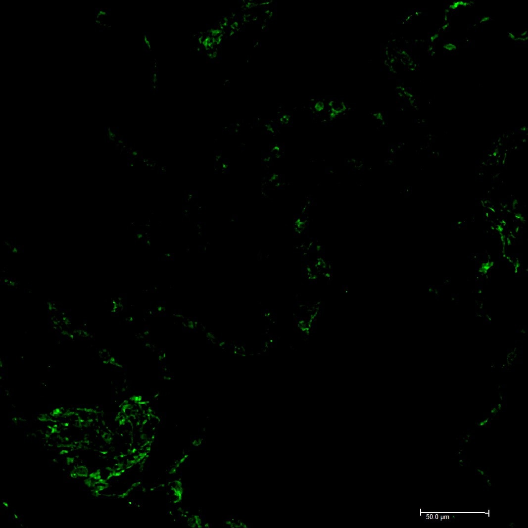
IF staining of IPAH patient CD31/PECAM-1 lung slice. Image from a verified customer review.Applications for CD31/PECAM-1 Antibody (JC/70A) - BSA Free
Application
Recommended Usage
Flow (Cell Surface)
1:100-1:250
Flow Cytometry
1:100-1:250
Immunohistochemistry
1:10-1:25
Immunohistochemistry-Paraffin
1:10-1:25
Western Blot
1:100-1:500
Application Notes
This CD31/PECAM1 Antibody (JC/70A) is useful for IHC on paraffin-embedded sections, Flow cytometry and Western blot. It reacts with an ~100 kDa glycoprotein expressed by endothelial cells and ~130 kDa glycoprotein present in platelets. It stains endothelial cells in normal as well as malignant tissues. Prior to immunostaining paraffin tissues, antigen retrieval with sodium citrate buffer (pH 6.0) is recommended. Use in ELISA reported in scientific literature (PMID: 8307606).
Reviewed Applications
Read 5 reviews rated 4.4 using NB600-562 in the following applications:
Flow Cytometry Panel Builder
Bio-Techne Knows Flow Cytometry
Save time and reduce costly mistakes by quickly finding compatible reagents using the Panel Builder Tool.
Advanced Features
- Spectra Viewer - Custom analysis of spectra from multiple fluorochromes
- Spillover Popups - Visualize the spectra of individual fluorochromes
- Antigen Density Selector - Match fluorochrome brightness with antigen density

Formulation, Preparation, and Storage
Purification
Protein G purified
Formulation
PBS
Format
BSA Free
Preservative
0.02% Sodium Azide
Concentration
1.0 mg/ml
Shipping
The product is shipped with polar packs. Upon receipt, store it immediately at the temperature recommended below.
Stability & Storage
Store at 4C short term. Aliquot and store at -20C long term. Avoid freeze-thaw cycles. After opening store under sterile conditions.
Background: CD31/PECAM-1
PECAM's intracellular cytoplasmic domain consists of a sequence of 118 amino acids and contains serine and tyrosine (also referred to as immunoreceptor tyrosine-based inhibitory motifs-ITIMs) residues, which may be phosphorylated upon cellular stimulation (3). ITIMs are phosphorylated by Src-family kinases and non-Src family kinases (e.g., Csk), leading to a conformational change which supports interactions with Src homology 2 (SH2) domain containing proteins such as protein-tyrosine phosphatase, SHP-2 (1,2). Formation of SHP-2/PECAM-1 complexes induces endothelial cell migration through the dephosphorylation of focal adhesion kinase and regulation of RhoA activity (1). Signaling downstream of ITIM tyrosine phosphorylations also plays a role in PECAM's anti-apoptotic activity, a function which is independent of its interaction with SHP-2. In platelets and leukocytes, phosphorylation of PECAM's cytosolic domain is inhibitory, preventing their activation.
References
1. Lertkiatmongkol, P., Liao, D., Mei, H., Hu, Y., & Newman, P. J. (2016). Endothelial functions of PECAM-1 (CD31). Current Opinion in Hematology. https://doi.org/10.1097/MOH.0000000000000239.Endothelial
2. Privratsky, J. R., & Newman, P. J. (2014). PECAM-1: Regulator of endothelial junctional integrity. Cell and Tissue Research. https://doi.org/10.1007/s00441-013-1779-3
3. Newman, P. J., & Newman, D. K. (2003). Signal transduction pathways mediated by PECAM-1: New roles for an old molecule in platelet and vascular cell biology. Arteriosclerosis, Thrombosis, and Vascular Biology. https://doi.org/10.1161/01.ATV.0000071347.69358.D9
Long Name
Platelet Endothelial Cell Adhesion Molecule 1
Alternate Names
CD31, EndoCAM, PECA1, PECAM-1, PECAM1, JC70A CD31, JC70A Clone
Entrez Gene IDs
5175 (Human)
Gene Symbol
PECAM1
UniProt
Additional CD31/PECAM-1 Products
Product Documents for CD31/PECAM-1 Antibody (JC/70A) - BSA Free
Certificate of Analysis
To download a Certificate of Analysis, please enter a lot or batch number in the search box below.
Product Specific Notices for CD31/PECAM-1 Antibody (JC/70A) - BSA Free
This product is for research use only and is not approved for use in humans or in clinical diagnosis. Primary Antibodies are guaranteed for 1 year from date of receipt.
Related Research Areas
Citations for CD31/PECAM-1 Antibody (JC/70A) - BSA Free
 Powered by Bioz
Customer Reviews for CD31/PECAM-1 Antibody (JC/70A) - BSA Free (5)
4.4 out of 5
5 Customer Ratings
Have you used CD31/PECAM-1 Antibody (JC/70A) - BSA Free?
Submit a review and receive an Amazon gift card!
$25/€18/£15/$25CAN/¥2500 Yen for a review with an image
$10/€7/£6/$10CAN/¥1110 Yen for a review without an image
Submit a review
Customer Images

-(05-ml)_NB600-562_7056.PNG)
Showing
1
-
5 of
5 reviews
Showing All
Filter By:
-
Application: ImmunofluorescenceSample Tested: Adult lungSpecies: HumanVerified Customer | Posted 09/10/2025cd31/pecam1 IF staining in IPAH lung sliceIF staining of IPAH patient cd31/pecam1 lung slice

-
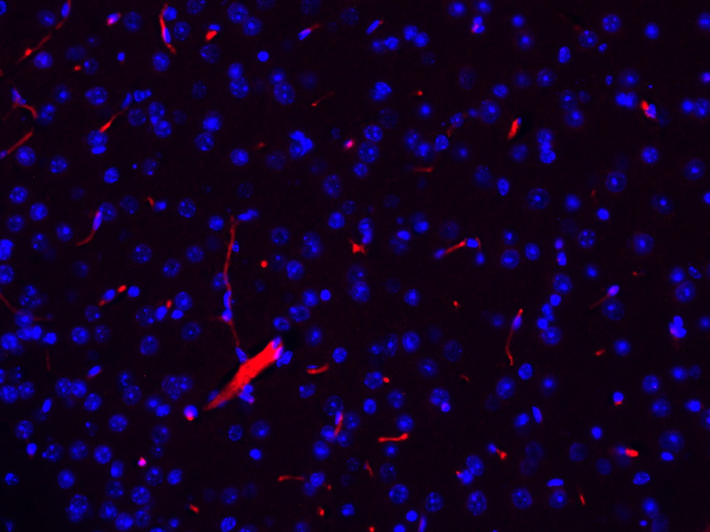
Application: Immunohistochemistry-FrozenSample Tested: Mouse brainSpecies: MouseVerified Customer | Posted 05/09/2019Mouse brain cryo sections were stained with anti-Pecam (1:50) using anti goat Alexa-Fluor 555 for detection. Magnification 20x. Although staining is a bit faint (dilution 1:50!), antibody seems to be specific.
-
Application: ImmunocytochemistrySample Tested: EndothelialSpecies: FelineVerified Customer | Posted 05/26/2017NB600-562 antibody dilution: 1:25 (Red), DAPI (Blue)
-
Application: Immunohistochemistry-ParaffinSample Tested: non-ischemic and ischemic gastrocnemius muscleSpecies: MouseVerified Customer | Posted 08/12/2016Representations of Capillary density and neovascularization of non-ischemic and ischemic muscle sections
-
Application: Immunohistochemistry-FrozenSample Tested: Rabbit HeartSpecies: OtherVerified Customer | Posted 04/28/2014Rabbit Heart, CD31 Stained Red.
There are no reviews that match your criteria.
Protocols
Find general support by application which include: protocols, troubleshooting, illustrated assays, videos and webinars.
- 7-Amino Actinomycin D (7-AAD) Cell Viability Flow Cytometry Protocol
- Antigen Retrieval Protocol (PIER)
- Antigen Retrieval for Frozen Sections Protocol
- Appropriate Fixation of IHC/ICC Samples
- Cellular Response to Hypoxia Protocols
- Chromogenic IHC Staining of Formalin-Fixed Paraffin-Embedded (FFPE) Tissue Protocol
- Chromogenic Immunohistochemistry Staining of Frozen Tissue
- ClariTSA™ Fluorophore Kits
- Detection & Visualization of Antibody Binding
- ELISA Sample Preparation & Collection Guide
- ELISA Troubleshooting Guide
- Extracellular Membrane Flow Cytometry Protocol
- Flow Cytometry Protocol for Cell Surface Markers
- Flow Cytometry Protocol for Staining Membrane Associated Proteins
- Flow Cytometry Staining Protocols
- Flow Cytometry Troubleshooting Guide
- Fluorescent IHC Staining of Frozen Tissue Protocol
- Graphic Protocol for Heat-induced Epitope Retrieval
- Graphic Protocol for the Preparation and Fluorescent IHC Staining of Frozen Tissue Sections
- Graphic Protocol for the Preparation and Fluorescent IHC Staining of Paraffin-embedded Tissue Sections
- Graphic Protocol for the Preparation of Gelatin-coated Slides for Histological Tissue Sections
- How to Run an R&D Systems DuoSet ELISA
- How to Run an R&D Systems Quantikine ELISA
- How to Run an R&D Systems Quantikine™ QuicKit™ ELISA
- ICC Cell Smear Protocol for Suspension Cells
- ICC Immunocytochemistry Protocol Videos
- ICC for Adherent Cells
- IHC Sample Preparation (Frozen sections vs Paraffin)
- ISH-IHC Protocol for Chromogenic Detection on Formalin Fixed Paraffin Embedded (FFPE) Tissue
- Immunocytochemistry (ICC) Protocol
- Immunocytochemistry Troubleshooting
- Immunofluorescence of Organoids Embedded in Cultrex Basement Membrane Extract
- Immunofluorescent IHC Staining of Formalin-Fixed Paraffin-Embedded (FFPE) Tissue Protocol
- Immunohistochemistry (IHC) and Immunocytochemistry (ICC) Protocols
- Immunohistochemistry Frozen Troubleshooting
- Immunohistochemistry Paraffin Troubleshooting
- Intracellular Flow Cytometry Protocol Using Alcohol (Methanol)
- Intracellular Flow Cytometry Protocol Using Detergents
- Intracellular Nuclear Staining Flow Cytometry Protocol Using Detergents
- Intracellular Staining Flow Cytometry Protocol Using Alcohol Permeabilization
- Intracellular Staining Flow Cytometry Protocol Using Detergents to Permeabilize Cells
- Preparing Samples for IHC/ICC Experiments
- Preventing Non-Specific Staining (Non-Specific Binding)
- Primary Antibody Selection & Optimization
- Propidium Iodide Cell Viability Flow Cytometry Protocol
- Protocol for Heat-Induced Epitope Retrieval (HIER)
- Protocol for Liperfluo
- Protocol for Making a 4% Formaldehyde Solution in PBS
- Protocol for VisUCyte™ HRP Polymer Detection Reagent
- Protocol for the Characterization of Human Th22 Cells
- Protocol for the Characterization of Human Th9 Cells
- Protocol for the Fluorescent ICC Staining of Cell Smears - Graphic
- Protocol for the Fluorescent ICC Staining of Cultured Cells on Coverslips - Graphic
- Protocol for the Preparation & Fixation of Cells on Coverslips
- Protocol for the Preparation and Chromogenic IHC Staining of Frozen Tissue Sections
- Protocol for the Preparation and Chromogenic IHC Staining of Frozen Tissue Sections - Graphic
- Protocol for the Preparation and Chromogenic IHC Staining of Paraffin-embedded Tissue Sections
- Protocol for the Preparation and Chromogenic IHC Staining of Paraffin-embedded Tissue Sections - Graphic
- Protocol for the Preparation and Fluorescent ICC Staining of Cells on Coverslips
- Protocol for the Preparation and Fluorescent ICC Staining of Non-adherent Cells
- Protocol for the Preparation and Fluorescent ICC Staining of Stem Cells on Coverslips
- Protocol for the Preparation and Fluorescent IHC Staining of Frozen Tissue Sections
- Protocol for the Preparation and Fluorescent IHC Staining of Paraffin-embedded Tissue Sections
- Protocol for the Preparation of Gelatin-coated Slides for Histological Tissue Sections
- Protocol for the Preparation of a Cell Smear for Non-adherent Cell ICC - Graphic
- Protocol: Annexin V and PI Staining by Flow Cytometry
- Protocol: Annexin V and PI Staining for Apoptosis by Flow Cytometry
- Quantikine HS ELISA Kit Assay Principle, Alkaline Phosphatase
- Quantikine HS ELISA Kit Principle, Streptavidin-HRP Polymer
- R&D Systems Quality Control Western Blot Protocol
- Sandwich ELISA (Colorimetric) – Biotin/Streptavidin Detection Protocol
- Sandwich ELISA (Colorimetric) – Direct Detection Protocol
- TUNEL and Active Caspase-3 Detection by IHC/ICC Protocol
- The Importance of IHC/ICC Controls
- Troubleshooting Guide: ELISA
- Troubleshooting Guide: Fluorokine Flow Cytometry Kits
- Troubleshooting Guide: Immunohistochemistry
- Troubleshooting Guide: Western Blot Figures
- Western Blot Conditions
- Western Blot Protocol
- Western Blot Protocol for Cell Lysates
- Western Blot Troubleshooting
- Western Blot Troubleshooting Guide
- View all Protocols, Troubleshooting, Illustrated assays and Webinars
FAQs for CD31/PECAM-1 Antibody (JC/70A) - BSA Free
Showing
1
-
3 of
3 FAQs
Showing All
-
Q: I have an inquiry about #NB600-562, Anti-CD31/PECAM1 (JC/70A), and wanted to know the recommended IHC-P antigen retrieval protocol?
A: Please see our protocols for antigen retrieval. There are different methods of antigen retrieval as you will see; our lab typically recommends the heat induced citrate buffer pH 6.0 antigen retrieval method as the most commonly used in our lab.
-
Q: What's the possibility of using this antibody for IHC with frozen sections?
A: Although this product has only been confirmed for use in IHC-P, it most likely will work with frozen sections as well.
-
Q: Would this antibody react with samples from Rabbit or not? Because only Hu is listed as its Species, but it is also mentioned that Cross-reacts with Human and Rabbit.
A: Yes, this antibody should react with rabbit and will be covered as such in our 100% guarantee.
-
Q: I have an inquiry about #NB600-562, Anti-CD31/PECAM1 (JC/70A), and wanted to know the recommended IHC-P antigen retrieval protocol?
A: Please see our protocols for antigen retrieval. There are different methods of antigen retrieval as you will see; our lab typically recommends the heat induced citrate buffer pH 6.0 antigen retrieval method as the most commonly used in our lab.
-
Q: What's the possibility of using this antibody for IHC with frozen sections?
A: Although this product has only been confirmed for use in IHC-P, it most likely will work with frozen sections as well.
-
Q: Would this antibody react with samples from Rabbit or not? Because only Hu is listed as its Species, but it is also mentioned that Cross-reacts with Human and Rabbit.
A: Yes, this antibody should react with rabbit and will be covered as such in our 100% guarantee.
Loading...