![Western Blot: CD38 Antibody (6E12D)BSA Free [NBP1-47462]](https://resources.rndsystems.com/images/products/CD38-Antibody-6E12D-Western-Blot-NBP1-47462-img0007.jpg "Western Blot: CD38 Antibody (6E12D)BSA Free [NBP1-47462]")
Loading...
Key Product Details
Species Reactivity
Validated:
Human
Cited:
Human
Applications
Validated:
Immunohistochemistry, Immunohistochemistry-Paraffin, Western Blot, ELISA, Immunocytochemistry/ Immunofluorescence
Cited:
Western Blot, Immunocytochemistry/ Immunofluorescence
Label
Unconjugated
Antibody Source
Monoclonal Mouse IgG1 Clone # 6E12D
Loading...
Product Specifications
Immunogen
Purified recombinant fragment of human CD38 expressed in E. Coli.
Clonality
Monoclonal
Host
Mouse
Isotype
IgG1
Theoretical MW
34.3 kDa.
Disclaimer note: The observed molecular weight of the protein may vary from the listed predicted molecular weight due to post translational modifications, post translation cleavages, relative charges, and other experimental factors.
Disclaimer note: The observed molecular weight of the protein may vary from the listed predicted molecular weight due to post translational modifications, post translation cleavages, relative charges, and other experimental factors.
Scientific Data Images for CD38 Antibody (6E12D)
Western Blot: CD38 Antibody (6E12D)BSA Free [NBP1-47462]
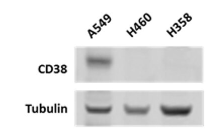
Western Blot: CD38 Antibody (6E12D) [NBP1-47462] - Detection of CD38 in three non-small cell lung cancer cell lines. Western blot image submitted by a verified customer review.![Immunohistochemistry-Paraffin: CD38 Antibody (6E12D) - BSA Free [NBP1-47462]](https://resources.rndsystems.com/images/products/CD38-Antibody-6E12D-Immunohistochemistry-Paraffin-NBP1-47462-img0004.jpg "Immunohistochemistry-Paraffin: CD38 Antibody (6E12D) - BSA Free [NBP1-47462]")
Immunohistochemistry-Paraffin: CD38 Antibody (6E12D) - BSA Free [NBP1-47462]
Immunohistochemistry-Paraffin: CD38 Antibody (6E12D) [NBP1-47462] - Analysis of human lung cancer (A), lymphonodus tissue (B),showing cytomembrane localization using CD38 mouse mAb with DAB staining.![Western Blot: CD38 Antibody (6E12D)BSA Free [NBP1-47462]](https://resources.rndsystems.com/images/products/CD38-Antibody-6E12D-Western-Blot-NBP1-47462-img0006.jpg "Western Blot: CD38 Antibody (6E12D)BSA Free [NBP1-47462]")
Western Blot: CD38 Antibody (6E12D)BSA Free [NBP1-47462]
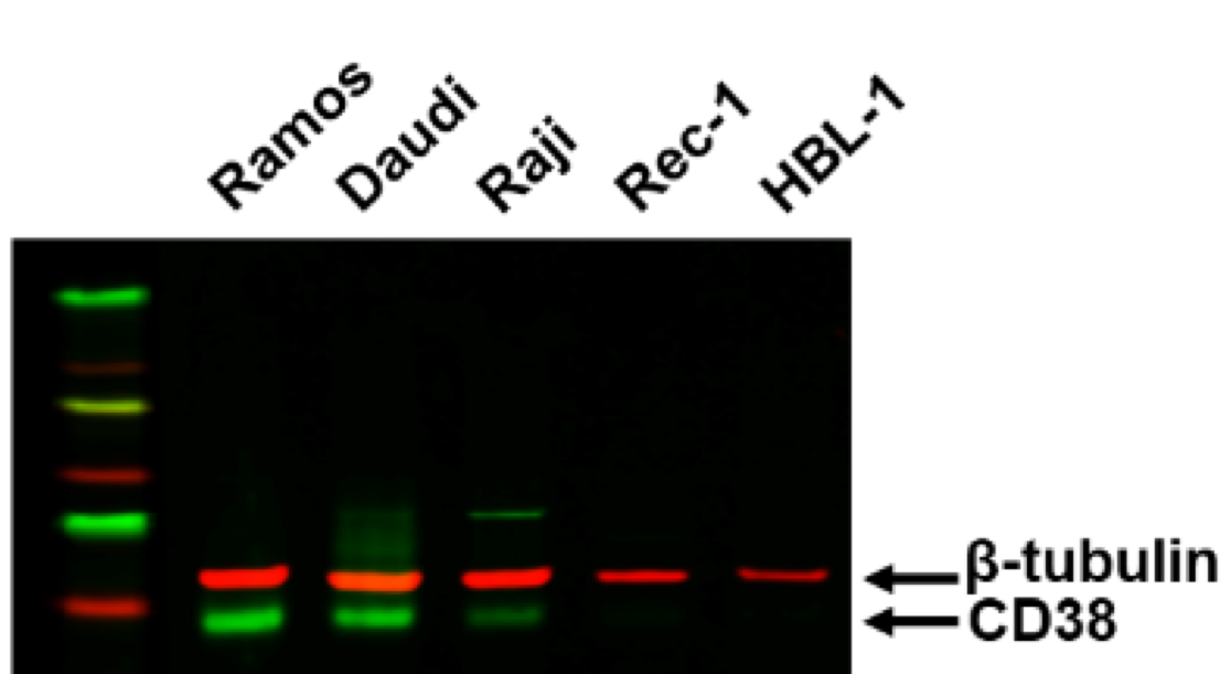
Western Blot: CD38 Antibody (6E12D) [NBP1-47462] - CD38 expression in 5 B cell lymphoma cell lines. Western blot image submitted by a verified customer review.![Western Blot: CD38 Antibody (6E12D)BSA Free [NBP1-47462]](https://resources.rndsystems.com/images/products/CD38-Antibody-6E12D-Western-Blot-NBP1-47462-img0005.jpg "Western Blot: CD38 Antibody (6E12D)BSA Free [NBP1-47462]")
Western Blot: CD38 Antibody (6E12D)BSA Free [NBP1-47462]
Western Blot: CD38 Antibody (6E12D) [NBP1-47462] - Analysis using CD38 mouse mouse mAb against CD38-hIgGFc transfected HEK293 cell lysate.Applications for CD38 Antibody (6E12D)
Application
Recommended Usage
ELISA
1:10000
Immunohistochemistry
1:200 - 1:1000
Immunohistochemistry-Paraffin
1:200 - 1:1000
Western Blot
1:500-1:2000
Application Notes
Use in ICC/IF reported in scientific literature (PMID: 28573863).
Reviewed Applications
Read 2 reviews rated 4.5 using NBP1-47462 in the following applications:
Formulation, Preparation, and Storage
Purification
Ascites
Formulation
Ascites
Preservative
0.03% Sodium Azide
Concentration
This product is unpurified. The exact concentration of antibody is not quantifiable.
Shipping
The product is shipped with polar packs. Upon receipt, store it immediately at the temperature recommended below.
Stability & Storage
Store at 4C short term. Aliquot and store at -20C long term. Avoid freeze-thaw cycles.
Background: CD38
As described above, CD38 is highly expressed in plasma cells and, as a result, is a target for treating multiple myeloma (MM), the cancer of white blood cells (4,6). The anti-CD38 monoclonal antibody daratumumab is one specific treatment for MM (4,6). Daratumumab has been shown to target MM cells through antibody-dependent cellular cytotoxicity and antibody dependent cellular phagocytosis (4). Additionally, CD38 has a potential role in neurodegenerative disorders and neuroinflammation as elucidated CD38's high expression in neurons, astrocytes, and microglia along with its enzymatic role in NAD degradation (3). Reduced NAD levels is a consequence of aging and occurs during neurodegeneration (3). Furthermore, murine studies have found that CD38 deletion inhibits neuroinflammation and neurodegeneration and therefore might be a potential therapeutic target (3). Similarly, CD38 inhibitors, like quercetin and luteolin, are used to treat age-related diseases and metabolic disorders (7).
References
1. Malavasi, F., Funaro, A., Alessio, M., DeMonte, L. B., Ausiello, C. M., Dianzani, U., Lanza, F., Magrini, E., Momo, M., & Roggero, S. (1992). CD38: a multi-lineage cell activation molecule with a split personality. International journal of clinical & laboratory research. https://doi.org/10.1007/BF02591400
2. Malavasi, F., Deaglio, S., Funaro, A., Ferrero, E., Horenstein, A. L., Ortolan, E., Vaisitti, T., & Aydin, S. (2008). Evolution and function of the ADP ribosyl cyclase/CD38 gene family in physiology and pathology. Physiological reviews. https://doi.org/10.1152/physrev.00035.2007
3. Guerreiro, S., Privat, A. L., Bressac, L., & Toulorge, D. (2020). CD38 in Neurodegeneration and Neuroinflammation. Cells. https://doi.org/10.3390/cells9020471
4. van de Donk, N., Richardson, P. G., & Malavasi, F. (2018). CD38 antibodies in multiple myeloma: back to the future. Blood. https://doi.org/10.1182/blood-2017-06-740944
5. Lund, F. E., Cockayne, D. A., Randall, T. D., Solvason, N., Schuber, F., & Howard, M. C. (1998). CD38: a new paradigm in lymphocyte activation and signal transduction. Immunological reviews. https://doi.org/10.1111/j.1600-065x.1998.tb01573.x
6. Glaria, E., & Valledor, A. F. (2020). Roles of CD38 in the Immune Response to Infection. Cells. https://doi.org/10.3390/cells9010228
7. Rajman, L., Chwalek, K., & Sinclair, D. A. (2018). Therapeutic Potential of NAD-Boosting Molecules: The In Vivo Evidence. Cell metabolism. https://doi.org/10.1016/j.cmet.2018.02.011
Long Name
Cluster of Differentiation 38
Alternate Names
ADP-ribosyl Cyclase, CD38, Cyclic ADP-ribose Hydrolase
Entrez Gene IDs
952 (Human)
Gene Symbol
CD38
UniProt
Additional CD38 Products
Product Documents for CD38 Antibody (6E12D)
Certificate of Analysis
To download a Certificate of Analysis, please enter a lot or batch number in the search box below.
Product Specific Notices for CD38 Antibody (6E12D)
This product is for research use only and is not approved for use in humans or in clinical diagnosis. Primary Antibodies are guaranteed for 1 year from date of receipt.
Citations for CD38 Antibody (6E12D)
 Powered by Bioz
Customer Reviews for CD38 Antibody (6E12D) (2)
4.5 out of 5
2 Customer Ratings
Have you used CD38 Antibody (6E12D)?
Submit a review and receive an Amazon gift card!
$25/€18/£15/$25CAN/¥2500 Yen for a review with an image
$10/€7/£6/$10CAN/¥1110 Yen for a review without an image
Submit a review
Customer Images
Showing
1
-
2 of
2 reviews
Showing All
Filter By:
-
Application: Western BlotSample Tested: B cell lymphomaSpecies: HumanVerified Customer | Posted 08/27/2017CD38 expression in 5 B cell lymphoma cell lines.

-
Application: Western BlotSample Tested: Human lung cancer cell linesSpecies: HumanVerified Customer | Posted 03/20/2017Detection of CD38 in three non-small cell lung cancer cell lines.
There are no reviews that match your criteria.
Protocols
Find general support by application which include: protocols, troubleshooting, illustrated assays, videos and webinars.
- Antigen Retrieval Protocol (PIER)
- Antigen Retrieval for Frozen Sections Protocol
- Appropriate Fixation of IHC/ICC Samples
- Cellular Response to Hypoxia Protocols
- Chromogenic IHC Staining of Formalin-Fixed Paraffin-Embedded (FFPE) Tissue Protocol
- Chromogenic Immunohistochemistry Staining of Frozen Tissue
- ClariTSA™ Fluorophore Kits
- Detection & Visualization of Antibody Binding
- ELISA Sample Preparation & Collection Guide
- ELISA Troubleshooting Guide
- Fluorescent IHC Staining of Frozen Tissue Protocol
- Graphic Protocol for Heat-induced Epitope Retrieval
- Graphic Protocol for the Preparation and Fluorescent IHC Staining of Frozen Tissue Sections
- Graphic Protocol for the Preparation and Fluorescent IHC Staining of Paraffin-embedded Tissue Sections
- Graphic Protocol for the Preparation of Gelatin-coated Slides for Histological Tissue Sections
- How to Run an R&D Systems DuoSet ELISA
- How to Run an R&D Systems Quantikine ELISA
- How to Run an R&D Systems Quantikine™ QuicKit™ ELISA
- ICC Cell Smear Protocol for Suspension Cells
- ICC Immunocytochemistry Protocol Videos
- ICC for Adherent Cells
- IHC Sample Preparation (Frozen sections vs Paraffin)
- Immunocytochemistry (ICC) Protocol
- Immunocytochemistry Troubleshooting
- Immunofluorescence of Organoids Embedded in Cultrex Basement Membrane Extract
- Immunofluorescent IHC Staining of Formalin-Fixed Paraffin-Embedded (FFPE) Tissue Protocol
- Immunohistochemistry (IHC) and Immunocytochemistry (ICC) Protocols
- Immunohistochemistry Frozen Troubleshooting
- Immunohistochemistry Paraffin Troubleshooting
- Preparing Samples for IHC/ICC Experiments
- Preventing Non-Specific Staining (Non-Specific Binding)
- Primary Antibody Selection & Optimization
- Protocol for Heat-Induced Epitope Retrieval (HIER)
- Protocol for Making a 4% Formaldehyde Solution in PBS
- Protocol for VisUCyte™ HRP Polymer Detection Reagent
- Protocol for the Fluorescent ICC Staining of Cell Smears - Graphic
- Protocol for the Fluorescent ICC Staining of Cultured Cells on Coverslips - Graphic
- Protocol for the Preparation & Fixation of Cells on Coverslips
- Protocol for the Preparation and Chromogenic IHC Staining of Frozen Tissue Sections
- Protocol for the Preparation and Chromogenic IHC Staining of Frozen Tissue Sections - Graphic
- Protocol for the Preparation and Chromogenic IHC Staining of Paraffin-embedded Tissue Sections
- Protocol for the Preparation and Chromogenic IHC Staining of Paraffin-embedded Tissue Sections - Graphic
- Protocol for the Preparation and Fluorescent ICC Staining of Cells on Coverslips
- Protocol for the Preparation and Fluorescent ICC Staining of Non-adherent Cells
- Protocol for the Preparation and Fluorescent ICC Staining of Stem Cells on Coverslips
- Protocol for the Preparation and Fluorescent IHC Staining of Frozen Tissue Sections
- Protocol for the Preparation and Fluorescent IHC Staining of Paraffin-embedded Tissue Sections
- Protocol for the Preparation of Gelatin-coated Slides for Histological Tissue Sections
- Protocol for the Preparation of a Cell Smear for Non-adherent Cell ICC - Graphic
- Quantikine HS ELISA Kit Assay Principle, Alkaline Phosphatase
- Quantikine HS ELISA Kit Principle, Streptavidin-HRP Polymer
- R&D Systems Quality Control Western Blot Protocol
- Sandwich ELISA (Colorimetric) – Biotin/Streptavidin Detection Protocol
- Sandwich ELISA (Colorimetric) – Direct Detection Protocol
- TUNEL and Active Caspase-3 Detection by IHC/ICC Protocol
- The Importance of IHC/ICC Controls
- Troubleshooting Guide: ELISA
- Troubleshooting Guide: Immunohistochemistry
- Troubleshooting Guide: Western Blot Figures
- Western Blot Conditions
- Western Blot Protocol
- Western Blot Protocol for Cell Lysates
- Western Blot Troubleshooting
- Western Blot Troubleshooting Guide
- View all Protocols, Troubleshooting, Illustrated assays and Webinars
FAQs for CD38 Antibody (6E12D)
Showing
1
-
1 of
1 FAQ
Showing All
-
Q: Which is your best antibody anti-human CD38 for IHC-P?
A:
We do have a number of CD38 antibodies validated in IHC-P, please use the filters on the left side of the search page to help find the product that mostly suits to your experimental design.
Loading...
Associated Pathways

